Abstract
Background
Reconstruction of the interosseous membrane (IOM) may play a role in the treatment of acute and chronic longitudinal forearm instability. Several reconstruction techniques have been proposed. Suture-button reconstruction is attractive because it obviates donor site morbidity and is relatively easy to perform. How this method compares to its alternatives, however, is unknown.
Materials and Methods
We review literature describing reconstruction of the forearm axis. We describe how we perform suture-button reconstruction of the IOM, summarize our previously published biomechanical data on the subject, and offer a case report.
Description of Technique
A suture-button is implanted so as to approximate the course of the interosseous ligament. This may be accomplished percutaneously, or when grafting is desired, through an open approach.
Results
Data informing the choice of one reconstruction technique over another consist mostly of biomechanical studies and a small number of case reports.
Conclusions
Suture-button reconstruction of the IOM may encourage anatomic healing of acute forearm axis injuries especially as an adjunct to radial head replacement or repair. Chronic injuries may benefit from a combination suture-button graft construct and ulnar shortening osteotomy.
Keywords: Suture-button, interosseous membrane, interosseous ligament, forearm, instability
Axial loads at the hand are borne mostly by the distal radius. This force is transferred in part to the ulna by the interosseous membrane (IOM) so that longitudinal forces are more evenly distributed between the forearm bones at the elbow. Despite this sharing of forces, the radial head remains a primary stabilizer of the forearm axis.1
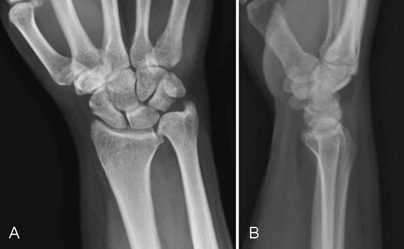
Injuries to the forearm axis are likely to involve fractures of the radial head or neck that are readily identified on radiographs. Although common, injuries to the IOM, as in the Essex–Lopresti injury, are more difficult to diagnose and are frequently missed. If the radial head is resected and an injury to the IOM is neglected, proximal translation of the radius and painful ulnar impaction at the wrist may occur (Fig. 1).2 3 Even if the radial head is replaced in the setting of a compromised IOM, increased radiocapitellar pressures may predispose to radiocarpal athrosis.4 5 As such, reconstruction of the IOM may play a role in the management of acute injuries to the forearm axis. In chronic cases, IOM reconstruction may supplement radial head replacement or may be the only feasible, motion-sparing, salvage procedure.
Fig. 1.

Interosseous membrane rupture has led to distal migration of the ulna and ulnocarpal impaction, (A) posteroanterior view; (B) lateral view.
Creation of a one-bone forearm restores longitudinal stability to the forearm but sacrifices all forearm rotation.1 Tendon autograft, bone-patellar tendon-bone autograft, and suture button-based procedures have been described for motion-sparing reconstruction of the IOM. English literature aiding in the choice of one technique over the other is limited to a series of patients treated with bone-patellar tendon-bone graft,6 a report of clinical success one year after suture-button reconstruction of the IOM,7 two biomechanical investigations of suture-button reconstruction8 9 and several biomechanical studies of graft-based techniques.4 5 10 11 12 13 14 15 16
Suture-button reconstruction is of particular interest because it obviates donor site morbidity and requires minimal dissection, making it relatively easy to perform. How suture button-based reconstruction compares to other motion-sparing reconstructive techniques is unknown. We therefore undertook a literature review of motion-sparing IOM reconstruction techniques. We additionally present our surgical technique for suture-button reconstruction and present a patient treated with this method at our institution.
Methods
We performed a literature review using the PubMed database. Manuscripts not published in English were excluded.
Results
Bone-Patellar Tendon-Bone Autograft
Marcotte and Osterman treated 16 patients with chronic longitudinal forearm instability with bone-patellar tendon-bone autograft and ulnar shortening osteotomy (USO). Most patients in this series presented within 2 years of radial head excision. A variety of concomitant procedures were performed including removal of radial head prostheses and wrist arthroscopy. Fifteen of 16 patients had improved wrist pain, grip strength improved, and no patients required additional surgery. Non or delayed healing of the ulna osteotomy was observed in 2 patients, and 4 patients complained of donor site pain.6
Jones et al performed a biomechanical investigation of bone-patellar tendon-bone onlay reconstruction of the IOM. This technique reduced radioulnar translation of sectioned specimens by half. When combined with radial head replacement, stability was similar to that of intact specimens.10
Tendon-Only Allograft
No clinical data exist describing tendon-only reconstruction of the IOM.
Skahen et al showed, in a cadaver model after radial head excision and sectioning of the interosseous ligament (IOL), that reconstruction with a single flexor carpi radialis (FCR) tendon graft improved, but did not normalize longitudinal force transfer in the forearm.13 Pfaeffle and colleagues, in two studies in which the radial head was left intact, noted that reconstruction of the IOL with two FCR tendon grafts completely restored physiologic transfer of longitudinal forces in the forearm.4 11 Achilles tendon and palmaris longus grafts have also been studied as tendon-only options for IOM reconstruction. A hierarchy emerges: bone-patellar tendon-bone is likely the stiffest of the graft materials, followed by single FCR, Achilles tendon, and finally palmaris longus.14 16
Suture-Button Reconstruction
Published clinical evidence informing suture-button reconstruction of the IOL is limited to the report of a single patient so treated for an Essex–Lopresti injury. The radial head was not replaced in this case. The authors report good clinical and radiographic results at 1 year. An ultrasound examination revealed “a continuous IOM with a scar”7 In an as-yet unpublished case series, Gaspar et al describe a small group of heterogeneous patients who improved subjectively after reconstructive procedures involving suture-button constructs.17
Kam et al tested the effect of isolated radial head replacement and radial head replacement plus reconstruction of the IOL with a suture-button construct. These authors found that reconstruction of the IOL increased forearm stability, but not to the native state. Stability added by the suture-button was similar to that achieved with radial head replacement. Kam et al also investigated force transmission of the distal ulna in several scenarios. Isolated radial head replacement, isolated suture-button reconstruction, and combined radial head replacement and suture-button reconstruction all reduced distal ulnar force transmission to approximately that of the uninjured state. The authors noted that forearm musculature often herniated through the sectioned IOM in their specimens implying a barrier to healing of the IOM.9
Previously, we performed a biomechanical study of forearm stability in seven cadaver forearms by excising the radial head, sectioning the interosseous membrane, and then reconstructing the IOL with a percutaneously inserted suture-button construct. Four conditions were tested: 1) intact forearm, 2) radial head excised/IOM intact, 3) radial head excised, IOM sectioned, and 4) radial head excised, IOM sectioned, and suture-button implanted and tensioned.8 The suture-button restored stability equal to that of the forearm with the radial head excised but not to the stability of the forearm in its intact state. No specimens lost forearm rotation, and no injuries to neurovascular structures were noted. We suggest a role for this technique in acute IOM disruption. In this scenario, the suture-button might splint the forearm and allow the IOM to heal anatomically. We expressed concern, as did Kam et al, that if healing of the IOM does not occur after splinting, the suture-button construct would impart initial stability and then break.3 8 9
Surgical Technique
The patient is positioned supine with the arm on a hand table. The ulna is felt and a point is marked at the junction of the bone's middle and distal third. A line is drawn, on either surface of the forearm, from this point proximally toward a point on the radius, at approximately three-fifths of its length from the radial styloid. (Fig. 2). This line approximates the course of the IOL, the stoutest portion of the IOM.13 18 19
Fig. 2.

The position of the graft (approximating the position of the IOL) is depicted between a point two-third the length of the ulna from the ulnar styloid and three-fifth the length of the radius from the radial styloid.
At this point, our technique varies depending on the acuity of the injury being treated. We believe that an acutely torn IOM may heal anatomically if the longitudinal forearm axis is stabilized. Chronic IOM injuries are less likely to heal and may be best treated with both stabilization and grafting.
Acute Injuries
A 4 cm longitudinal skin incision is made at the mark on the radius. Superficial sensory nerve branches are identified and retracted, and blunt dissection of muscle is used to expose the radius, taking care not to endanger the posterior interosseous nerve or sensory branches of the radial nerve. The ulna is likewise exposed where marked. With the forearm in pronation, two cortices of the radius and ulna are drilled obliquely using the 2.7 mm drill bit included with the device, along the line describing the IOL. These holes may be made individually or with a single pass from the radius to the ulna. The TightRope (Arthrex, Naples, FL) device is introduced though the hole in the radius and into the hole in the ulna using the device's aiming guide and fluoroscopic control. It is then tensioned and fixed as reduction of the forearm axis is assessed fluoroscopically. The incisions are sutured, and a soft dressing is applied. Active range of motion exercises are begun at 4 weeks, passive range of motion exercise at 6 weeks, and strengthening at 8 weeks.7 8
Chronic Injuries
An USO is performed first. From the ulnar incision, dorsal tissues are elevated off of the ulna, the interosseous membrane, and then the radius in a distal-to-proximal direction. A second incision is made along the radius where previously marked. The radius is exposed while protecting the posterior interosseous nerve and sensory branches of the radial nerve. In this way, a tunnel is created along the axis of the IOL on top of the interosseous membrane. The suture-button device may be placed as in acute injuries, although in this setting, with the aid of direct visualization. Graft material (Graftfacket, KCI, San Antonio) is thawed, hydrated, pretensioned, and drawn through the tunnel before being fixed to either the periosteum of the forearm bones or the bones themselves (via bone tunnels or suture anchors) with stout nonabsorbable suture. Closure and postoperative care are similar to that for acute injuries.
Case Report
This patient was involved in a motorcycle accident as a teenager and had been treated with right radial head resection. He developed right elbow arthritis and underwent elbow capsulectomy, radiocapitellar arthroplasty, and lateral complex reconstruction at the age of 47.
In the following 5 years, the patient developed recurrent elbow pain and stiffness. His radial head implant dislocated posteriorly, and he developed arthrosis of his proximal radioulnar joint. At age 49, the patient underwent explantation of his arthroplasty components, debridement of heterotopic bone from the proximal radioulnar joint, interposition arthroplasty of the proximal radioulnar joint, and reconstruction of the lateral collateral ligament.
Most recently, this now 52-year-old contractor complained of progressive ulnar-sided wrist pain and limited wrist and forearm range of motion refractory to bracing and anti-inflammatory medication. He did not have elbow pain. The patient had a prominent, tender distal ulna and pain with ulnar deviation of the wrist. The DRUJ was stable. X-rays taken at this time showed 8 mm of positive ulnar variance and ulnocarpal abutment. (Fig. 1)
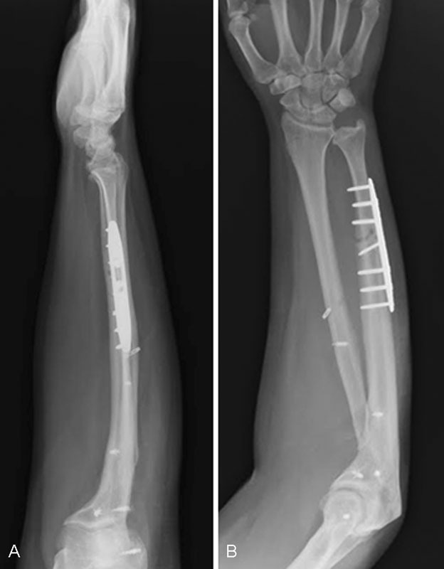
We performed a forearm reconstruction consisting of an 11 mm USO, IOM reconstruction with a suture-button construct, and grafting (Fig. 2).
At his first postoperative visit, the patient was capable of 40 degrees of pronation and supination (Fig. 3). He was placed in a Munster-type splint. Three weeks later he was transitioned to a volar splint. Approximately 10 weeks after surgery, the patient was free to return to light activities. At his most recent follow-up, approximately 4 months from surgery, the patient had no pain, improved cosmesis at the wrist, and 10 degrees of forearm pronation and supination.
Fig. 3.

Lateral (A) and posteroanterior (B) postoperative radiographs demonstrating an ulnar shortening osteotomy and suture button reconstruction of the interosseous membrane are shown. Suture anchors were placed around the elbow in a previous procedure.
Discussion
The IOM plays an important, if secondary, role in longitudinal forearm stability. Intuitively, reconstruction of the IOM may be useful acutely in the management of Essex–Lopresti injuries, especially as an adjunct to radial head fixation or replacement. IOM reconstruction may be appropriate for the same purpose in chronic injuries, or may be used in isolation as the only feasible salvage technique. Chronic IOM tears may be less likely to heal spontaneously,20 and we chose to augment suture-button reconstruction of these injuries with graft material.
Published clinical experience with the variety of described forearm reconstruction techniques is scarce. Marcotte and Osterman have provided the only relevant case series. Although their study may recommend use of bone-patellar tendon-bone reconstruction in general for patients with chronic forearm instability, their results are confounded by the diversity of injuries treated and by the variety of concomitant procedures performed. Donor site pain is a concern.6
A single case report7 of successful suture-button reconstruction of the IOM exists, and we have added our own experience with this technique to the literature.
Remaining evidence is in the form of biomechanical studies that adhere to no universal protocol and are therefore difficult to compare. Further confusing an appraisal of various reconstruction techniques is the fact that, rather than directly measuring inter-articular pressures, previous investigators have gathered different proxy data, for example, radioulnar translation,4 8 9 10 11 13 14 forces within the forearm bones,4 5 9 10 11 15 and/or forces within the IOM13 14 to make conclusions about pressures at the wrist or elbow. The ultimate measure of any forearm reconstruction is the effect of the technique on intra-articular pressures, particularly at the wrist, as painful ulnocarpal impaction is most often the impetus for surgery. We recently used thin film load cells (6900 Quad Sensor, TekScan, Boston) to minimize dissection of forearm specimens and to directly record intra-articular pressures after reconstruction of the IOM. We recommend this technique to other investigators in the interest of collecting important data and facilitating comparison.
Suture-button reconstruction of the IOM is an attractive alternative to autologous grafting because it obviates donor site morbidity and is relatively easy to perform. It appears to have certain limitations, however. We have shown previously that suture-button reconstruction restores stability to that of a forearm with an intact IOM and absent radial head.8 This finding was at odds with the work of Kam et al who reported that a suture-button construct could completely restore stability without a radial head.9 Our in vivo experience with this technique is limited, but does corroborate the clinical success of other authors.7
Especially until better, more readily synthesized data are available, we speculate, as other authors have,4 5 that suture-button reconstruction of the IOM is likely most appropriate as an adjunct to radial head replacement or repair in the acute setting. We acknowledge additionally, that because the suture-button is inert, it may fatigue and break in time.8 If the suture-button does only offer temporary stability, it is nonetheless likely to provide a longer period of splinting than a percutaneous Kirschner wire, which is often placed across the distal radioulnar joint for this purpose. A hybrid construct consisting of a suture-button and biologic graft material, as illustrated in our case example, might provide both immediate internal splinting and long-term stability. We have limited our use of this combination construct, to date, however, to chronic injuries.
Footnotes
Conflict of Interest None.
References
- 1.Means K J, Graham T. London: Elsevier; 2011. Disorders of the forearm axis. [Google Scholar]
- 2.Sowa D T, Hotchkiss R N, Weiland A J. Symptomatic proximal translation of the radius following radial head resection. Clin Orthop Relat Res. 1995;(317):106–113. [PubMed] [Google Scholar]
- 3.McGinley J C, Kozin S H. Interosseous membrane anatomy and functional mechanics. Clin Orthop Relat Res. 2001;(383):108–122. doi: 10.1097/00003086-200102000-00013. [DOI] [PubMed] [Google Scholar]
- 4.Pfaeffle H J, Stabile K J, Li Z M, Tomaino M M. Reconstruction of the interosseous ligament unloads metallic radial head arthroplasty and the distal ulna in cadavers. J Hand Surg Am. 2006;31(2):269–278. doi: 10.1016/j.jhsa.2005.09.022. [DOI] [PubMed] [Google Scholar]
- 5.Tomaino M M, Pfaeffle J, Stabile K, Li Z M. Reconstruction of the interosseous ligament of the forearm reduces load on the radial head in cadavers. J Hand Surg [Br] 2003;28(3):267–270. doi: 10.1016/s0266-7681(03)00012-3. [DOI] [PubMed] [Google Scholar]
- 6.Marcotte A L Osterman A L Longitudinal radioulnar dissociation: identification and treatment of acute and chronic injuries Hand Clin 2007232195–208., vi vi [DOI] [PubMed] [Google Scholar]
- 7.Brin Y S, Palmanovich E, Bivas A. et al. Treating acute Essex-Lopresti injury with the TightRope device: a case study. Tech Hand Up Extrem Surg. 2014;18(1):51–55. doi: 10.1097/BTH.0000000000000036. [DOI] [PubMed] [Google Scholar]
- 8.Drake M L, Farber G L, White K L, Parks B G, Segalman K A. Restoration of longitudinal forearm stability using a suture button construct. J Hand Surg Am. 2010;35(12):1981–1985. doi: 10.1016/j.jhsa.2010.09.009. [DOI] [PubMed] [Google Scholar]
- 9.Kam C C, Jones C M, Fennema J L, Latta L L, Ouellette E A, Evans P J. Suture-button construct for interosseous ligament reconstruction in longitudinal radioulnar dissociations: a biomechanical study. J Hand Surg Am. 2010;35(10):1626–1632. doi: 10.1016/j.jhsa.2010.07.020. [DOI] [PubMed] [Google Scholar]
- 10.Jones C M, Kam C C, Ouellette E A, Milne E L, Kaimrajh D, Latta L L. Comparison of 2 forearm reconstructions for longitudinal radioulnar dissociation: a cadaver study. J Hand Surg Am. 2012;37(4):741–747. doi: 10.1016/j.jhsa.2012.01.025. [DOI] [PubMed] [Google Scholar]
- 11.Pfaeffle H J, Stabile K J, Li Z M, Tomaino M M. Reconstruction of the interosseous ligament restores normal forearm compressive load transfer in cadavers. J Hand Surg Am. 2005;30(2):319–325. doi: 10.1016/j.jhsa.2004.10.005. [DOI] [PubMed] [Google Scholar]
- 12.Sellman D C, Seitz W H Jr, Postak P D, Greenwald A S. Reconstructive strategies for radioulnar dissociation: a biomechanical study. J Orthop Trauma. 1995;9(6):516–522. doi: 10.1097/00005131-199509060-00010. [DOI] [PubMed] [Google Scholar]
- 13.Skahen J R III, Palmer A K, Werner F W, Fortino M D. Reconstruction of the interosseous membrane of the forearm in cadavers. J Hand Surg Am. 1997;22(6):986–994. doi: 10.1016/S0363-5023(97)80037-8. [DOI] [PubMed] [Google Scholar]
- 14.Stabile K J, Pfaeffle J, Saris I, Li Z M, Tomaino M M. Structural properties of reconstruction constructs for the interosseous ligament of the forearm. J Hand Surg Am. 2005;30(2):312–318. doi: 10.1016/j.jhsa.2004.11.018. [DOI] [PubMed] [Google Scholar]
- 15.Tejwani S G, Markolf K L, Benhaim P. Graft reconstruction of the interosseous membrane in conjunction with metallic radial head replacement: a cadaveric study. J Hand Surg Am. 2005;30(2):335–342. doi: 10.1016/j.jhsa.2004.07.022. [DOI] [PubMed] [Google Scholar]
- 16.Tejwani S G, Markolf K L, Benhaim P. Reconstruction of the interosseous membrane of the forearm with a graft substitute: a cadaveric study. J Hand Surg Am. 2005;30(2):326–334. doi: 10.1016/j.jhsa.2004.05.017. [DOI] [PubMed] [Google Scholar]
- 17.Gaspar M Osterman A Culp R Short-to intermediate term outcomes following interosseous membrane reconstruction using Tightrope suture-button suspensionplasty system. American Association of Hand Surgery http://meeting.handsurgery.org/files/2016/ePoster-Abstracts.pdf. Accessed May 10, 2016
- 18.Chandler J W, Stabile K J, Pfaeffle H J, Li Z M, Woo S L, Tomaino M M. Anatomic parameters for planning of interosseous ligament reconstruction using computer-assisted techniques. J Hand Surg Am. 2003;28(1):111–116. doi: 10.1053/jhsu.2003.50033. [DOI] [PubMed] [Google Scholar]
- 19.Skahen J R III, Palmer A K, Werner F W, Fortino M D. The interosseous membrane of the forearm: anatomy and function. J Hand Surg Am. 1997;22(6):981–985. doi: 10.1016/S0363-5023(97)80036-6. [DOI] [PubMed] [Google Scholar]
- 20.Hotchkiss R N An K N Sowa D T Basta S Weiland A J An anatomic and mechanical study of the interosseous membrane of the forearm: pathomechanics of proximal migration of the radius J Hand Surg Am 198914(2 Pt 1):256–261. [DOI] [PubMed] [Google Scholar]


