

Abstract
Introduction
Due to a higher risk for implant loosening, particularly of the distal component, patients with physically demanding lifestyles are infrequently considered for total wrist arthroplasty (TWA). A distal radius hemiarthroplasty may obviate the need for the strict restrictions recommended for patients treated by TWA, thus providing another surgical option for active patients with severe wrist arthritis, especially those with articular degeneration of the lunate facet of the radius, capitate head, or combination of both, who are not typically candidates for traditional motion-preserving procedures.
Materials and Methods
Eight fresh-frozen cadaver limbs (age range, 43–82 years) with no history of rheumatoid arthritis or upper extremity trauma were used. Radiodense markers were inserted in the radius and hand. Posteroanterior (PA) fluoroscopic images with the wrist in neutral, radial deviation, and ulnar deviation, and lateral images with the wrist in neutral, flexion, and extension were obtained for each specimen before implantation, after distal radius hemiarthroplasty, and after combined hemiarthroplasty and PRC.
Results
On the PA images, the capitate remained within 1.42 and 2.21 mm of its native radial-ulnar position following hemiarthroplasty and hemiarthroplasty with PRC, respectively. Lateral images showed the capitate remained within 1.06 mm of its native dorsal-volar position following hemiarthroplasty and within 4.69 mm following hemiarthroplasty with PRC. Following hemiarthroplasty, capitate alignment changed 2.33 and 2.59 mm compared with its native longitudinal alignment on PA and lateral films, respectively. These changes did not reach statistical significance. As expected, significant shortening in longitudinal alignment was seen on both PA and lateral films for hemiarthroplasty with PRC.
Conclusion
A distal radius implant hemiarthroplasty with or without a PRC provides good static alignment of the wrist in a cadaver model and thus supports the concept as potential treatment alternatives for advanced wrist arthritis; however, combined hemiarthroplasty with a PRC has more clinical relevance because it avoids the risk of proximal carpal row instability and eliminates the commonly arthritic radioscaphoid joint.
Keywords: wrist arthroplasty, wrist hemiarthroplasty, wrist replacement, distal radius arthroplasty, distal radius replacement
Although total wrist arthroplasty has been used in selective patients with severe arthritis, patients with physically demanding lifestyles are infrequently considered to be candidates due primarily to the high risk for implant loosening, particularly of the distal component. A distal radius hemiarthroplasty may obviate the need for the strict restrictions required of patients treated by total wrist arthroplasty and thus provide another motion-preserving surgical option for active patients with severe wrist arthritis, especially those with distal radius articular degeneration from osteoarthritis or those with arthritis and posttraumatic deformity.
The purpose of this laboratory study was to evaluate the radiographic alignment of the wrist following distal radius hemiarthroplasty and after hemiarthroplasty combined with proximal row carpectomy (PRC) using the Universal 2 radius component (Integra Life Sciences, Plainsboro, NJ).
Materials and Methods
Institutional review board approval was obtained prior to beginning the study. Eight fresh-frozen cadaveric specimens from eight different donors that were amputated through the level of the proximal forearm were used. Five were left and three were right. Six were female and two were male. None had a history of rheumatoid arthritis or upper extremity trauma, which was confirmed by radiographs. The average age of the specimen was 71 years (range, 43–82 years). The same testing protocol was used for each specimen.
Implantation Technique
An Esmarch bandage was applied circumferentially to the proximal forearm to help maintain soft tissue tension in the flexor and extensor muscles. A dorsal longitudinal incision was made over the wrist in line with the third metacarpal. The skin and subcutaneous tissues were elevated from the extensor retinaculum. Three radiographic markers were inserted on specific bone sites described below.
A radially based extensor retinaculum flap and a distally based wrist capsular flap were raised over the dorsal wrist to expose the radiocarpal joint. The distal radioulnar joint was not opened and the triangular fibrocartilage complex was not violated. A radial component of the Universal 2 implant was inserted using the standard technique described for this system. In this technique, the medullary canal of the radius is entered through the distal radius articular surface with an awl. The opening is widened with a countersink tool and the guide rod is inserted; its central position within the canal is confirmed fluoroscopically. The cutting guide is mounted on the rod and a small oscillating saw is used to trim the distal radius into a generally flat surface by resecting its dorsal rim; however, the depths of the lunate and scaphoid fossa remain. The broach is applied over the guide rod to prepare the metaphysis of the distal radius. A press-fit insertion of the implant was used. When using this system for total wrist arthroplasty, the implant size for the radial component is chosen based on the fit of the carpal component using radiographic parameters whereby the carpal component stem would lie in the central portion of the capitate and the ulnar screw would enter the proximal pole of the hamate. Although this same method for determining implant sizing was used to make an initial estimate of implant size, if there was a choice between two sizes the larger component was chosen to ensure the capitate was well seated. The capsule, retinaculum, and skin were closed securely with multiple sutures. The fluoroscopic images described below were obtained.
The sutures were cut and the wrist joint reopened. The capsular flap was extended distally just enough to perform a PRC using both sharp dissection and piecemeal excision of the carpal bones. The capsule, retinaculum, and skin were closed securely again and repeat fluoroscopic evaluation performed.
Data Collection
Radiographic markers (size 8 birdshot, 0.0055 mm in diameter) were placed in the base of the third metacarpal, in the capitate, and in the distal radius to efficiently track changes in position of the capitate with respect to the radius. These sites were chosen for specific reasons: the base of the third metacarpal is a fixed point in the hand and its relationship to the capitate is unchanged during wrist motion because the third carpometacarpal joint is not mobile. The capitate is near the idealized center of rotation of the normal wrist and remains unchanged with respect to the third metacarpal after PRC.1 2
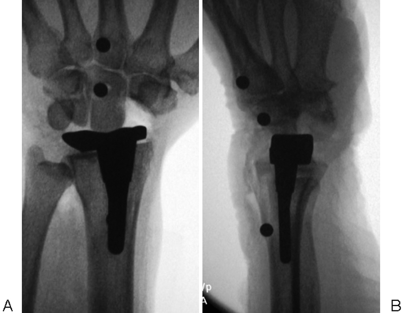
To ensure that proper images were obtained, fluoroscopic imaging was used. The specific wrist positions were obtained by manually aligning the hand with a goniometer and repeating the image until the proper view was seen. The same six images were obtained before component implantation, after implantation (Fig. 1A,B), and after the addition of a PRC (Fig. 2A,B): (1) posteroanterior (PA) with the wrist in neutral position, (2) PA with the wrist in radial deviation, (3) PA with the wrist in ulnar deviation, (4) lateral with the wrist in neutral position, (5) lateral with the wrist in ∼45 degrees of wrist flexion, and (6) in ∼45 degrees of wrist extension.
Fig. 1.

(A, B) Postoperative posteroanterior and lateral views of specimen following distal radius hemiarthroplasty.
Fig. 2.
(A, B) Posteroanterior and lateral views of specimen after undergoing hemiarthroplasty and PRC.
The fluoroscopic images were imported into AutoCAD 2002 (Autodesk, Inc, San Rafael, CA) for analysis. A pixilated two-dimensional Cartesian coordinate system was constructed on each image by rotating each image until the third metacarpal marker and the capitate marker were aligned. A straight line was drawn through the center of these two markers to serve as the y-axis. A line perpendicular to this axis and passing through the center of the capitate marker was then drawn and served as the x-axis (Figs. 3 and 4). Each marker was assigned x and y coordinates, which were converted to millimeters using a radiolucent ruler included on all images.
Fig. 3.

Posteroanterior view showing coordinate system used to perform measurements.
Fig. 4.

Lateral view showing coordinate system used to perform measurements.
Using these coordinates, three distances were measured on each image: (1) third metacarpal marker to capitate marker, (2) x-axis component of capitate marker to distal radius marker, and (3) y-axis component of capitate marker to distal radius marker. The distance from the y-axis component of the capitate marker to the distal radius marker was used to measure changes in longitudinal position between images, while the x-axis component was used to measure changes in radial-ulnar position on the PA images and dorsal-volar changes on lateral images. The changes in these three distances were measured in all six fluoroscopic views and compared between the native and hemiarthroplasty wrists and between the native and hemiarthroplasty with PRC wrists. The distance from the third metacarpal marker to capitate marker was used to assess projection error between images, with the initial PA and initial lateral images used as the baselines for subsequent measurements.
Statistical Analysis
Paired t-tests were used to calculate the probability of the absolute value of the mean differences in these three measured distances being greater than zero by chance alone. All statistical analysis was completed using SAS v 9.1 (SAS Institute, Cary, NC). Statistical significance was set at a p-value < 0.05.
Results
Average change in capitate position and statistical results are summarized in Tables 1 2 3. On the PA images, changes in radial-ulnar alignment of the capitate between the native and hemiarthroplasty wrists ranged from 0.78 to 1.42 mm. Radial-ulnar alignment differences ranged from 0.21 to 2.21 mm in the hemiarthroplasty with PRC when compared with native. No changes in radial-ulnar alignment reached statistical significance for either procedure. Changes in longitudinal alignment seen on PA images between the capitate and the radius ranged from 1.94 to 2.33 mm in the hemiarthroplasty wrist when compared with the native. This change did not reach statistical significance. As would be expected due to the PRC, significant changes (range, 5.72–6.10 mm) were seen in longitudinal alignment following hemiarthroplasty with PRC (p < 0.01 in all positions).
Table 1. Mean capitate position as measured in posteroanterior fluoroscopic views.
| Hemi | Hemi + PRC | ||||
|---|---|---|---|---|---|
| Position | Axis | Change from native | p-Value | Change from native | p-Value |
| Neutral | x | −0.78 | 0.52 | 0.21 | 0.88 |
| y | 1.94 | 0.80 | −5.72 | <0.01 | |
| Radial deviation | x | −1.42 | 0.35 | 0.22 | 0.87 |
| y | 2.12 | 0.39 | −6.10 | <0.01 | |
| Ulnar deviation | x | −1.42 | 0.34 | 2.21 | 0.14 |
| y | 2.33 | 0.10 | 5.90 | <0.01 | |
Abbreviations: Hemi, hemiarthroplasty; PRC, proximal row carpectomy.
Table 2. Mean capitate position as measured in lateral fluoroscopic views.
| Hemi | Hemi + PRC | ||||
|---|---|---|---|---|---|
| Position | Axis | Change from native | p-Value | Change from native | p-Value |
| Neutral | x | 0.37 | 0.66 | −0.64 | 0.32 |
| y | 2.26 | 0.13 | −4.26 | <0.01 | |
| Flexion | x | −1.06 | 0.49 | −4.69 | 0.13 |
| y | 2.57 | 0.17 | −5.37 | <0.01 | |
| Extension | x | 0.84 | 0.34 | 0.21 | 0.81 |
| y | 2.59 | 0.70 | −5.43 | <0.01 | |
Abbreviations: Hemi, hemiarthroplasty; PRC, proximal row carpectomy.
Table 3. Mean distance between capitate and metacarpal.
| Radial deviation | Ulnar deviation | |||
|---|---|---|---|---|
| Group | Change from neutral | p-Value | Change from neutral | p-Value |
| Native | 0.164 (0.01–0.94) | 0.37 | 0.305 (0.0–1.43) | 0.14 |
| Hemi | 0.104 (0.15–0.84) | 0.56 | 0.233 (0.11–1.13) | 0.33 |
| Hemi + PRC | 0.184 (0.04–0.93) | 0.42 | 0.116 (0.04–1.11) | 0.42 |
Abbreviations: Hemi, hemiarthroplasty; PRC, proximal row carpectomy.
Note: Data are presented as mean (range). N = 8 for each group.
On lateral images, changes in volar-dorsal alignment of the capitate ranged from 0.37 to 1.06 mm and 0.21 to 4.69 mm for the hemiarthroplasty and hemiarthroplasty with PRC, respectively, when compared with native; these changes did not reach statistical significance (p > 0.05 in all positions). Longitudinal changes in capitate alignment on lateral films ranged from 2.26 to 2.59 mm in the hemiarthroplasty group when compared with native; this change did not reach statistical significance. As expected, longitudinal alignment changes were significant for hemiarthroplasty and PRC on lateral films when compared with native (range, 4.36–5.43 mm).
Projection error (see Table 3) averaged 0.16 mm (range, 0.01–0.94) for radial deviation and 0.31 mm (range, 0.0–1.43) for ulnar deviation in the native wrist. Change in this distance for the hemiarthroplasty wrist was 0.10 mm (range, 0.15–0.84) and 0.23 mm (range, 0.11–1.13) for radial and ulnar deviation, respectively. Average change was 0.18 mm (range, 0.04–0.93) for radial deviation and 0.12 mm (range, 0.04–1.11) for ulnar deviation in the hemiarthroplasty wrist with PRC.
Although joint stability was not quantitatively assessed, none of the specimens were unstable after the procedures during manipulation, including after combined hemiarthroplasty and PRC.
Discussion
The objective of wrist arthroplasty is to provide functional wrist motion while relieving pain and correcting deformity. Patients with generalized arthritis who have had a fusion of one wrist and total wrist arthroplasty on the other side prefer the side with the arthroplasty,3 4 primarily because basic activities of daily living such as perineal care, fastening buttons, combing hair, and writing are made easier if some wrist motion is preserved.5 6 Similarly, active individuals often desire to maintain wrist motion to better perform tasks and activities that are important to their lifestyle. However, patients with posttraumatic or degenerative arthritis who typically lead physically demanding lifestyles are infrequently considered for total wrist replacement due to the limitations of activities recommended following total wrist arthroplasty. In these individuals, there is believed to be a higher risk for implant loosening, particularly of the distal component. A distal radius hemiarthroplasty may obviate the need for strict physical restrictions and allow implant arthroplasty to be considered in select patients.
Several criteria need to be satisfied before distal radius implant hemiarthroplasty is used clinically. One structural criterion is to provide reproducible, proper alignment of the wrist. The static measurements found in this study demonstrated good wrist alignment in both the coronal and sagittal planes in all wrist positions following hemiarthroplasty alone and after combined hemiarthroplasty and PRC. However, because many patients with advanced wrist arthritis also have malalignment of the proximal carpal row, including a PRC increases the potential application of an implant hemiarthroplasty.
Other structural criteria that are important to consider include joint stability and motion. Joint stability is influenced by the shape of the implant's articular surface, integrity of the joint capsule, preexisting deformity of the joint, and muscle control. Although joint stability was not quantitatively assessed, none of the specimens were unstable throughout the range of motion tested, including compensating for the laxity created by the PRC. The shape of this implant's articular surface conforms well to the head of the capitate and its volar and dorsal rims are prominent. This implant does not necessarily reflect the performance of other radial implant designs. Furthermore, all of the specimens were free of advanced arthritis and deformity, and thus the influence of possible anatomical differences associated with wrist arthritis that could affect stability and motion was not assessed.
Erosion of the carpus due to prolonged contact against the cobalt chrome surface is likely, but the rate of erosion will be difficult to predict. Erosion will depend on several factors including the initial condition of the capitate surface, bone density, and activity of the patient. Pain relief will also likely vary among patients due to the variation in response of the articular surface against cobalt chrome.
Despite the potential disadvantages of hemiarthroplasty, it could be an alternative for patients with advanced wrist arthritis who would normally only be indicated for total wrist fusion, especially those with articular degeneration of the lunate facet of the radius, capitate head, or combination of both, who are not typically candidates for traditional motion-preserving procedures. Combined distal radius hemiarthroplasty and PRC has more clinical relevance than hemiarthroplasty without PRC because it avoids the risk of proximal carpal row instability and eliminates the arthritic radioscaphoid joint present in most patients with advanced wrist arthritis.
Footnotes
Conflict of Interest Brian D. Adams serves as a consultant and receives royalties from Integra Life Sciences, Plainsboro, NJ.
References
- 1.Kobayashi M, Berger R A, Nagy L. et al. Normal kinematics of carpal bones: a three-dimensional analysis of carpal bone motion relative to the radius. J Biomech. 1997;30(8):787–793. doi: 10.1016/s0021-9290(97)00026-2. [DOI] [PubMed] [Google Scholar]
- 2.Blankenhorn B D, Pfaeffle H J, Tang P, Robertson D, Imbriglia J, Goitz R J. Carpal kinematics after proximal row carpectomy. J Hand Surg Am. 2007;32(1):37–46. doi: 10.1016/j.jhsa.2006.10.014. [DOI] [PubMed] [Google Scholar]
- 3.Vicar A J, Burton R I. Surgical management of the rheumatoid wrist—fusion or arthroplasty. J Hand Surg Am. 1986;11(6):790–797. doi: 10.1016/s0363-5023(86)80224-6. [DOI] [PubMed] [Google Scholar]
- 4.Murray P M. Current status of wrist arthrodesis and wrist arthroplasty. Clin Plast Surg. 1996;23(3):385–394. [PubMed] [Google Scholar]
- 5.Kobus R J, Turner R H. Wrist arthrodesis for treatment of rheumatoid arthritis. J Hand Surg Am. 1990;15(4):541–546. doi: 10.1016/s0363-5023(09)90012-0. [DOI] [PubMed] [Google Scholar]
- 6.Weiss A C, Wiedeman G Jr, Quenzer D, Hanington K R, Hastings H II, Strickland J W. Upper extremity function after wrist arthrodesis. J Hand Surg Am. 1995;20(5):813–817. doi: 10.1016/s0363-5023(05)80437-x. [DOI] [PubMed] [Google Scholar]