Abstract
Recent exploitation of the avian immune system has highlighted its suitability for the generation of high-quality, high-affinity antibodies to a wide range of antigens for a number of therapeutic and biotechnological applications. The glycosylation profile of potential immunoglobulin therapeutics is species specific and is heavily influenced by the cell-line/culture conditions used for production. Hence, knowledge of the carbohydrate moieties present on immunoglobulins is essential as certain glycan structures can adversely impact their physicochemical and biological properties. This study describes the detailed N-glycan profile of IgY polyclonal antibodies from the serum of leghorn chickens using a fully quantitative high-throughput N-glycan analysis approach, based on ultra-performance liquid chromatography (UPLC) separation of released glycans. Structural assignments revealed serum IgY to contain complex bi-, tri- and tetra-antennary glycans with or without core fucose and bisects, hybrid and high mannose glycans. High sialic acid content was also observed, with the presence of rare sialic acid structures, likely polysialic acids. It is concluded that IgY is heavily decorated with complex glycans; however, no known non-human or immunogenic glycans were identified. Thus, IgY is a potentially promising candidate for immunoglobulin-based therapies for the treatment of various infectious diseases.
Introduction
Antibodies are at the forefront of the field of targeted therapeutics and diagnostics due to their natural high affinity and excellent half-life properties [1]. These molecules can be readily manipulated using standard molecular biology techniques into specialised antibodies that are tailored to perform efficiently in their chosen end-point application [2]. The biopharmaceutical industry has heavily invested in antibody-based therapeutics, which currently represents the largest and fastest growing class of biopharmaceuticals [3].
Polyclonal and recombinant antibodies are developed in many different species. However, a large number of protein targets are highly conserved in mammalian evolution and commonly used mammalian species, such as rabbits and mice, are thus inclined to render a somewhat limited immune response due to immunological tolerance invoked during foetal development [4]. The use of a species more phylogenetically distant from humans such as chickens, who diverged from mammalian genomes some 310 million years ago [5], are ideal alternatives for immunisation and selection of antibodies against highly conserved human proteins [4,6].
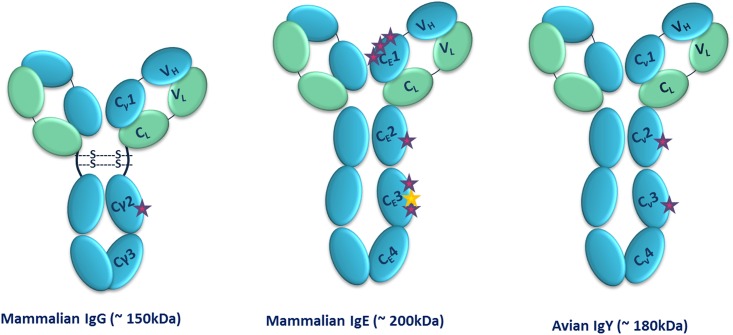
IgY is the predominant serum immunoglobulin in birds, reptiles and amphibians and is considered to be evolutionary ancestor of uniquely mammalian IgG and IgE antibodies [6]. Although IgY has characteristics and functions similar to its mammalian counterpart, IgG, with 2 heavy (67–70 kDa each) and two light (25 kDa each) chains (Fig 1), structural differences exist in the number of constant heavy domains, as IgY has an additional constant heavy domain resulting in its higher molecular mass (180 kDa). Furthermore, IgY lacks of a hinge region and has significantly reduced flexibility in comparison to IgG. This limited flexibility is derived from proline-glycine rich regions around the Cν1-Cν2 and Cν2-Cν3 domains [6]. These structural differences provide IgY with distinct biochemical properties and behaviour (Table 1).
Fig 1. Structures of Immunoglobulin’s G, E and Y and their N-glycosylation sites.
IgG (left) is composed of 2 identical heavy chains that each comprise a variable domain (VH) and three constant domains (Cγ1, Cγ2 and Cγ3) with a single carbohydrate site (purple star). In contrast, the additional constant domain in IgE (middle) is much more heavily glycosylated than IgG, with 7 N-glycosylation sites, however, one site (Asn 264) is unoccupied (yellow star) [7]. IgY is also comprised of four constant domains per heavy chain (Cv1-Cv4), with two carbohydrate sites (right). The flexible hinge region found in IgG is absent in IgE and IgY and thus may restrict their flexibility in comparison to IgG.
Table 1. Comparison of the different properties of IgG and IgY.
| IgG | IgE | IgY | |
|---|---|---|---|
| Species | Mammals | Mammals | Birds, reptiles, amphibians and lungfish |
| Molecular weight (kDa) | 150 | 200 | 180 |
| Isoelectric point (pI) | 6.4–9.0 | 5.2–5.8 | 5.7–7.6 |
| Concentration (mg/mL) | 10–12 (serum) | 10−4 | 8–10 (Serum) 15–25 (Egg Yolk) |
| Number of constant domains | 4 (3 H and 1 L) | 5 (4 H and 1 L) | 5 (4 H and 1 L) |
| Hinge | Yes | No | No |
| Complement binding | Yes | No | No |
| Rheumatoid factor binding | Yes | No | No |
| Fc receptor binding | Yes | No | No |
| Mediates anaphylaxis | No | Yes | Yes |
| Binding to protein A/G | Yes | No | No |
IgY is more heavily glycosylated than its mammalian counterpart as it contains two potential N-glycosylation sites. One is located in Cv3 domain, that is absent in the mammalian IgG, and the other is located in the Cv3 domain which corresponds to the CH2 (Cγ2) domain of mammalian IgG (Fig 1) [6,8]. Structural characterisation of N-glycans present on antibody therapeutics is a regulatory requirement as the nature of these glycans can decisively influence the therapeutic performance of an antibody [9]. The linked carbohydrate moieties of therapeutic antibodies affect both their thermal stability and physicochemical properties, along with other crucial features like receptor-binding activity, circulating half-life and immunogenicity [10]. N-glycan profiling of therapeutic antibodies with good reproducibility is vital to fulfil the needs of the both the biopharmaceutical industry and national regulatory agency requirements [11].
There is now considerable awareness of the therapeutic value of IgY antibodies with respect to a variety of pathologies including, but not limited to, pulmonary or gastrointestinal infections. For a detailed review of the current IgY therapeutic approaches in both animal studies and clinical trials in human cohorts see Spillner et al., 2012 [1].
The N-glycosylation pattern of avian IgY was previously shown to be more analogous to that in mammalian IgE than IgG, presumably reflecting the structural similarity to mammalian IgE [7]. While previous studies have elucidated the IgY N-glycan profile in detail from serum, egg yolk and various other expression vehicles [8,12,13], in this study, the chromatography technique was significantly improved through the use of hydrophilic interaction chromatography ultra-performance liquid chromatography (HILIC UPLC) which allows for shorter run times and greatly increased resolution. In addition, the bioinformatics tool, GlycoBase, was used to greatly assist the analyses. GlycoBase consists of a database with HILIC and mass spectrometry data for over 460 2-AB-labeled N-linked, 68 O-linked, and 71 free glycan structures. This reliable and robust method facilitates detailed analysis of femtomolar quantities of N-linked sugars released from glycoproteins
This study describes the detailed N-glycan profile of IgY polyclonal antibodies from the serum of Leghorn chickens using a fully quantitative high-throughput N-glycan analysis based on ultra-performance liquid chromatography (UPLC) separation of released glycans.
Materials and Methods
Ethics Statement
This study was approved by the Research Ethics Committee of Dublin City University (Approval Ref. DCUREC/2011/016) and all procedures in which animals were used were carried out in accordance with licence B100/2705 granted by the Minister for Health of Ireland under the Cruelty to Animals Act 1876 (as amended). The minimum number of animals was used, and that chicken was socially housed under environmentally controlled conditions for the duration of the study. The procedures were classified as "mild", and all procedures were carried out by highly trained, competent, personnel.
Immunoglobulin Y purification
Polyclonal IgY antibodies were purified from the serum of an adult female leghorn chicken using the Pierce® Thiophilic Adsorption Kit (Thermo Scientific, Ireland). This protocol was carried out as per manufacturer’s guidelines and the eluted protein fractions were pooled and concentrated in a 10kDa MWCO Vivaspin column (Sartorius, Ireland). Total protein concentration was measured by spectrophotometry at 280 nm (NanoDrop 1000). The purified IgY polyclonal antibody sample was subjected to sodium dodecyl sulfate-polyacrylamide gel electrophoresis (SDS-PAGE) in a 12% gel and stained with InstantBlue (MyBio, Ireland). The sample was also subjected to SDS-PAGE in a 12% gel and subsequently blotted onto a nitrocellulose membrane using the Pierce G2 Fast Blotter system (Thermo Scientific, Ireland). The membrane was blocked for 1 hour at room temperature with PBS containing 5% (w/v) skim milk. To detect the heavy and light chains of the IgY, the sample was probed with a horse radish peroxidase (HRP) labelled-donkey anti- IgY (H+L)-specific antibody (Gallus Immunotech, Canada) (1:2,000 dilution) in PBS-Tween 20 (0.05%, v/v)–skim milk (1%, w/v) for 2 hours at room temperature. Specific bands were visualised using liquid TMB as a substrate for HRP.
Glycan release
N-glycans were released from the dissolved samples (1 mg/mL) using PNGase F, (100 mU/mL; Prozyme, San Leandro, CA, USA) in accordance with methods previously described by Royle et al., 2006 and Royle et al,. 2008 [14,15]. Briefly, samples were reduced and alkylated, immobilized in SDS-gel blocks in 96 well plates and then washed. Released glycans were then fluorescently labelled with 2-aminobenzamide (2AB) by reductive amination [16]. Excess 2AB reagent was removed by ascending chromatography on Whatman 3MM paper in acetonitrile.
Ultra-Performance Liquid Chromatography (UPLC)
2AB derivatized N-glycans were separated by UPLC with fluorescence detection on a Waters Acquity UPLC H-Class instrument consisting of a binary solvent manager, sample manager and fluorescence detector under the control of Empower 3 chromatography workstation software (Waters, Milford, MA, USA). The hydrophilic interaction liquid chromatography (HILIC) separations were performed using a Waters Ethylene Bridged Hybrid (BEH) Glycan column, 150 x 2.1 mm i.d., 1.7 μm BEH particles, with 50 mM ammonium formate, pH 4.4, as solvent A and acetonitrile (MeCN) as solvent B. The 30 minute method was used with a linear gradient of 30–47% (v/v) with buffer A at 0.56 mL/minute flow rate for 23 minutes followed by 47–70% (v/v) A and finally reverting back to 30% (v/v) A to complete the run [17]. An injection volume of 10 μL sample prepared in 70% (v/v) MeCN was used throughout. Samples were maintained at 5°C prior to injection, while separation was carried out at 40°C. The fluorescence detection excitation/emission wavelengths were λexcitation = 330 and λemission = 420 nm, respectively. The system was calibrated using an external standard of hydrolysed and 2AB-labeled glucose oligomers to create a dextran ladder, as described previously [14].
Weak Anion-Exchange High-Performance Liquid Chromatography (WAX HPLC)
Weak anion exchange (WAX) HPLC to separate the N-glycans by charge was carried out as detailed in Royle et al., 2006, with a fetuin N-glycan standard as reference. WAX HPLC was performed using a Vydac 301VHP575 7.5 × 50 mm column (Anachem) on a 2695 Alliance separations module with a 2475 fluorescence detector (Waters), which was set with detection excitation/emission wavelengths of λexcitation = 330 and λemission = 420 nm, respectively [15]. Solvent A was 0.5 M formic acid adjusted to pH 9.0 with ammonia solution, and solvent B was 10% (v/v) methanol in water. Gradient conditions were as follows: a linear gradient of 0 to 5% (v/v) A over 12 minutes at a flow rate of 1 mL/minute, followed by 5–21% (v/v) A over 13 minutes and then 2–50% (v/v) A over 25 minutes, 80–100% (v/v) A over 5 minutes followed by 5 minutes at 100% A. Samples were prepared in water and a fetuin N-glycan standard was used for calibration [15].
Exoglycosidase digestions
Exoglycosidase digestions were carried out on aliquots of the total labelled N-glycan pools in accordance with methods previously described by Royle et al., 2006 [14] and according to manufacturer’s instructions. All exoglycosidase enzymes were obtained from Prozyme, San Leandro, CA, USA. The 2AB-labeled glycans were digested in a volume of 10 μL for 18 hours at 37°C in 50 mM sodium acetate buffer, pH 5.5 (except in the case of jack bean α-mannosidase (JBM) where the buffer was 100 mM sodium acetate, 2 mM Zn2+, pH 5.0), using arrays of the following enzymes: Arthrobacter ureafaciens sialidase (ABS), 0.5 U/mL; Streptococcus pneumoniae sialidase (NAN1), 1 U/mL; bovine testes β-galactosidase (BTG), 1 U/mL; Streptococcus pneumoniae β-galactosidase (SPG), 0.4 U/mL; bovine kidney α-fucosidase (BKF), 1 U/mL; β-N-Acetylglucosaminidase cloned from S. pneumonia, expressed in Escherichia coli (GUH), 8 U/mL and JBM, 60 U/mL. After incubation, enzymes were removed by filtration through a 10 kDa protein-binding EZ filters (Millipore Corporation) [15]. N-Glycans were then analysed by HILIC UPLC and WAX HPLC.
Ultra-Performance Liquid Chromatography -Fluorescence-Mass Spectrometry (UPLC-FLR-MS)
For UPLC-FLR-QTOF MS analysis lyophilised IgY samples were reconstituted in 3 μl of water and 9 μl acetonitrile. Online coupled fluorescence (FLR)-mass spectrometry detection was performed using a Waters Xevo G2 QTof—with Acquity® UPLC (Waters Corporation, Milford, MA, USA) and BEH Glycan column (1.0 x 150mm, 1.7 μm particle size).
For MS acquisition data the instrument was operated in negative-sensitivity mode with a capillary voltage of 1.80 kV. The ion source block and nitrogen desolvation gas temperatures were set at 120°C and 400°C, respectively. The desolvation gas was set to a flow rate of 600 L/h. The cone voltage was maintained at 50V. Full-scan data for glycans were acquired over m/z range of 450 to 2500. Data collection and processing were controlled by MassLynx 4.1 software (Waters Corporation, Milford, MA, USA).
The fluorescence detector settings were as follows: λexcitation: 330 nm, λemission: 420 nm; data rate was 1pts/second and a PMT gain = 10. Sample injection volume was 10 μL. The flow rate was 0.150 mL/minute and column temperature was maintained at 60°C; solvent A was 50 mM ammonium formate in water (pH 4.4) and solvent B was acetonitrile. A 40 minute linear gradient was used and was as follows: 28% (v/v) A for 1 minute, 28–43% (v/v) A for 30 minutes, 43–70% (v/v) A for 1 minute, 70% (v/v) A for 3 minutes, 70–28% (v/v) solvent A for 1 minute and finally 28% (v/v) A for 4 minutes.
Samples were diluted in 75% (v/v) acetonitrile prior to analysis. The weak wash solvent was 80% (v/v) acetonitrile and the strong wash solvent was 20% (v/v) acetonitrile. To avoid contamination of Mass Spec system, flow was sent to waste for the first 1.2 minutes and after 32 minutes.
Molecular Modelling of IgY
Molecular modelling of IgY was performed on a Silicon Graphics Fuel workstation using InsightII and Discover software (Accelrys Inc., San Diego, USA). Figures were produced using the program Pymol [18]. Protein structures used for modelling were obtained from the Protein Data Bank (PDB) database [19]. The peptide structure of chicken IgY was based on the crystal structures of human IgE domains Cε2–4[20] and human IgG Fab domain [21]. Sequence alignment and methods for generation of homology model are provided in S1 File.
Results
IgY Purification
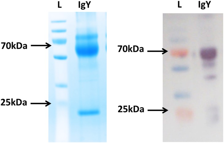
Immunoglobulin Y differs from most of the other immunoglobulins as it does not bind protein A or protein G [22]. Here, IgY was successfully recovered from the serum of chickens using thiophilic adsorption, which is based on the principles of hydrophobic interaction chromatography. Many proteins, particularly immunoglobulins will bind to an immobilised ligand that contains a sulfone group neighbouring a thioether. Addition of salts such as potassium sulphate will promote binding by encouraging the protein into close proximity of the ligand [23,24]. Total protein concentration was determined to be 11.5 mg/mL by spectrophotometry at 280 nm (NanoDrop 1000). The heavy and light chains of the purified IgY were visualized by Western Blot analysis (Fig 2).
Fig 2. IgY Purification.
IgY purified from chicken serum was resolved on 12% SDS-PAGE gels and visualised by staining with InstantBlue (left). The resolved proteins were also transferred to nitrocellulose membranes and the presence of the heavy chains at approximately 65–68 kDa and the light chains at 25 kDa can be seen after probing with an anti-IgY H+L-HRP-tagged antibody (right). L: PageRuler Plus Prestained protein ladder.
IgY N-glycan profiling
The N-glycans released from the purified IgY were analysed by WAX HPLC and HILIC UPLC in combination with exoglycosidase digestions and structural assignments using established methods [15] and the software tool GlycoBase (https://glycobase.nibrt.ie). UPLC-FLR-QTOF MS analysis was also carried out for comparative analysis. Annotation of the N-glycans present in each chromatographic peak was based upon the oligosaccharide composition as derived from the m/z value.
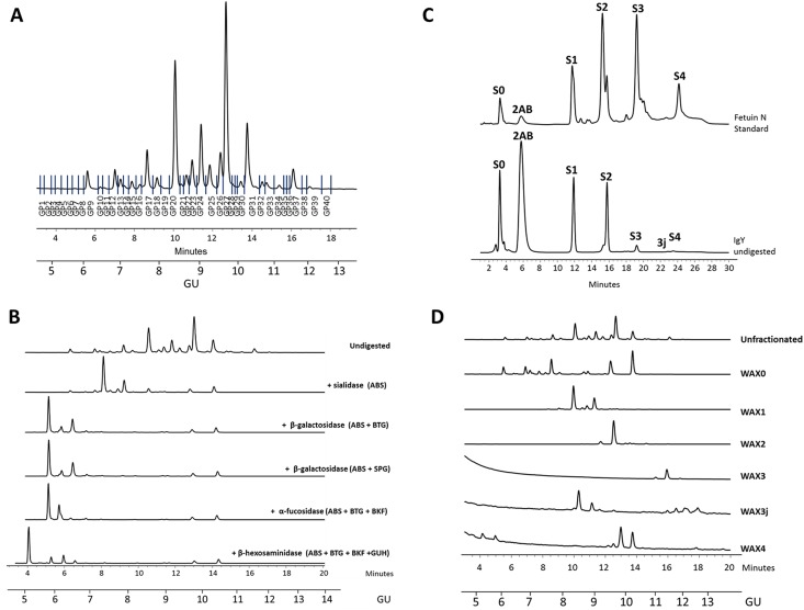
Over 80 different glycans structures were assigned to 40 peaks (each peak contains one or more glycans) (Figs 3 and 4 and S1 Table). N-glycan structures annotated include high mannose, hybrid and complex glycans with variable degrees of core fucosylation, galactosylation and sialylation. To assign the complex sialylation properly, the N-glycome was separated by WAX HPLC according to the number of sialic acids and then each WAX fraction was subjected to an array of sialidases for assignment of sialic acids linkages (S1 Table). The resulting HILIC UPLC profiles were combined with other exoglycosidase digests and indicated the presence of complex glycans, with more sialic acids than branches (Fig 4, S1 Table).
Fig 3. IgY N-glycan assignment.
(A) HILIC UPLC profile of undigested N-glycans from serum IgY. Profiles are standardised against a dextran hydrolysate (GU). The HILIC chromatogram was separated into 40 peaks. (B) Unfractionated IgY profile was subjected to exoglycosidase digestions. (C) IgY glycans were separated according number of sialic acids on WAX HPLC and (D) each WAX fraction was then subjected to HILIC UPLC (Final IgY Structural assignments are listed S1 Table).
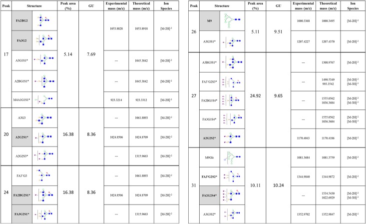
Fig 4. Summary of N-glycans identified from IgY purified form avian serum.
Summary of N-glycans released from IgY purified from serum. The HILIC chromatogram was separated into 40 peaks and structural assignment carried made using established methods (Royle et al., 2008) and the software tool GlycoBase (https://glycobase.nibrt.ie). Nomenclature used is according to Royle et al., 2008 and Harvey et al., 2009 [15,25]. Shown here are the most abundant glycans identified–glycans assigned to peaks with % area great than 5%- highlighted in grey is the most abundant glycan(s) within that particular peak. For full glycan assignment see S1 Table. *Glycan nomenclature.
*Sialic acid linkages; WAX fractions were separated out into 5 fractions; S1: Monosialylated, S2: Disialylated, S3: Trisialylated, S3J: Trisialylated and S4: Tetrasialylated. All monosialylated glycans are linked by α2–3 and α2–6; Disialylated glycans have all combinations of α2–3 and α2–6 linkages [i.e. (3, 3), (3, 6) and (6, 6)]; Trisialylated glycans have all combinations of α2–3 and α2–6 linkages except (6, 6, 6) [i.e. (3, 3, 3), (3, 3, 6) and (3, 6, 6)]; and Tetrasialylated glycans have all combinations of α2–3 and α2–6 linkages [i.e. (3,3,3,3), (3,3,3,6), (3,3,6,6) and (6,6,6,6)]. Structure abbreviations: all N-glycans have two core GlcNAcs; F at the start of the abbreviation indicates a core-fucose α1,6-linked to the inner GlcNAc; Mx, number (x) of mannose on core GlcNAcs; Ax, number of antenna (GlcNAc) on trimannosyl core; A2, biantennary with both GlcNAcs as β1,2-linked; A3, triantennary with a GlcNAc linked β1,2 to both mannose and the third GlcNAc linked β1,4 to the α1,3 linked mannose; A3’, isomer with the third GlcNAc linked β1–6 to the α1–6 linked mannose; A4, GlcNAcs linked as A3 with additional GlcNAc β1,6 linked to α1,6 mannose; B, bisecting GlcNAc linked β1,4 to β1,3 mannose; Gx, number (x) of β1,4 linked galactose on antenna; F(x), number (x) of fucose linked α1,3 to antenna GlcNAc; Sx, number (x) of sialic acids linked mostly to galactose (Structures with more sialic acids then galactoses may have some sialic acids linked to GlcNAc or another sialic acid in form of polysialic acid.).
Molecular Modelling of IgY
The precise glycans chosen for sites N390 (M9Glc/peak 31 and A2G2S1/peak 20) were based on the site specific glycan analysis of IgY [8]. The glycans chosen for sites N292 (A2G2S2/peak 27 and FA3G3/peak 24) were representatives from the other largest peaks. Glycan structures were generated using the database of glycosidic linkage conformations [26] and in vacuo energy minimisation to relieve unfavourable steric interactions. The Asn-GlcNAc linkage conformations were based on the observed range of crystallographic values [27], the torsion angles around the Asn Cα-Cβ and Cβ-Cγ bonds then being adjusted to eliminate unfavourable steric interactions between the glycans and the protein surface (Fig 5). The complete IgY sequence is provided in S1 Fig.
Fig 5. Molecular model of glycosylated IgY.
Magenta—heavy chain; pink—light chain; yellow—stick representation of cysteine residues; blue—glycans. The glycans are labelled according to the nomenclature in Fig 4.
Discussion
Chicken antibodies have several distinct biochemical advantages over mammalian antibodies and are widely utilised in the field of biotechnology. They do not activate the mammalian complement system nor interact with rheumatoid factors, or bacterial and human Fc receptors. Hence IgY antibodies make ideal regents for immunological assays as they can reduce assay interference in a mammalian serum sample, resulting in increased sensitivity as well as decreased background [28].
The use of chickens as hosts for the generation of therapeutic antibodies is becoming increasingly more prevalent with a greater understanding of the unique attributes of avian antibodies [2,29]. Polyclonal IgY represents an attractive approach to immunotherapy for the treatment of numerous diseases [1]. Notably, orally administered IgY preparations have been demonstrated as an alternative to antibiotics for the prevention of pulmonary Pseudomonas aeruginosa (PA) infections in a group of patients with cystic fibrosis [30,31]. In this study, the authors show that the IgY treated group had significantly less incidents of colonization with PA than the control group and none of the IgY-treated patients became chronically colonized with PA [30,31].
The robust, reliable methods employed in this study allow for shorter run times with increased resolution that enables identification of glycans that may not have been previously observed in other IgY glycoprofiling studies. Our structural assignments revealed serum IgY to contain mainly complex, bi-, tri- and tetraantennary glycans with or without core fucose and bisects, all with varying levels of galactosylation and sialylation, hybrid and high mannose glycans.
In investigating the site-specific N-glycosylation of IgY, Suzuki and Lee [8] noted the Fc portion of IgY possesses a N-glycosylation site which is structurally equivalent to conserved glycosylation sites of other Ig classes in mammals and is composed of predominantly high-mannose type oligosaccharides [8]. This uniquely avian glycosylation pattern at the conserved N-glycosylation site is thought to be attributed to the structural differences between IgG and IgY (IgY lacks the defined hinge region observed in IgG) [12]. The additional N-glycosylation site, located in Cv2 domain, was previously shown to contain exclusively complex-type oligosaccharides [8,12]. These distinct avian glycosylation patterns and structural differences provide IgY with unique biochemical properties and behaviour. A model of IgY with the glycans identified from this study was generated following glycan assignment (Fig 5). Characterisation of the individual glycans decorated on a protein is essential for detailed understanding of structure/function relationships and the design of potential therapeutic agents. The model generated from this study aims to enhance our understanding of the therapeutic potential of IgY. Computational modelling methods are universally accepted as central tools in the invention process for many biopharmaceuticals, facilitating drug development areas, such as optimising affinity for a target while minimising cross reactive effects, alongside optimising pharmacokinetic properties [32].
The oligosaccharide content of therapeutic immunoglobulins plays a significant role in its bioactivity and pharmacokinetic (PK) activity. Raju and colleagues (2000) examined at variations between the glycan content of IgG across several species. These authors highlighted the importance of choosing the right host in generating therapeutic IgG as the terminal sialylation of IgG is species specific [13]. In this study, LC-MS analysis of chicken IgY suggested the N-linked glycosylation of chicken IgY is considerably more heterogeneous than in human IgG. Our results are consistent with previous N-glycan studies of IgY from both serum and egg yolk [8,12,13] and also detect several previously unidentified structures.
In this study high sialic acid content was observed, with many sialic acid isomers (same composition but different sialic acid linkage arrangements resulting in a different GU from the original structure). The presence of unusual sialic acids was also noted, which is likely to be polysialic or Sialic acid linked on N-Acetylgalactosamine (GalNAc) as well as on Galactose. Sialic acids are most commonly α2–3 or α2–6 linked to galactose (Gal) or α 2–6 linked to GalNAc. However, Sialic acid can also be found linked to N-Acetylglucosamine (GlcNAc) or to another Sialic acid in α2–8 or α2–9 linkage [33]. Polysialic acids occupy internal positions within glycans, the most common being one Sialic acid residue attached to another, often at the C-8 position [34].
The high sialic acid content of IgY is very important when considering IgY as a therapeutic agent as the level of sialic acid can have a significant impact on the PK of therapeutic antibodies. A lower content of total sialic acids can significantly reduce the half-life of a drug [35]. Hence, the high sialic acid content of IgY that was observed suggests IgY-based biotherapeutics could have potentially extended circulating half-lives and are promising candidates against a variety of pathogens. High mannose glycans were also found on the IgY, which can be removed from circulation by mannose-binding receptor, therefore lowering its half-life [36]. However, the high mannose glycans on IgY are rather low in quantity in comparison to the highly sialylated complex glycans and should have no effect on the therapeutic application of IgY. Certain glycan structures have a direct impact on the immunogenicity of therapeutic proteins, that is, their presence can affect protein structure in such a way that the protein becomes immunogenic. However the glycan structure itself can also induce an immune response. The sialic acid N-Glycolylneuraminic acid (Neu5Gc) and terminal galactose-α-1,3-galactose are examples of such structures that are not naturally present in humans and are known to be immunogenic when used as therapeutics [37]. These non-human antigenic structures could promote clearance of a biopharmaceutical preparation from circulation [38–40]. The chimeric mouse–human IgG1 monoclonal antibody, Cetuximab, is an anti-human epidermal growth factor receptor (EGFR) antibody used for the treatment several cancers [38]. High incidences of hypersensitivity reactions to Cetuximab were reported and a study by Chung and colleagues showed that the majority of patients who had a hypersensitivity reaction to Cetuximab also had circulating IgE antibodies against Cetuximab before therapy was initiated. These antibodies were specific for the glycan structure galactose-α-1,- 3-galactose, which is present on the Fab portion of the Cetuximab heavy chain [38]. In order to overcome these severe hypersensitivity reactions which are observed in many immunoglobulin-based biotherapeutic agents it is of primary importance to ensure the oligosaccharide content will not elicit such reactions. Recently, a glyco-engineered anti-EGFR monoclonal antibody with a lower α-Gal content than Cetuximab was developed [41], highlighting the importance of these structures to the biopharma industry in the development of novel biotherapeutics.
In conclusion, while IgY is heavily decorated with complex glycans, no non-human immunogenic structures were identified. These results were determined using highly robust methods and are in accordance with previous IgY glycosylation studies from chicken serum [8]. The results from this study, combined with other known advantages of chicken antibodies, such as increased stability over IgG and phylogenetic distance from man [1], makes chickens ideal hosts for the generation of novel oral therapeutic interventions for the treatment of numerous infectious diseases.
Supporting Information
(TIF)
(DOCX)
(DOCX)
(XLSX)
Abbreviations
- 2AB
2-aminobenzamide
- ABS
Arthrobacter ureafaciens sialidase
- BEH
ethylenebridged hybrid
- BKF
bovine kidney α-fucosidase
- BTG
bovine testes β-galactosidase
- EGFR
epidermal growth factor receptor
- FLR
fluorescence
- GU
glucose unit
- GUH
β-N-Acetylglucosaminidase cloned from Streptococcus pneumoniae, expressed in Escherichia coli
- HILIC
hydrophilic interaction liquid chromatography
- HPLC
high-performance liquid chromatography
- HRP
horse radish peroxidase
- JBM
jack bean α-mannosidase
- MS
mass spectrometry
- MWCO
molecular weight cut-off
- NAN1
Streptococcus pneumoniae sialidase
- PA
Pseudomonas aeruginosa
- PDB
protein data bank
- PK
pharmacokinetic
- PMT
photomultiplier
- QTOF
quadrupole time of flight
- SDS-PAGE
sodium dodecyl sulphate polyacrylamide gel electrophoresis
- SPG
Streptococcus pneumoniae β-galactosidase
- TMB
3, 3', 5, 5'- tetramethylbenzidine
- UPLC
ultra-performance liquid chromatography
- WAX
weak anion exchange chromatography
Data Availability
All relevant data are within the paper and its Supporting Information files.
Funding Statement
SG, RS, RJO’K and PMR are funded by the Irish Cancer Society Programme grant PCI11WAT as part of the Prostate Cancer Research Consortium, Dublin, Ireland. RS acknowledges funding from the European Union Seventh Framework Programme (FP7/2007-2013) under grant agreement number 260600 (“GlycoHIT”) and funding from the Science foundation Ireland Starting Investigator Research grant (SFI SIRG) under grant number 13/SIRG/2164. The funders had no role in study design, data collection and analysis, decision to publish, or preparation of the manuscript.
References
- 1.Spillner E, Braren I, Greunke K, Seismann H, Blank S, du Plessis D. Avian IgY antibodies and their recombinant equivalents in research, diagnostics and therapy. Biologicals. 2012. September;40(5):313–22. 10.1016/j.biologicals.2012.05.003 [DOI] [PMC free article] [PubMed] [Google Scholar]
- 2.Conroy PJ, Law RHP, Gilgunn S, Hearty S, Caradoc-Davies TT, Lloyd G, et al. Reconciling the structural attributes of avian antibodies. J Biol Chem. 2014;289:15384–92. 10.1074/jbc.M114.562470 [DOI] [PMC free article] [PubMed] [Google Scholar]
- 3.Venetz D, Hess C, Lin C, Aebi M, Neri D. Glycosylation profiles determine extravasation and disease-targeting properties of armed antibodies. Proc Natl Acad Sci. 2015;112(7):201416694. [DOI] [PMC free article] [PubMed] [Google Scholar]
- 4.Andris-Widhopf J, Rader C, Steinberger P, Fuller R, Barbas CF. Methods for the generation of chicken monoclonal antibody fragments by phage display. J Immunol Methods. 2000. August 28;242(1–2):159–81. [DOI] [PubMed] [Google Scholar]
- 5.International Chicken Genome Sequencing Consortium. Sequence and comparative analysis of the chicken genome provide unique perspectives on vertebrate evolution. Nature. 2004. December 9;432(7018):695–716. [DOI] [PubMed] [Google Scholar]
- 6.Narat M. Production of Antibodies in Chickens. Food Technol Biotechnol. 2003;41(3):259–67. [Google Scholar]
- 7.Plomp R, Hensbergen PJ, Rombouts Y, Zauner G, Dragan I, Koeleman CAM, et al. Site-specific N-glycosylation analysis of human immunoglobulin. J Proteome Res. 2014. February 7;13(2):536–46. 10.1021/pr400714w [DOI] [PubMed] [Google Scholar]
- 8.Suzuki N, Lee YC. Site-specific N-glycosylation of chicken serum IgG. Glycobiology. 2004;14:275–92. [DOI] [PubMed] [Google Scholar]
- 9.Millán Martín S, Delporte C, Farrell A, Navas Iglesias N, McLoughlin N, Bones J. Comparative analysis of monoclonal antibody N-glycosylation using stable isotope labelling and UPLC-fluorescence-MS. Analyst. 2015;140:1442–7. 10.1039/c4an02345e [DOI] [PubMed] [Google Scholar]
- 10.Szekrényes A, Partyka J, Varadi C, Krenkova J, Foret F, Guttman A. Microchip Capillary Electrophoresis Protocols. Methods Mol Biol. 2015;1274:183–95. 10.1007/978-1-4939-2353-3_16 [DOI] [PubMed] [Google Scholar]
- 11.Hristodorov D, Fischer R, Linden L. With or without sugar? (A)glycosylation of therapeutic antibodies. Mol Biotechnol. 2013;54:1056–68. 10.1007/s12033-012-9612-x [DOI] [PubMed] [Google Scholar]
- 12.Kondo S, Yagi H, Kamiya Y, Ito A, Kuhara M, Kudoh A, et al. N-Glycosylation Profiles of Chicken Immunoglobulin Y Glycoproteins Expressed by Different Production Vehicles. J Glycomics Lipidomics 2012; S5:002. [Google Scholar]
- 13.Raju TS, Briggs JB, Borge SM, Jones AJ. Species-specific variation in glycosylation of IgG: evidence for the species-specific sialylation and branch-specific galactosylation and importance for engineering recombinant glycoprotein therapeutics. Glycobiology. 2000;10:477–86. [DOI] [PubMed] [Google Scholar]
- 14.Royle L, Radcliffe CM, Dwek RA, Rudd PM. Detailed structural analysis of N-glycans released from glycoproteins in SDS-PAGE gel bands using HPLC combined with exoglycosidase array digestions. Methods Mol Biol. 2006;347:125–43. [DOI] [PubMed] [Google Scholar]
- 15.Royle L, Campbell MP, Radcliffe CM, White DM, Harvey DJ, Abrahams JL, et al. HPLC-based analysis of serum N-glycans on a 96-well plate platform with dedicated database software. Anal Biochem. 2008;376(1):1–12. 10.1016/j.ab.2007.12.012 [DOI] [PubMed] [Google Scholar]
- 16.Bigge JC, Patel TP, Bruce JA, Goulding PN, Charles SM, Parekh RB. Nonselective and efficient fluorescent labeling of glycans using 2-amino benzamide and anthranilic acid. Anal Biochem. 1995;230:229–38. [DOI] [PubMed] [Google Scholar]
- 17.Saldova R, Asadi Shehni A, Haakensen VD, Steinfeld I, Hilliard M, Kifer I, et al. Association of N-glycosylation with breast carcinoma and systemic features using high-resolution quantitative UPLC. J Proteome Res. 2014;13:2314–27. 10.1021/pr401092y [DOI] [PubMed] [Google Scholar]
- 18.Schrodinger LLC. The PyMOL Molecular Graphics System, 2010. 2010.
- 19.Berman HM, Westbrook J, Feng Z, Gilliland G, Bhat TN, Weissig H, et al. The Protein Data Bank. Nucleic Acids Res. 2000;28(1):235–42. [DOI] [PMC free article] [PubMed] [Google Scholar]
- 20.Wan T, Beavil RL, Fabiane SM, Beavil AJ, Sohi MK, Keown M, et al. The crystal structure of IgE Fc reveals an asymmetrically bent conformation. Nat Immunol. 2002;3(7):681–6. [DOI] [PubMed] [Google Scholar]
- 21.Stanfield RL, Fieser TM, Lerner RA, Wilson IA. Crystal structures of an antibody to a peptide and its complex with peptide antigen at 2.8 A. Science. 1990;248(4956):712–9. [DOI] [PubMed] [Google Scholar]
- 22.Constantinoiu CC, Molloy JB, Jorgensen WK, Coleman GT. Purification of immunoglobulins from chicken sera by thiophilic gel chromatography. Poult Sci. 2007;86:1910–4. [DOI] [PubMed] [Google Scholar]
- 23.Porath J, Carlsson J, Olsson I, Belfrage G. Metal chelate affinity chromatography, a new approach to protein fractionation. Nature. 1975;258(5536):598–9. [DOI] [PubMed] [Google Scholar]
- 24.Hutchens TW, Porath J. Thiophilic adsorption: a comparison of model protein behavior. Biochemistry. 1987;26:7199–204. [DOI] [PubMed] [Google Scholar]
- 25.Harvey DJ, Merry AH, Royle L, Campbell MP, Dwek RA, Rudd PM. Proposal for a standard system for drawing structural diagrams of N- and O-linked carbohydrates and related compounds. Proteomics. 2009;(15):3796–801. 10.1002/pmic.200900096 [DOI] [PubMed] [Google Scholar]
- 26.Wormald MR, Petrescu AJ, Pao YL, Glithero A, Elliott T, Dwek RA. Conformational studies of oligosaccharides and glycopeptides: Complementarity of NMR, X-ray crystallography, and molecular modelling. Chem Rev. 2002;102(2):371–86. [DOI] [PubMed] [Google Scholar]
- 27.Petrescu AJ, Milac AL, Petrescu SM, Dwek RA, Wormald MR. Statistical analysis of the protein environment of N-glycosylation sites: Implications for occupancy, structure, and folding. Glycobiology. 2004;14(2):103–14. [DOI] [PubMed] [Google Scholar]
- 28.Dias da Silva W, Tambourgi D V. IgY: a promising antibody for use in immunodiagnostic and in immunotherapy. Vet Immunol Immunopathol. 2010;135(3–4):173–80. 10.1016/j.vetimm.2009.12.011 [DOI] [PMC free article] [PubMed] [Google Scholar]
- 29.Shih HH, Tu C, Cao W, Klein A, Ramsey R, Fennell BJ, et al. An ultra-specific avian antibody to phosphorylated tau protein reveals a unique mechanism for phosphoepitope recognition. J Biol Chem. 2012;287(53):44425–34. 10.1074/jbc.M112.415935 [DOI] [PMC free article] [PubMed] [Google Scholar]
- 30.Nilsson E, Kollberg H, Johannesson M, Wejåker P-E, Carlander D, Larsson A. More than 10 years’ continuous oral treatment with specific immunoglobulin Y for the prevention of Pseudomonas aeruginosa infections: a case report. J Med Food. 2007;10:375–8. [DOI] [PubMed] [Google Scholar]
- 31.Nilsson E, Larsson A, Olesen H V., Wejåker PE, Kollberg H. Good effect of IgY against Pseudomonas aeruginosa infections in cystic fibrosis patients. Pediatr Pulmonol. 2008;43:892–9. 10.1002/ppul.20875 [DOI] [PubMed] [Google Scholar]
- 32.Kuroda D, Shirai H, Jacobson MP, Nakamura H. Computer-aided antibody design. Protein Eng Des Sel. 2012;25(10):507–22. [DOI] [PMC free article] [PubMed] [Google Scholar]
- 33.Cohen M, Varki A. The sialome—far more than the sum of its parts. OMICS. 2010;14(4):455–64. 10.1089/omi.2009.0148 [DOI] [PubMed] [Google Scholar]
- 34.Varki A, Cummings R, Esko J, Freeze HH, Stanley P, Bertozzi R. Essentials of Glycobiology. 2nd ed Cold Spring Harbor, New York: Cold Spring Harbor Laboratory Press; 2009. [PubMed] [Google Scholar]
- 35.Liu L. Antibody Glycosylation and Its Impact on the Pharmacokinetics and Pharmacodynamics of Monoclonal Antibodies and Fc-Fusion Proteins. J Pharm Sci. 2015;104(6):1866–84 10.1002/jps.24444 [DOI] [PubMed] [Google Scholar]
- 36.Goetze AM, Liu YD, Zhang Z, Shah B, Lee E, Bondarenko P V, et al. High-mannose glycans on the Fc region of therapeutic IgG antibodies increase serum clearance in humans. Glycobiology. 2011;21(7):949–59. 10.1093/glycob/cwr027 [DOI] [PubMed] [Google Scholar]
- 37.Van Beers MMC, Bardor M. Minimizing immunogenicity of biopharmaceuticals by controlling critical quality attributes of proteins. Biotechnology Journal. 2012. (12):1473–84 10.1002/biot.201200065 [DOI] [PubMed] [Google Scholar]
- 38.Chung CH, Mirakhur B, Chan E, Le Q-T, Berlin J, Morse M, et al. Cetuximab-induced anaphylaxis and IgE specific for galactose-alpha-1,3-galactose. N Engl J Med. 2008;358(11):1109–17. 10.1056/NEJMoa074943 [DOI] [PMC free article] [PubMed] [Google Scholar]
- 39.Tangvoranuntakul P, Gagneux P, Diaz S, Bardor M, Varki N, Varki A, et al. Human uptake and incorporation of an immunogenic nonhuman dietary sialic acid. Proc Natl Acad Sci U S A. 2003;100(21):12045–50. [DOI] [PMC free article] [PubMed] [Google Scholar]
- 40.Padler-Karavani V, Yu H, Cao H, Chokhawala H, Karp F, Varki N, et al. Diversity in specificity, abundance, and composition of anti-Neu5Gc antibodies in normal humans: Potential implications for disease. Glycobiology. 2008;18(10):818–30. 10.1093/glycob/cwn072 [DOI] [PMC free article] [PubMed] [Google Scholar]
- 41.Yi C, Ruan C, Wang H, Xu X, Zhao Y, Fang M, et al. Function characterization of a glyco-engineered anti-EGFR monoclonal antibody cetuximab in vitro. Acta Pharmacol Sin. 2014;35(11):1439–46. 10.1038/aps.2014.77 [DOI] [PMC free article] [PubMed] [Google Scholar]
- 42.Parvari R, Avivi A, Lentner F, Ziv E, Tel-Or S, Burstein Y, et al. Chicken immunoglobulin gamma-heavy chains: limited VH gene repertoire, combinatorial diversification by D gene segments and evolution of the heavy chain locus. EMBO J. 1988;7(3):739–44. [DOI] [PMC free article] [PubMed] [Google Scholar]
- 43.Reynaud CA, Dahan A, Weill JC. Complete sequence of a chicken lambda light chain immunoglobulin derived from the nucleotide sequence of its mRNA. Proc Natl Acad Sci U S A. 1983;80(13):4099–103. [DOI] [PMC free article] [PubMed] [Google Scholar]
Associated Data
This section collects any data citations, data availability statements, or supplementary materials included in this article.
Supplementary Materials
(TIF)
(DOCX)
(DOCX)
(XLSX)
Data Availability Statement
All relevant data are within the paper and its Supporting Information files.


