

Abstract
The urachus is an intra-abdominal fibrous remnant of the allantois. The non-involution of the allantois can result in urachal anomalies. The abnormal appearance of the umbilicus may be a sign of such anomalies. We have observed 3 cases of term neonates with atypical appearance of the umbilical stump, all of which manifested urachal anomalies, as documented by ultrasound scan. These appearances are rarely described in the literature, and seem to regress at around 2 months. Therefore, it is important that healthcare professionals should be aware of the possible implications of atypical umbilical stumps, evaluate each case accordingly and, if an urachal anomaly is diagnosed, refer the patient to a paediatric surgery centre, as such malformations carry an underlying risk of infection or malignancy.
Background
The allantois appears at day 16 of gestation, as a tiny finger-like outpouching on the caudal wall of the yolk sac, which is contiguous with the ventral cloaca (figure 1).1 The involution of the allantois results in a thick fibrous cord—the urachus.1–5 The exact timing of urachal closure is uncertain, but it progressively narrows to a small, epithelised, fibromuscular strand by the 20–24th week of gestation.2 6 By the time of birth, the tubular urachus normally changes into a thin fibrous cord—the median umbilical ligament.1 2 4
Figure 1.

Embryology of the urachus and urachal anomalies. In human embryos, the allantois is a vestigial structure which extends from the ventral region of cloaca to the umbilicus (A). Between the fourth and seventh weeks of gestation the cloaca divides into the urogenital sinus, anteriorly and the anal canal, posteriorly. The larger and upper part of the urogenital sinus is the urinary bladder, which is continuous with the allantois. As the bladder enlarges, the allantois obliterates, becoming a thick fibrous cord—the urachus. Between the fourth and fifth month of gestation, the urachus narrows into a small tube lined by transitional epithelium (B). When the allantois’ lumen persists, urachal anomalies can be present: urachal diverticulum (C), patent urachus (D), urachal sinus (E) and urachal cyst (F). Illustrated by Clauso Neves.
The non-involution of the allantois may result in several types of urachal anomalies (figure 1), most notably: patent urachus, urachal diverticulum, urachal sinus and urachal cyst.2 6 In the case of patent urachus (10–50%)1 7 8 there is a communication between the umbilicus and the bladder; in the case of urachal diverticulum (3–12.6%)1 7 8 the bladder end fails to close; urachal sinus (11–49%)1 3 4 6–8 occurs when the umbilical end of the urachal structure fails to close; and, finally, a urachal cyst (31–61%)1 3 6–8 is formed when both ends close, but a central lumen remains open and is filled with fluid.
Although urachal anomalies can be asymptomatic, their presence may also be indicated by symptoms such as urine drainage (42–68.9%),3 6–9 omphalitis (5–43%),3 9 abdominal mass (14–33%),6 8 abdominal pain (3.9–30%),3 6–8 recurrent urinary symptoms (1.9–14%)3 7–9 or abnormal appearance of umbilicus (9.7%).6 In rare circumstances, these anomalies may result in life-threatening infections, such as peritonitis or sepsis.1 5 Development of malignant neoplastic changes in urachal anomalies have been reported in later life.2–4 10 In light of these potential complications, an early diagnosis is essential for close follow-up in a reference centre.2
We describe three cases in which the atypical appearance of the umbilical stump led to the diagnosis of urachal anomalies.
Case presentation
Case 1
Female term neonate with normal prenatal ultrasound scan and first physical examination. On the eighth day of life, an unpleasant smell was noticed and traced to the umbilical cord stump. On observation, the presence of umbilical serous drainage, periumbilical erythema and a fibrous structure adjacent to the umbilical stump (3 mm thick and 2 cm long) were noticed (figure 2). Laboratory evaluation revealed negative infection parameters. Microbiology of the exudate revealed the presence of Staphylococcus aureus, Enterobacter cloacae and Acinetobacter baumanni. Empirical antibiotics (flucloxacillin and gentamicin) were administered for 7 days. After 3 days, signs of umbilical inflammation decreased, and the adjacent fibrous structure had regressed. An abdominal ultrasound scan revealed the presence of an urachal cyst, along with an occluded distal remnant of the urachus in the bladder (figure 3). The neonate was referred to a paediatric surgery centre. At 1 month the umbilical scar was normal. At 2 months the abdominal ultrasound scan still showed a remnant of the urachus. At 9 months the ultrasound scan was normal.
Figure 2.

Fibrous adjacent structure to the umbilical stump at presentation.
Figure 3.

Abdominal ultrasound scan—bladder with an occluded distal remnant of the urachus and urachal cyst.
Case 2
Male term neonate with normal prenatal ultrasound scans and first physical examination. On the ninth day of life, a tubular structure appeared in the umbilical stump (2 mm thick and 1 cm long), without inflammatory signs (figure 4). The abdominal ultrasound scan revealed a persistent urachus with liquid content (figure 5). At the sixth month follow-up, the ultrasound scan and umbilical scar were normal.
Figure 4.
Tubular structure in the umbilical stump at day 8 of life.
Figure 5.

Abdominal ultrasound scan—persistent urachus with liquid content.
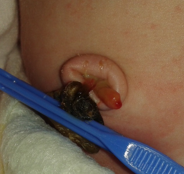
Case 3
Female neonate with normal prenatal ultrasound scans, showing an umbilical stump tumefaction (figure 6) and a supernumerary finger in each hand on first physical examination. On the first day, a serous fluid discharge from the umbilical stump was observed. On the seventh day of life, a 1 cm long gelatinous structure was observed in the umbilical cord stump, draining serous-hematic fluid at days 14 and 28 of life. An abdominal ultrasound scan was performed, revealing a continuous tubular structure between the bladder and the umbilicus, suggesting a patent urachus (figure 7). At day 56, the umbilicus appeared normal. Nevertheless, the child has since been under close monitoring in a paediatric surgery centre.
Figure 6.

Umbilical stump tumefaction at birth.
Figure 7.

Abdominal ultrasound scan—patent urachus.
Differential diagnosis
The differential diagnosis includes: omphalitis, omphalomesenteric duct remnants, anomalies of vitelline ducts, umbilical granulomas and abdominal defects.
Discussion
The true incidence of urachal anomalies is unknown.9 Urachal anomalies were thought to be rare, with an incidence between 0.3 and 20 cases in every 100 000 hospital admissions.1 11 12 However, paediatric autopsy studies have shown an incidence of 1 in every 7 610 cases of patent urachus and 1 in every 5 000 cases of urachal cyst.1 More recently, Gleason et al13 analysed abdominal imaging of 64 803 children and found urachal anomalies in 1% of cases. This difference may be explained by the frequency of asymptomatic urachal anomalies (93%),13 which therefore go underdiagnosed.
Three cases were recorded within a 1-year period, in a hospital where ∼2 500 births take place annually. Although congenital urachal anomalies have been shown to be twice as common in males as in females,4 of the three aforementioned cases there was a female predominance. All presented an atypical umbilical stump during the first days of life. Only the third case showed changes on the first physical examination, presenting a common symptom of urachal anomalies (fluid drainage). In all cases, the appearance of the umbilicus was normal within 1–2 months. Such atypical umbilical shapes are rarely described in the literature, and seem to be present only for a limited period of time. As such, it is important that healthcare professionals check for atypical umbilical stumps, as these may suggest urachal anomalies. Besides, urachal anomalies might develop concomitantly with other genitourinary defects—the prevalence of vesicoureteral reflux is 13–17% greater in these cases than among the general paediatric population,6 9 which further underlies the importance of promptly referring the child to a paediatric surgery centre for specialised evaluation.
Information is lacking as to how to manage paediatric urachal anomalies. The classic treatment consisted in a surgical approach, applied to all patients with urachal anomalies in an effort to prevent complications. Nowadays, it is known that many anomalies may spontaneously recede. Nogueras-Ocaña et al12 observed spontaneous regressions in 61.5% of 13 patients within 16.5 months (median time) since the diagnosis. Galati et al6 reported a complete resolution of urachal anomalies in 80% of patients younger than 6 months. However, should symptoms persist or the urachal anomaly fail to recede after 6 months, the authors recommend its excision as a means of preventing recurrent infections.6 Therefore, as in the cases presently under study, close monitoring of the anomaly with periodic ultrasound scans in a reference centre seems to be the more reasonable therapeutic option.
Learning points.
Atypical appearance of the umbilicus should be understood to possibly indicate urachal anomalies.
It is important to identify urachal anomalies for they have an underlying risk of recurrent infections and adenocarcinomas in adulthood.
Routine procedure used to be the surgical excision of the entire lesion on diagnosis, even in asymptomatic patients. More recently, however, surgeons have expressed a preference towards non-surgical approaches, which have commonly produced favourable outcomes, namely a complete regression of the urachal anomaly.
Acknowledgments
The authors would like to acknowledge Dr Ana Vaz and Dr Rosário Cancella de Abreu.
Footnotes
Contributors: RES, MA and AT conducted the analysis of the described cases. RES, MA and SRA drafted the manuscript. AT critically reviewed the manuscript. All authors read and approved the final version.
Competing interests: None declared.
Patient consent: Obtained.
Provenance and peer review: Not commissioned; externally peer reviewed.
References
- 1.Choi YJ, Kim JM, Ahn SY et al. Urachal anomalies in children: a single center experience. Yonsei Med J 2006;47: 782–6. 10.3349/ymj.2006.47.6.782 [DOI] [PMC free article] [PubMed] [Google Scholar]
- 2.Shima Y, Hayashida M, Hayashi T et al. Characteristic prenatal ultrasonographic findings of patent urachus: a case report. J Nippon Med Sch 2003;70:172–4. 10.1272/jnms.70.172 [DOI] [PubMed] [Google Scholar]
- 3.Yiee JH, Garcia N, Baker LA et al. A diagnostic algorithm for urachal anomalies. J Pediatr Urol 2007;3:500–4. 10.1016/j.jpurol.2007.07.010 [DOI] [PubMed] [Google Scholar]
- 4.Yu JS, Kim KW, Lee HJ et al. Urachal remnant diseases: spectrum of CT and US findings. Radiographics 2001;21:451–61. 10.1148/radiographics.21.2.g01mr02451 [DOI] [PubMed] [Google Scholar]
- 5.Tazi F, Ahsaini M, Khalouk A et al. Abscess of urachal remnants presenting with acute abdomen: a case series. J Med Case Rep 2012;6:226 10.1186/1752-1947-6-226 [DOI] [PMC free article] [PubMed] [Google Scholar]
- 6.Galati V, Donovan B, Ramji F et al. Management of urachal remnants in early childhood. J Urol 2008;180:1824–6. 10.1016/j.juro.2008.03.105 [DOI] [PubMed] [Google Scholar]
- 7.Naiditch JA, Radhakrishnan J, Chin AC. Current diagnosis and management of urachal remnants. J Pediatr Surg 2013;48:2148–52. 10.1016/j.jpedsurg.2013.02.069 [DOI] [PubMed] [Google Scholar]
- 8.Sreepadma S, Rao BR, Ratkal J et al. A rare case of urachal sinus. J Clin Diagn Res 2015;9:PD01–2. 10.7860/JCDR/2015/13243.6185 [DOI] [PMC free article] [PubMed] [Google Scholar]
- 9.Copp HL, Wong IY, Krishnan C et al. Clinical presentation and urachal remnant pathology: implications for treatment. J Urol 2009;182:1921–4. 10.1016/j.juro.2009.03.026 [DOI] [PubMed] [Google Scholar]
- 10.Lee SR, Kang H, Kang MH et al. The youngest Korean case of urachal carcinoma. Case Rep Urol 2015;2015:707456 10.1155/2015/707456 [DOI] [PMC free article] [PubMed] [Google Scholar]
- 11.Wildhaber BE, Antonelli E, Pfister RE. The giant umbilical cord. Arch Dis Child Fetal Neonatal 2005;90:F535–6. 10.1136/adc.2005.076380 [DOI] [PMC free article] [PubMed] [Google Scholar]
- 12.Nogueras-Ocaña M, Rodríguez-Belmonte R, Uberos-Fernández J et al. Urachal anomalies in children: surgical or conservative treatment? J Pediatr Urol 2014;10:522–6. 10.1016/j.jpurol.2013.11.010 [DOI] [PubMed] [Google Scholar]
- 13.Gleason JM, Bowlin PR, Bagli DJ et al. A comprehensive review of paediatric urachal anomalies and predictive analysis for adult urachal adenocarcinoma. J Urol 2015;193:632–6. 10.1016/j.juro.2014.09.004 [DOI] [PubMed] [Google Scholar]