Abstract
Modified TiO2 nanomaterials are considered to be promising in energy conversion and ferrites modification may be one of the most efficient modifications. In this research, various ferrites, incorporated with various cations (MFe2O4, M = Ni, Co, Zn, and Sr), are utilized to modify the well aligned TiO2 nanorod arrays (NRAs), which is synthesized by hydrothermal method. It is found that all MFe2O4/TiO2 NRAs show obvious red shift into the visible light region compared with the TiO2 NRAs. In particular, NiFe2O4 modification is demonstrated to be the best way to enhance the photoelectrochemical and photocatalytic activity of TiO2 NRAs. Furthermore, the separation and transfer of charge carriers after MFe2O4 modification are clarified by electrochemical impedance spectroscopy measurements. Finally, the underlying mechanism accounting for the enhanced photocatalytic activity of MFe2O4/TiO2 NRAs is proposed. Through comparison among different transition metals modified TiO2 with the same synthesis process and under the same evaluating condition, this work may provide new insight in designing modified TiO2 nanomaterials as visible light active photocatalysts.
In recent years, titanium oxide (TiO2), as a “green” photocatalyst, has attracted lots of attention worldwide due to its low cost, nontoxicity and excellent photochemical stability1. However, due to the rapid recombination of photogenerated electron-hole pairs as well as the lack of visible light absorption2, the photocatalytic and photoelectrochemical efficiency of pure TiO2 is quite limited. Pure TiO2, with bandgap of ca. 3.0 to 3.2 eV, can only be excited under UV light irradiation, which comprises only ~5% of the total solar radiation, so, application of the unmodified TiO2 in solar energy conversion is far limited3. Besides, the excited charge carriers in TiO2 can recombine quickly and more than 90% of the recombination processes take place in 10 ns4, therefore, only a little fraction of the excited carriers can transfer to the surface of TiO2 and take part in the following photocatalytic process. To make full use of solar energy and reduce the recombination rate, many attempts have been made, such as doping with metal/nonmetal atoms and deposition of metals. Though the aforementioned methods can partly improve the photocatalytic as well as photoelectrochemical activity of TiO2, some problems remain unresolved. For example, doped TiO2 may suffer from thermal instability, photo corrosion, lattice distortion, while metal loading may result in an increase in the carrier-recombination probability5. In this case, one of the promising strategies is to couple TiO2 with other narrow band gap semiconductors, which are capable of harvesting photons in the visible light region. Recently, CdS with a low band gap (ca. 2.4 eV) was studied intensively to improve the visible light uitilizition of TiO2. However, its application is also hindered due to the low photostability6. In view of this, transition metal ferrites with molecular formula of MFe2O4 (M = Zn, Co, Ni, etc.) go into the vision of researchers considering its outstanding attributes. Firstly, these materials possess an important characteristic of narrow band gap, which could absorb the visible light efficiently7 and thus promote the photocatalytic reactions. Secondly, MFe2O4 has a good electrical conductivity due to the electron hopping process between different valence states of metals in O-sites, which is beneficial for the transfer of charge carriers8,9. Furthermore, transition metal ferrites have many intriguing advantages such as well stability against photocorrosion, good superparamagnetic properties, low toxicity, easy preparation, high adsorption ability, low cost, and abundant resources10,11,12. So far, most of the researches are focused on separation of TiO2 powders from treated water by employing its magnetic property of transition metal ferrites13,14,15. It is scarce on study of the visible responsiveness of MFe2O4 to increase utilization of solar energy as well as to enhance the photoelectrochemical and photocatalytic performance of TiO2.
ZnFe2O4, with a relatively small band-gap (ca. 1.9 eV)16, is the most frequently studied to modify TiO2 in enhancing the photoelectrochemical capacity. Yuan et.al observed that the ZnFe2O4/TiO2 nanocomposite is more effective as a photocatalyst in the phenol degradation than pure TiO2. However the mechanism of the enhanced photoactivity of the ZnFe2O4/TiO2 composite is still needed to be further understood17. Furthermore, the following researches proposed similar theory to explain the role of ZnFe2O4 in enhancing photoactivity of TiO218,19, that is, the adoption of ZnFe2O4 makes the ZnFe2O4/TiO2 composite could use visible light, and the good match of band edges between ZnFe2O4 and TiO2 is in favor of charge carriers separating effectively. Reports about other MFe2O4 modified TiO2, such as NiFe2O4/TiO2, Mn0.5Zn0.5Fe2O4/TiO2, MgFe2O4/TiO2 and CuFe2O4/TiO2 all show higher photoelectrochemical and photocatalytic performance20,21,22,23. It seems that MFe2O4 is such a promising material to improve the photoelectrochemical and photocatalytic performance of TiO2. However, the comparison among the photoelectrochemical and photocatalytic performances of transition metal ferrites modified TiO2 reported in the literatures is extremely difficult, because the experimental conditions were very different, such as catalysts synthesis process, light irradiation wavelength, reactor geometric configuration, catalyst loading and so on. Moreover, the origin and the crystalline structure of TiO2, which strongly affect its electronic and photoactivity, are also different. Therefore, the same condition should be taken into consideration when assessing the real effect of transition metal ferrites on the photoactivity of TiO2, such as using the same bare TiO2 as the starting material, taking the same procedure to modify TiO2 by transition metal ferrites, and finally evaluating their performances with unified standards. NiFe2O4, CoFe2O4, ZnFe2O4 and SrFe2O4 are four common transition metal ferrites which have been frequently studied with their magnetism, but except for ZnFe2O4, the other three are not common in modifying TiO2 to enhance its photoactivity, therefore, we select the four as research objects, making a comparision between the common one (ZnFe2O4/TiO2) and the uncommon ones (CoFe2O4, ZnFe2O4 and SrFe2O4 modified TiO2).
In addition to incorporate other materials to modify TiO2, structural design is another important method to enhance the photoactivity of TiO2. One-dimensional (1D) nanostructure such as nanowire, nanotube, nanorod have attracted lots of attention due to the unique physical and chemical properties. 1D TiO2 nanomaterials possess all the typical features of TiO2 nanoparticles24. Electron diffusion length (up to ~100 μm) can be prolonged by using vertically aligned 1D nanostructures and excited electrons can easily pass along 1D nanostructure to the transparent conducting oxide electrode25,26, which facilitate charge transfer and promote charge separating efficiently20,27. However, the relatively low specific surface area on a smooth surface of 1D nanostructures may decrease the absorption ability and a single crystal phase of 1D nanostructures may pose certain constraints on the photoelectrochemical performance24,28. Fortunately, these disadvantages can be surmouned by introducing the second phase, i.e., doping metals/nonmetals or forming heterjunctions. Among1D nanostructures, 1D nanorod arrays with large area can be easily obtained by hydrothermal method, which is facile, economic and controllable29. Therefore, coupling the traits of one-dimensional TiO2 nanorods (TiO2 NRAs) and visible light responsive MFe2O4 nanoparticles seems to be a promising way to enhance the solar energy conversion efficiency of TiO2.
To the best of our knowledge, there is few systematic research on the photoelectrochemical and photocatalytic capacity of various MFe2O4 modified one-dimensional TiO2 NRAs so far. In this study, large area uniform TiO2 NRAs were synthesized hydrothermally and ferrites containing vaious cations (MFe2O4, M = Ni, Co, Zn, and Sr) were utilized to modify the as-prepared TiO2 NRAs. The morphology, crystalline structures and optical properties as well as photoelectrochemical performances of TiO2 NRAs and MFe2O4/TiO2 NRAs were investigated. Moreover, the photocatalytic activities of the MFe2O4/TiO2 NRAs were evaluated in the degradation of Cr(VI) aqueous solution under visible light irradiation. Finally, the underlying photcatalytic mechanism was discussed.
Results and Discussion
Figure 1(a) displays the XRD patterns collected from the TiO2 NRAs and MFe2O4/TiO2 NRAs. It can be seen that the TiO2 NRAs and MFe2O4/TiO2 NRAs all features the characteristic peaks at 2θ = 36.078°, 62.750°, 69.010° and 69.795°, indicative of rutile TiO2 (PDF NO. 21–1276). Other peaks can be attributed to the diffraction of FTO substrate. There is no typical diffraction peaks of MFe2O4 after modification, which may be owing to the low content of MFe2O4. The content of MFe2O4 will be discussed in the following SEM characterization.
Figure 1.
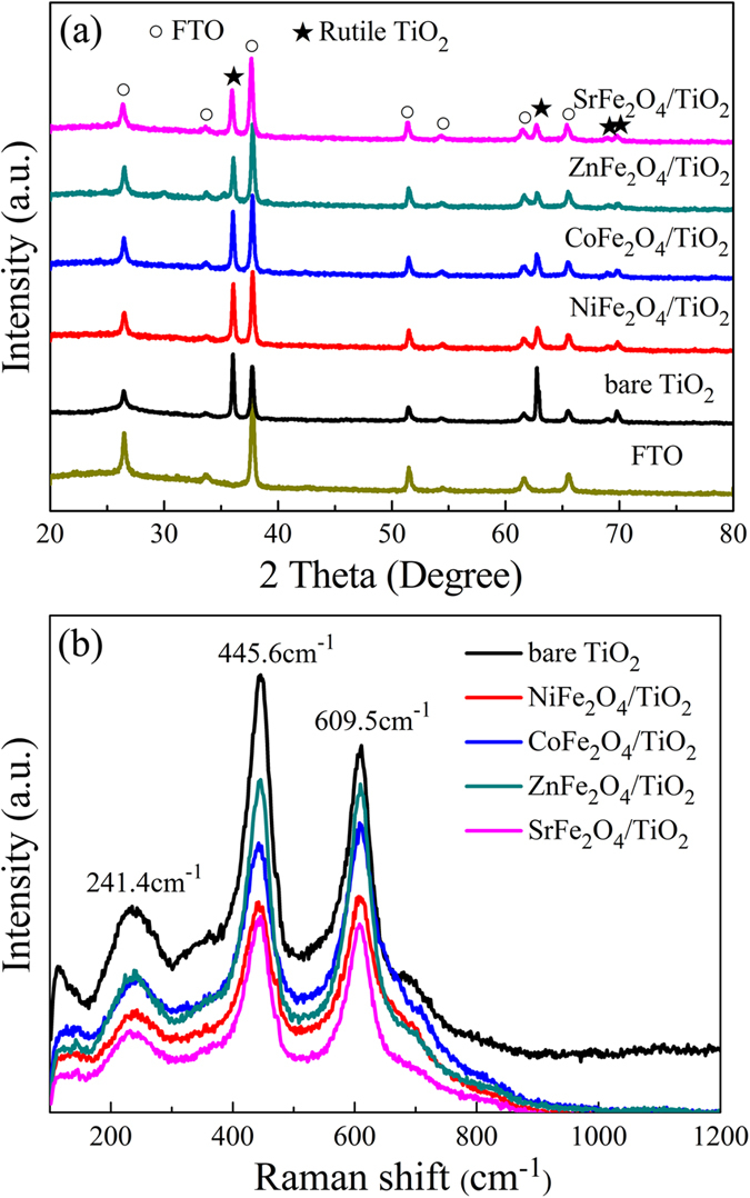
(a) XRD patterns and (b) Raman spectra for TiO2 NRAs and MFe2O4/TiO2 NRAs.
In order to further examine the phase composition of the samples and confirm the existence of MFe2O4, Raman spectroscopy was employed. As is shown clearly in Fig. 1(b), there are three Raman peaks at 241.4, 445.6 and 609.5 cm−1 for all samples, which are assigned to the Raman active modes of rutile30. This result indicates that the rutile phase dominates the crystalline structure of the samples, which is in accordance with the XRD result. However, the Raman peak corresponding to MFe2O4 is not discernable due to the low content of MFe2O4. The peak at 117 cm−1 is due to plasma emission of the Ar+ laser31.
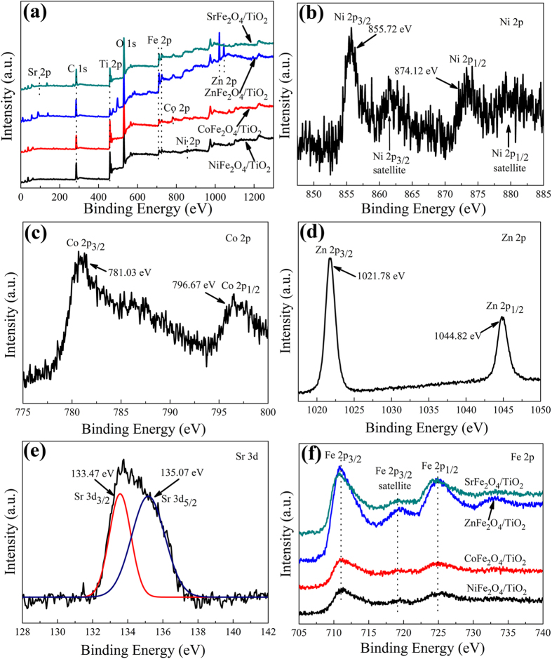
In order to further confirm the existence of MFe2O4, XPS measurement was carried out. The XPS survey spectra are shown in Fig. 2(a). The peaks located at the binding energies of ca. 458–464 eV, 529–531 eV, 711–725 eV and 284–288 eV in all samples are ascribed to the Ti 2p, O 1s, Fe 2p, and C 1s, respectively. On the other hand, these MFe2O4/TiO2 NRAs materials also show their characteristic peaks located between 850 and 875 eV (Ni 2p) for NiFe2O4/TiO2 NRAs, 781 and 796 eV (Co 2p) for CoFe2O4/TiO2 NRAs, 1021 and 1044 eV (Zn 2p) for ZnFe2O4/TiO2 NRAs, and 134 eV (Sr 2p) for SrFe2O4/TiO2 NRAs.
Figure 2.
(a) XPS survey spectra of MFe2O4/TiO2 NRAs and high-resolution XPS spectra of (b) Ni 2p, (c) Co 2p, (d) Zn 2p, (e) Sr 2p and (f) Fe 2p.
As is shown in Fig. 2(b), the Ni 2p peaks of NiFe2O4/TiO2 NRAs consist of two characteristics of Ni 2p3/2 (855.72 eV) and Ni 2p1/2 (874.12 eV)32, indicative of the presence of Ni2+. Similar to the Ni 2p peaks in NiFe2O4/TiO2 NRAs, the Co 2p XPS spectra recorded from the CoFe2O4/TiO2 NRAs sample, containing Co 2p3/2 (781.03 eV, Co2+ in Tet-site) and Co 2p1/2 (796.67 eV, Co2+ in Tet-site), indicate that Co2+ exists in the CoFe2O4/TiO2 NRAs according to the literature reports33. For the ZnFe2O4/TiO2 NRAs, the recorded Zn 2p XPS spectra indicate that Zn2+ exists in the ZnFe2O4/TiO2 NRAs, which is also consistent with literature reports34,35. Furthermore, the manganese valences were determined by the position of the multiplet splitting of Sr 2p peaks, the positions of Sr 2p3/2 and Sr 2p1/2 were all assigned to Sr2+. As for high-resolution XPS spectra of Fe 2p in Fig. 2(d), one can see that the peaks at ca. 711.6 eV and ca. 724.9 eV can be attributed to Fe 2p3/2 and Fe 2p1/2 for Fe3+, respectively, which reveals the oxidation state of Fe3+ in the MFe2O4/TiO2 heterostructure33,36.
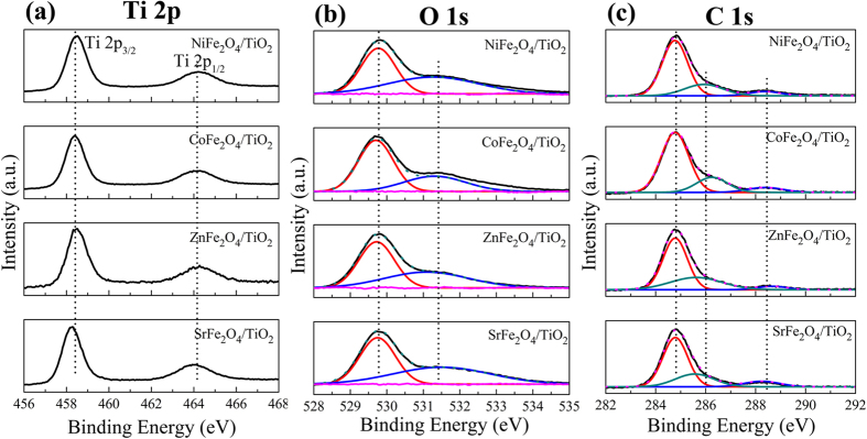
The high resolution XPS spectra of Ti 2p, O 1s, and C 1s are shown in Fig. 3. The Ti 2p spectra, as presented in Fig. 3(a), all show the main peak located at ca. 458.5 eV and ca. 464.2 eV, which can be attributed to Ti 2p3/2 and Ti 2p1/2 in TiO2, respectively37. It is clear that the O 1s spectra of these MFe2O4/TiO2 NRAs samples can be deconvoluted into two components centered at ca. 529.8 eV and ca. 531.4 eV using two Gaussian curve fittings {Fig. 3(b)}, The components at the lower and higher binding energy side can be assigned to the crystal lattice oxygen of TiO2 and MFe2O4 and chemisorbed oxygen in a defective lattice site (i.e.–OH), respectively32,38,39,40,41. It is suggested that the hydroxyl group can capture the photogenerated holes and form highly reactive hydroxyl free radicals, which plays an important role in enhancing photocatalytic activity18. The high resolution XPS spectrum of C 1s is shown in Fig. 3(c). The primary peak located at ca. 284.6 eV is assigned to C–C/C–H bonds from adventitious carbon42, while the peaks at ca. 286.2 eV and ca. 288.4 eV can be attributed to the formation of carbonate species, resulting mainly from CO2 adsorption38,43,44,45. Especially, the peak at 288.4 eV can be ascribed to the Ti–O–C structure in carbon doped TiO2 by substituting some of the lattice titanium atoms46,47,48. Interestingly, carbon doping is beneficial to light absorption capability as well as absorption of organic molecules to some extent24,25.
Figure 3.
High-resolution XPS spectra of (a) Ti 2p, (b) O 1s and (c) C 1s.
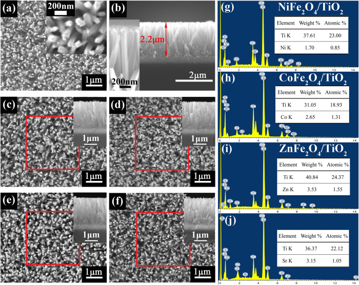
The SEM images of the bare TiO2 NRAs and MFe2O4/TiO2 NRAs are shown in Fig. 4. It is noteworthy that, after MFe2O4 modification as shown in Fig. 4(c–f) from the top view images, the samples have no obvious changes in morphology compared with the bare TiO2 NRAs in Fig. 4(a), which indicates that the deposited MFe2O4 nanoparticles are of extremely fine size. The vertically or slantingly aligned TiO2 nanorods arrays, with diameter of 60~120 nm and length of 2.2 μm, are grown homogeneously on FTO substrate with rectangular cross section. In order to measure the content of MFe2O4 in MFe2O4/TiO2 NRAs heterjunction, energy dispersive x-ray spectrum (EDS) analysis was carried out. The results, shown in Fig. 4(g~j), are obtained from collecting the EDS data in red square region of the MFe2O4/TiO2 NRAs in Fig. 4(c–f), respectively. It is confirmed that Ni, Co, Zn and Sr are present in NiFe2O4, CoFe2O4, ZnFe2O4 and SrFe2O4 modified TiO2 NRAs, respectively. Indeed, only a trace amount of Ni, Co, Zn and Sr can be observed in these samples.
Figure 4.
SEM images of (a) the bare TiO2 NRAs, (b) the cross section image of the bare TiO2 NRAs, (c) NiFe2O4/TiO2 NRAs, (d) CoFe2O4/TiO2 NRAs, (e) ZnFe2O4/TiO2 NRAs and (f) SrFe2O4/TiO2 NRAs. The insets of (c–f) are the corresponding cross section images. (h,i,g,k) are the EDS results of the red square region in (c–f), respectively.
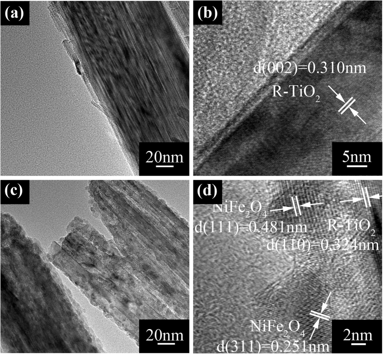
Furthermore, structural characterizations of the MFe2O4 modified TiO2 nanorods were investigated by TEM. Figure 5(a) shows the TEM image of the bare TiO2 nanorod. Essentially, the diameter of the bare TiO2 nanorod under TEM observation is consistent with the SEM result. It can be seen clearly that the bare TiO2 nanorod is very smooth. After MFe2O4 modification shown in Fig. 5(c), the nanorod surface becomes rough, and the ultrafine NiFe2O4 particles, with diameter of ca. 3~5 nm as shown in Fig. 5(d), are uniformly deposited on the nanorod. In addition, the high resolution HRTEM image gives lattice fringes of about 0.481 nm and 0.251 nm, corresponding to the d (111) and d (311) space of NiFe2O4, respectively. Analysis of TEM was also applied to CoFe2O4, ZnFe2O4 and SrFe2O4 modified TiO2 NRAs (shown in Supplementary Fig. S1), and all show the same morphology. i.e., the smooth surface of TiO2 nanorod become rough after MFe2O4 modification. The corresponding lattice fringes of CoFe2O4, ZnFe2O4 and SrFe2O4 are shown in Figure S1(b,d,f), respectively.
Figure 5.
(a,b) TEM images of TiO2 NRAs, (c,d) TEM images of NiFe2O4/TiO2 NRAs.
The optical absorption spectra of TiO2 NRAs and MFe2O4/TiO2 NRAs are shown in Fig. 6. All samples exhibit typical UV absorption (λ < 380 nm). It is noteworthy that, compared with bare TiO2 NRAs, all MFe2O4/TiO2 samples exhibit strong light absorption in a wide region from 380 nm to 900 nm, which can be attributed to the intrinsic band gap absorption of MFe2O4. However, unlike other pure TiO2, tiny absorption of the as-prepared TiO2 sample in the visible light range can be observed. There are two reasons accounting for this abnormal phenomenon, one is the scattering of light caused by the nanorod arrays, and the other is the impurity doping during the hydrothermal and sintering process49,50,51. The absorption capacity of CoFe2O4/TiO2 NRAs is the biggest, followed by ZnFe2O4, SrFe2O4 and NiFe2O4 modified TiO2 NRAs sequentially. The corresponding band gaps are calculated from the plots of Eg = 1240/λ by extrapolating the linear portion of absorbance to the wavelength axis where absorbance is zero52. As is shown in Fig. 6, the steep absorption edge of the bare TiO2 NRAs locates at about 410 nm, corresponding to band gap (Eg) of about 3.02 eV. MFe2O4 modified TiO2 NRAs samples all exhibit red-shift with smaller band gaps compared with bare TiO2 NRAs, and the Eg is ca. 1.84 eV, 1.63 eV, 1.81 eV and 1.53 eV for NiFe2O4/TiO2 NRAs, ZnFe2O4/TiO2 NRAs, SrFe2O4/TiO2 NRAs, and CoFe2O4/TiO2 NRAs, respectively.
Figure 6. Absorption spectra of MFe2O4/TiO2 NRAs.
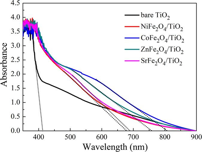
The dash lines are the extension of the linear portion of absorbance.
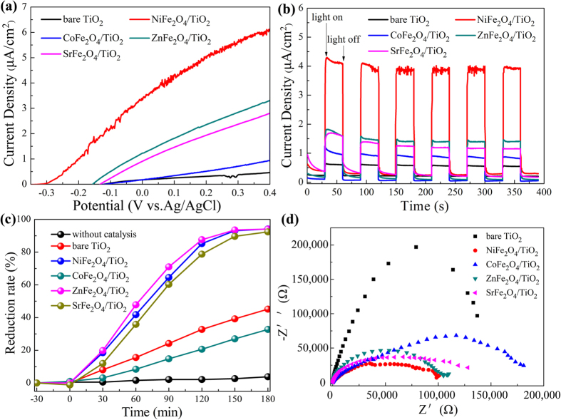
To evaluate the effect of MFe2O4 modification on the photoelectrochemical properties of TiO2 NRAs, the photocurrent intensity versus potential (I–V) and photocurrent density versus time (I-T) measurements of MFe2O4/TiO2 NRAs were performed. The I-V characteristics of MFe2O4/TiO2 NRAs are shown in Fig. 7(a). The photocurrent density in dark can be neglected for all samples. Under visible light irradiation, the photocurrent density of bare TiO2 NRAs varies little with increase in bias potential, while the photocurrent density of MFe2O4/TiO2 NRAs increases significantly at more positive bias potentials, except for CoFe2O4/TiO2 NRAs with only a slight increase. For example, at bias potential of 0.4 V vs. Ag/AgCl, the photocurrent density of NiFe2O4, ZnFe2O4 and SrFe2O4 modified TiO2 NRAs is 6.13, 3.31 and 2.81 μA/cm2, respectively, while the photocurrent density of CoFe2O4/TiO2 NRAs is only 0.95 μA/cm2, which is far lower than that of other MFe2O4 modified samples, and only a little higher than that of the bare TiO2 NRAs (0.46 μA/cm2 at 0.4 V vs. Ag/AgCl). It is reported that the more negative open circuit potential (Voc) means better charge carrier separation and electron accumulation in semiconductor-semiconductor heterojunctions53,54,55,56. After MFe2O4 modification, Voc for NiFe2O4/TiO2 NRAs, ZnFe2O4/TiO2 NRAs and SrFe2O4/TiO2 NRAs is −0.323, −0.156 and −0.133 V, respectively, which becomes more negative than that of the bare TiO2 NRAs (−0.121 V), except for CoFe2O4 modified one(−0.117 V). From the varying trend of Voc, one can see that MFe2O4/TiO2 NRAs (M = Ni, Zn and Sr) heterjunction facilitates the separation and transfer of the charge carriers, while CoFe2O4/TiO2 NRAs is not favourable for charge carriers separation. Figure 7(b) plots the I-T characteristics of the MFe2O4/TiO2 NRAs. It is observed that all the samples exhibit a quick response to the on/off of the incident light, and the current density of MFe2O4 modified TiO2 NRAs shows an enhancement compared with that of bare TiO2 NRAs. NiFe2O4/TiO2 NRAs displays the biggest photocurrent density of ca. 4.13 μA/cm2, followed by ZnFe2O4, SrFe2O4 and CoFe2O4 modified ones, with 1.73, 1.68 and 1.01 μA/cm2, respectively. The enhancement induced by CoFe2O4 modification is relatively low, only 0.4 μA/cm2 higher than that of bare TiO2 NRAs (0.61 μA/cm2). The changing trend of I-T result is consistent with the I-V characteristics of the MFe2O4/TiO2 NRAs.
Figure 7.
(a) Photocurrent density versus potential of the MFe2O4/TiO2 NRAs, (b) Photocurrent density versus time measurements of MFe2O4/TiO2 NRAs under 0 V versus Ag/AgCl bias, (c) Photocatalytic reduction of Cr(VI) by MFe2O4/TiO2 NRAs under visible light, (d) Nyquist plots of the EIS spectra of MFe2O4/TiO2 NRAs.
Though all MFe2O4 modified TiO2 NRAs samples exhibit a broader and stronger absorption than the bare TiO2 NRAs (see Fig. 6), only NiFe2O4/TiO2 NRAs possesses a significant enhancement in PEC performance. Very limited improvement for CoFe2O4 modification may result from the inefficient separation of photoexcited charge carriers. This phenomenon is due to the fact that the conduction band (CB) of CoFe2O4 is more positive than that of TiO2, while the valence band (VB) of CoFe2O4 is more negative than that of TiO241,57, which is not favour in carriers separating.
To investigate the photocatalytic capacity of the MFe2O4/TiO2 NRAs, experiments were carried out for Cr(VI) photoreduction under visible light irradiation. The concentration changes are detected by the absorption peak (365 nm) of Cr(VI) in the UV-vis spectrum. The photodegradation results are shown in Fig. 7(c). After irradiation for 180 minutes, little Cr(VI) was reduced without catalyst (the reduction rate is only 3.8%). Under the same condition, only 45.1% of Cr(VI) was reduced when bare TiO2 NRAs was used as a photocatalyst. However, the potoreduction capacity of NiFe2O4, ZnFe2O4 and SrFe2O4 modified TiO2 NRAs are enhanced greatly (94.18%, 94.086% and 92.39%, respectively), reaching the same level. This may be attributed to the function of citric acid serving as a sacrificial electron donator to quickly consume the photogenerated holes19, thus greatly promote charge separation and further improv photocatalytic reactions. Unfortunately, CoFe2O4 modification makes the photocatalytic degradation rate of Cr(VI) even lower. The following reason may account for this abnormal phenomenon. Eventhough CoFe2O4 modified TiO2 NRAs can be excited more easily under visible light irradiation, and then generates more charge carriers, the recombination rate of CoFe2O4/TiO2 NRAs seems to be higher than that of the bare TiO2 NRAs which can be deduced from the Voc changes, thus leading to the lower photocatalytic capacity of CoFe2O4/TiO2 NRAs.
In order to clarify the enhancement in the phototelectochemical and photocatalytic capacity of TiO2 NRAs after MFe2O4 modification, it is important to figure out the separating and transferring efficiency of the charge carriers, so electrochemical impedance spectroscopy (EIS) measurements were conducted. As shown in Fig. 7(d), except for CoFe2O4/TiO2 NRAs, other MFe2O4 modified TiO2 NRAs samples all have a smaller arc radius compared with that of the bare TiO2 NRAs. It is generally assumed that the smaller arc radius on the EIS Nyquist plot suggests a more effective separation of the photogenerated electron-hole pairs and a faster interfacial charge transfer38,58. From the EIS spectra, it can be seen clearly that NiFe2O4, ZnFe2O4 and SrFe2O4 modified TiO2 NRAs have a smaller arc radius than the bare TiO2 NRAs. It means that the charge carriers separate and transfer more effectively in NiFe2O4/TiO2 NRAs, ZnFe2O4/TiO2 NRAs and SrFe2O4/TiO2 NRAs, thus leading to the significant enhancement of the phototelectochemical and photocatalytic capacity of the modified TiO2 NRAs. While the arc radius of CoFe2O4/TiO2 NRAs is even bigger than that of the bare TiO2 NRAs, suggesting lower separating rate of charge carriers in CoFe2O4/TiO2 NRAs and thus resulting in the limited enhancement of the phototelectochemical capacity and even decrease in photocatalytic performance. This EIS result of CoFe2O4/TiO2 NRAs is in accordance with the Voc value of CoFe2O4/TiO2 NRAs in the I-V curves as well as the deduction from the band matching between CoFe2O4 and TiO2 in previous literature, that is, the CoFe2O4/TiO2 heterojunction is not conducive to effective separation of carriers.
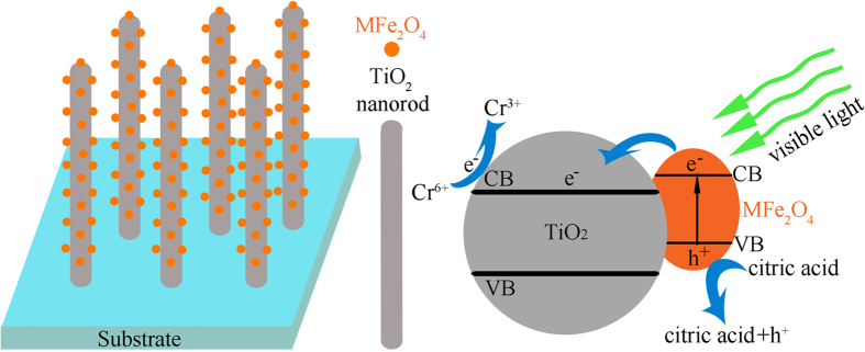
Photocatalytic schematic of Cr(VI) by MFe2O4/TiO2 NRAs is shown in Fig. 8. Under visible light illumination, MFe2O4 is effectively excited to generate electrons and holes. Because the conduction band of MFe2O4 is more positive than that of TiO2, the excited electrons can quickly transfer from MFe2O4 to the conduction band of TiO2, whereas the generated holes accumulate in the valence band of MFe2O4. Consequently, the excited electron/hole pairs could be separated effectively, which contributes to the improvement of photoelectrochemical properties of MFe2O4/TiO2 NRAs, except for CoFe2O4/TiO2 NRAs. Due to the efficient separation of the photogenerated electrons and holes by MFe2O4 modification, the lifetime of the charge carriers are prolonged, leading to an efficient oxidation-reduction reaction, so the photodegradation activity can be enhanced. When the photoreduction is carried out in the presence of citric acid, it can quickly consume the accumulated holes in the valence band, and thus the electrons in the conduction band have enough time to function with the Cr(VI) in the aqueous solution.
Figure 8. Photocatalytic schematic of Cr(VI) by MFe2O4/TiO2 NRAs.
Conclusions
The effect of different ferrits (MFe2O4, M = Ni, Co, Zn and Sr) modification on improving the photoelectrochemical and photocatalytic properties of TiO2 have been probed. By changing the incorporated cations in the MFe2O4, we have found that NiFe2O4 modification can greatly enhance the photoelectrochemical and photocatalytic performance of TiO2 NRAs, while CoFe2O4 has relative limited effect. Compared with the bare TiO2 NRAs, the photocurrent density of NiFe2O4/TiO2 NRAs is twelve-fold higher in the I-V curve at 0.4 V vs. Ag/AgCl. Under visible light irradiation, the Cr(VI) photoreduction rate of NiFe2O4/TiO2 NRAs achieves one-fold higher than that of the bare TiO2 NRAs. The EIS measurement provides a clearer understanding of the role that MFe2O4 have in photogenerated charge carriers effectively separating and transferring. Except for CoFe2O4/TiO2 NRAs, other MFe2O4 modified TiO2 NRAs have more effective separation and transfer of the charge carriers, thus leading to the difference in the photoelectrochemical and photocatalytic performance of MFe2O4 modified TiO2 NRAs. The obtained results point that the visible active MFe2O4 modification may be a promising way to improve TiO2 for applications in photocatalytic activity as well as in photoelectrochemical conversion with solar light.
Methods
Materials synthesis
All reagents used were analytical grade chemicals and used without further treatment.
Synthesis of MFe2O4 modified TiO2 nanorod arrays
Aligned TiO2 NRAs were vertically grown on transparent fluorine-doped tin oxide (FTO) substrates by the hydrothermal method. Deionized water (DI, 10 mL) was mixed with hydrochloric acid (36.8 wt%, 10 mL) and stirred for 10 min before tetrabutyl titanate (98%, 0.4 mL) was added. When the solution was stirred until clear clarification, it was transferred to a Teflon-lined stainless steel autoclave. Clean FTO substrates were immersed with the conducting side face down. The autoclave was put in an oven at a temperature of 150 °C and was taken out from the oven after 5 h. After the autoclave was cooled to room temperature, the FTO substrate was rinsed with DI water and dried naturally at room temperature. The final area of the nanorod arrays was approximately 4.5 cm2.
For the preparation of ZnFe2O4/TiO2 NRAs, briefly, zinc nitrate and iron nitrate were dissolved in DI water at room temperature to form a mixture, the as-prepared TiO2 NRAs were soaked in the Fe(NO3)3 and Zn(NO3)2 mixed solution (with concentrations of 0.25 M and 0.125 M, respectively) for 1 h, followed by dipping in DI water for 5s. Afterwards the nanorod arrays were dried in air for 24 h and then annealed at 500 °C in air for 2 h with heating and cooling rates of 5 °C·min−1. The MFe2O4/TiO2 NRs (M = Ni, Co and Sr) were prepared using the same method by replacing the zinc nitrate with other nitrate.
Characterization
The surface morphology was obtained with a scanning electron microscopy (SEM, VEDAIIXMUINCN) equipped with an energy dispersive X-ray spectroscopy (EDS) system. The film microstructure was further characterized by transmission electron microscopy (TEM). X-ray diffraction (XRD, PANalytical) with Cu-Ka (λ = 0.15401 nm) was operated at 40 kV and 40 mA in a 2θ range of 20–80° at a scanning speed of 5° min−1 to characterize the crystal structure. Raman spectra were recorded at room temperature using a inVia Reflex Raman spectrometer under Ar+ (532 nm) laser excitation. The optical properties were probed by a UV–vis spectrophotometer (UV1800, Shimadzu) with a FTO substrate as a blank. X-ray photoelectron spectroscopy (XPS) was obtained using a ESCALAB 250Xi (The binding energy of the XPS spectra was calibrated with the reference to the C 1s peak at 284.8 eV.)
Photoelectrochemical and photocatalytic measurement
photoelectrochemical measurements were performed in a 250 mL quartz cell using a three-electrode configuration, including the prepared samples as working electrode, a Pt foil as counter electrode, a saturated Ag/AgCl as reference electrode, and 0.5 M Na2SO4 aqueous solution as an electrolyte. The working electrode was illuminated within an area of about 1.5 cm2 at zero bias voltage versus the Ag/AgCl electrode under solar-simulated (AM 1.5 G filtered, 100 mW·cm−2, CEL-HXF300) light sources with a UV cutoff filter (providing visible light with λ ≥ 420 nm). The electrochemical impedance spectroscopy (EIS) measurements were recorded by employing an AC voltage of 5 mV amplitude with the initial potencial at 0.4 v (vs. Ag/AgCl) over the frequency range from 100 kHz to 100 mHz without light illumination.
The Cr(VI) photoreduction was performed in a quartz cell. In the photoreduction experiments, 15 mL of aqueous solution containing 20 mg·L−1 of K2Cr2O7 and 85 mg·L−1 of citric acid was used. The citric acid served as a sacrificial electron donator. Prior to irradiation, the photocatalyst (area about 6 cm2) was immersed into the Cr(VI) solution in the dark for 30 minutes to establish an adsorption/desorption equilibrium. The relative concentration of Cr(VI) in the solution was derived by comparing its UV–vis absorption intensity with that of the initial Cr(VI) solution at 365 nm. The light source was a 300 W xenon lamp with visible light illumination of 26.5 mW·cm−2.
Additional Information
How to cite this article: Gao, X. et al. Enhanced photoelectrochemical and photocatalytic behaviors of MFe2O4 (M = Ni, Co, Zn and Sr) modified TiO2 nanorod arrays. Sci. Rep. 6, 30543; doi: 10.1038/srep30543 (2016).
Supplementary Material
Acknowledgments
The authors are highly grateful to Mr Zhaobin She for the help during film characterization.
Footnotes
Author Contributions X.G. and Z.X. designed the study, proposed the mechanism, and wrote the manuscript. X.G., Z.X. and Z.Z. performed the experiments, analyzed the data, and prepared figures. X.L. and X.W. gave many suggestions during this work process. All authors reviewed the manuscript.
References
- Yuan R. et al. Surface Chlorination of TiO2-Based Photocatalysts: A Way to Remarkably Improve Photocatalytic Activity in Both UV and Visible Region. ACS Catalysis 1, 200–206 (2011). [Google Scholar]
- Wang H., You T., Shi W., Li J. & Guo L. Au/TiO2/Au as a Plasmonic Coupling Photocatalyst. Journal of Physical Chemistry C 116, 6490–6494 (2012). [Google Scholar]
- Desario P. A. et al. Plasmonic enhancement of visible-light water splitting with Au-TiO2 composite aerogels. Nanoscale 5, 8073–8083 (2013). [DOI] [PubMed] [Google Scholar]
- Chen X. & Mao S. S. Titanium Dioxide Nanomaterials: Synthesis, Properties, Modifications, and Applications. Chemical Reviews 38, 2891–2959 (2007). [DOI] [PubMed] [Google Scholar]
- R. A., T. M., T. O., K. A. & Y. T. Visible-light photocatalysis in nitrogen-doped titanium oxides. Science 293, 269–271 (2001). [DOI] [PubMed] [Google Scholar]
- Zheng X. et al. Enhanced photoelectrochemical and photocatalytic performance of TiO2 nanorod arrays/CdS quantum dots by coating TiO2 through atomic layer deposition. Nano Energy 11, 400–408 (2014). [Google Scholar]
- N. H. K., Bhojya N. H. S., Prashanth K. P. N. & R. V. Optical and Photocatalytic Properties of Solar Light Active Nd-Substituted Ni Ferrite Catalysts: For Environmental Protection. ACS Sustainable Chem Eng 1, 1143–1153 (2013). [Google Scholar]
- H. Z., S. Z., X. H. Y., L. W. & S. S. Monodisperse M(x)Fe(3−x)O4 (M = Fe, Cu, Co, Mn) nanoparticles and their electrocatalysis for oxygen reduction reaction. Nano Letters 13, 2947–2951 (2013). [DOI] [PubMed] [Google Scholar]
- Neburchilov V., Wang H., Martin J. J. & Wei Q. A review on air cathodes for zinc–air fuel cells. Journal of Power Sources 195, 1271–1291 (2010). [Google Scholar]
- Bai C. et al. Core– Ring Structured NiCo2O4 Nanoplatelets: Synthesis, Characterization, and Electrocatalytic Applications. Advanced Functional Materials 18, 1440–1447 (2008). [Google Scholar]
- Yanguang L., Panitat H. & Yiying W. Ni(x)Co(3-x)O4 nanowire arrays for electrocatalytic oxygen evolution. Advanced Materials 22, 1926–1929 (2010). [DOI] [PubMed] [Google Scholar]
- Scaife D. E. Oxide semiconductors in photoelectrochemical conversion of solar energy. Solar Energy 25, 41–54 (1980). [Google Scholar]
- Wuyou F. et al. Anatase TiO2 nanolayer coating on cobalt ferrite nanoparticles for magnetic photocatalyst. Materials Letters 59, 3530–3534 (2005). [Google Scholar]
- Li C. J., Wang J. N., Wang B., Gong J. R. & Lin Z. Direct formation of reusable TiO2/CoFe2O4 heterogeneous photocatalytic fibers via two-spinneret electrospinning. Journal of Nanoscience & Nanotechnology 12, 2496–2502 (2012). [DOI] [PubMed] [Google Scholar]
- Kim H. S. et al. Synthesis of magnetically separable core@shell structured NiFe2O4 @TiO2 nanomaterial and its use for photocatalytic hydrogen production by methanol/water splitting. Chemical Engineering Journal 243, 272–279 (2014). [Google Scholar]
- Yuan Z., You W., Jia J. & Zhang L. Optical Absorption Red Shift of Capped ZnFe2O4 Nanoparticle. Chinese Physics Letters 15, 535–536 (1998). [Google Scholar]
- Yuan Z. H. & Zhang L. D. Synthesis, characterization and photocatalyticactivity of ZnFe2O4/TiO2 nanocomposite. Journal of Materials Chemistry 11, 1265–1268 (2001). [Google Scholar]
- Hou Y., Li X., Zhao Q., Quan X. & Chen G. Electrochemically assisted photocatalytic degradation of 4-chlorophenol by ZnFe2O4-modified TiO2 nanotube array electrode under visible light irradiation. Environmental Science & Technology 44, 5098–5103 (2010). [DOI] [PubMed] [Google Scholar]
- Chen L. et al. Surface photovoltage phase spectra for analysing the photogenerated charge transfer and photocatalytic activity of ZnFe2O4-TiO2 nanotube arrays. Physical Chemistry Chemical Physics 15, 14262–14269, 10.1039/c3cp51850g (2013). [DOI] [PubMed] [Google Scholar]
- Pan J. et al. Construction of Mn0.5Zn0.5Fe2O4 modified TiO2 nanotube array nanocomposite electrodes and their photoelectrocatalytic performance in the degradation of 2,4-DCP. Journal of Materials Chemistry C 3 (2015). [Google Scholar]
- Kezzim A., Nasrallah N., Abdi A. & Trari M. Visible light induced hydrogen on the novel hetero-system CuFe2O4/TiO2. Energy Conversion & Management 52, 2800–2806 (2011). [Google Scholar]
- Zhang L., He Y., Wu Y. & Wu T. Photocatalytic degradation of RhB over MgFe2O4/TiO2 composite materials. Materials Science & Engineering B 176, 1497–1504 (2011). [Google Scholar]
- Šutka A. et al. Photocatalytic activity of anatase–nickel ferrite heterostructures. Physica Status Solidi Applications & Materials 212, 796–803 (2015). [Google Scholar]
- Zhou W. One-dimensional single-crystalline Ti–O based nanostructures: properties, synthesis, modifications and applications. Journal of Materials Chemistry 20, 5993–6008 (2010). [Google Scholar]
- Z. S., J. H. K., Y. Z., D. A. & S. X. D. Morphology-controllable 1D-3D nanostructured TiO2 bilayer photoanodes for dye-sensitized solar cells. Chemical Communications 49, 966–968 (2013). [DOI] [PubMed] [Google Scholar]
- Sun Z. Continually adjustable oriented 1D TiO2 nanostructure arrays with controlled growth of morphology and their application in dye-sensitized solar cells. Crystengcomm 14, 5472–5478 (2012). [Google Scholar]
- Zhou H., Qu Y., Zeid T. & Duan X. Towards highly efficient photocatalysts using semiconductor nanoarchitectures. Energy & Environmental Science 5, 6732–6743 (2012). [Google Scholar]
- Zhang J., Xiao F. X., Xiao G. & Liu B. Self-assembly of a Ag nanoparticle-modified and graphene-wrapped TiO2 nanobelt ternary heterostructure: surface charge tuning toward efficient photocatalysis. Nanoscale 6, 11293–11302 (2014). [DOI] [PubMed] [Google Scholar]
- Wang L., Zhang X., Ma Y., Yang M. & Qi Y. Rapid microwave-assisted hydrothermal synthesis of one-dimensional MoO3 nanobelts. Materials Letters 164, 623–626 (2015). [Google Scholar]
- Ma H. L. et al. Raman study of phase transformation of TiO2 rutile single crystal irradiated by infrared femtosecond laser. Applied surface science 253, 7497–7500 (2007). [Google Scholar]
- Robert T. D., Laude L. D., Geskin V. M., Lazzaroni R. & Gouttebaron R. Micro-Raman spectroscopy study of surface transformations induced by excimer laser irradiation of TiO2. Thin Solid Films 440, 268–277 (2003). [Google Scholar]
- Ji H. et al. Correction: Magnetic g-C3N4/NiFe2O4 hybrids with enhanced photocatalytic activity. RSC Adv. 5, 64299–64299 (2015). [Google Scholar]
- Hao J. et al. In situ controllable growth of CoFe2O4 ferrite nanocubes on graphene for colorimetric detection of hydrogen peroxide. Journal of Materials Chemistry A 1, 4352–4357 (2013). [Google Scholar]
- Wu S. Reduced graphene oxide anchored magnetic ZnFe2O4 nanoparticles with enhanced visible-light photocatalytic activity. RSC Advances 5, 9069–9074 (2015). [Google Scholar]
- Song H. Preparation of ZnFe2O4 nanostructures and highly efficient visible-light-driven hydrogen generation with the assistance of nanoheterostructures. Journal of Materials Chemistry A 3, 8353–8360 (2015). [Google Scholar]
- Fu M., Jiao Q. & Zhao Y. Preparation of NiFe2O4 nanorod–graphene composites via an ionic liquid assisted one-step hydrothermal approach and their microwave absorbing properties. Journal of Materials Chemistry A 1, 5577–5586 (2013). [Google Scholar]
- Wang S. et al. Facile synthesis of nitrogen self-doped rutile TiO2 nanorods. CrystEngComm 14, 7672–7679 (2012). [Google Scholar]
- Fang-Xing X. et al. Spatially branched hierarchical ZnO nanorod-TiO2 nanotube array heterostructures for versatile photocatalytic and photoelectrocatalytic applications: towards intimate integration of 1D-1D hybrid nanostructures. Nanoscale 6, 14950–14961 (2014). [DOI] [PubMed] [Google Scholar]
- Cheng X., Yu X. & Xing Z. Characterization and mechanism analysis of N doped TiO2 with visible light response and its enhanced visible activity. Applied surface science 258, 3244–3248 (2012). [Google Scholar]
- Yanli C. et al. Enhanced photoelectric performance of PbS/CdS quantum dot co-sensitized solar cells via hydrogenated TiO2 nanorod arrays. Chemical Communications 50, 9509–9512 (2014). [DOI] [PubMed] [Google Scholar]
- Singh S. Reduced Graphene Oxide Coupled CdS/CoFe2O4 Ternary Nanohybrid for Enhanced Photocatalytic Activity and Stability: A Potential Role of Reduced Graphene Oxide as a Visible Light Responsive Photosensitizer. Journal of Virology 77, 4139–4148 (2015). [Google Scholar]
- Hongwei B., Zhaoyang L. & Darren Delai S. Hierarchical ZnO/Cu “corn-like” materials with high photodegradation and antibacterial capability under visible light. Physical Chemistry Chemical Physics 13, 6205–6210 (2011). [DOI] [PubMed] [Google Scholar]
- Xiao F. X. et al. Spatially branched hierarchical ZnO nanorod-TiO2 nanotube array heterostructures for versatile photocatalytic and photoelectrocatalytic applications: towards intimate integration of 1D-1D hybrid nanostructures. Nanoscale 6, 14950–14961 (2014). [DOI] [PubMed] [Google Scholar]
- Zeng H. C., Xie F., Wong K. C. & Mitchell K. A. R. Insertion and Removal of Protons in Single-Crystal Orthorhombic Molybdenum Trioxide under H2S/H2 and O2/N2. Chemistry of Materials 14, 1788–1796 (2002). [Google Scholar]
- Xu M. W. & Hua C. Z. Sulfidation of Single Molecular Sheets of MoO3 Pillared by Bipyridine in Nanohybrid MoO3 (4,4′-bipyridyl)0.5. Chemistry of Materials 15, 433–442 (2003). [Google Scholar]
- Qi X. & Ouyang L. Photocatalytic activity and hydroxyl radical formation of carbon-doped TiO2 nanocrystalline: Effect of calcination temperature. Chemical Engineering Journal 148, 248–253 (2009). [Google Scholar]
- Li Y., Hwang D. S., Lee N. H. & Kim S. J. Synthesis and characterization of carbon-doped titania as an artificial solar light sensitive photocatalyst. Chemical Physics Letters 404, 25–29 (2005). [Google Scholar]
- Ren W. et al. Low temperature preparation and visible light photocatalytic activity of mesoporous carbon-doped crystalline TiO2. Applied Catalysis B Environmental 69, 138–144 (2007). [Google Scholar]
- Wei G., Shen Y., Boschloo G., Hagfeldt A. & Ma T. Influence of nitrogen dopants on N-doped TiO2 electrodes and their applications in dye-sensitized solar cells. Electrochimica Acta 56, 4611–4617 (2011). [Google Scholar]
- Dai G., Yu J. & Liu G. Synthesis and Enhanced Visible-Light Photoelectrocatalytic Activity of p–n Junction BiOI/TiO2 Nanotube Arrays. Journal of Physical Chemistry C 115, 7339–7346 (2011). [Google Scholar]
- Xu P. et al. Visible-light-driven photocatalytic S- and C- codoped meso/nanoporous TiO2. Energy Environ Sci 3, 1128–1134 (2010). [Google Scholar]
- Yang Y. C. et al. Electrospun nanofibers of p-type BiFeO3/n-type TiO2 hetero-junctions with enhanced visible-light photocatalytic activity. RSC Advances 4, 31941–31947 (2014). [Google Scholar]
- Baker D. R. & Kamat P. V. Photosensitization of TiO2 Nanostructures with CdS Quantum Dots: Particulate versus Tubular Support Architectures. Advanced Functional Materials 19, 805–811 (2009). [Google Scholar]
- Subramanian V., Wolf E. E. & Kamat P. V. Green emission to probe photoinduced charging events in ZnO-Au nanoparticles. Charge distribution and Fermi-level equilibration. Journal of Materials Chemistry B 107, 7479–7485 (2003). [Google Scholar]
- And M. J., Levanon H. & Kamat P. V. Charge Distribution between UV-Irradiated TiO2 and Gold Nanoparticles: Determination of Shift in the Fermi Level. Nano Letters 3, 353–358 (2003). [Google Scholar]
- Wood A., Giersig M. & Mulvaney P. Fermi Level Equilibration in Quantum Dot–Metal Nanojunctions†. Journal of Physical Chemistry B 105, 8810–8815 (2001). [Google Scholar]
- Kudo A. & Miseki Y. Heterogeneous photocatalyst materials for water splitting. Chemical Society Reviews 38, 253–278 (2009). [DOI] [PubMed] [Google Scholar]
- Xuan P. et al. Comparing Graphene-TiO2 Nanowire and Graphene-TiO2 Nanoparticle Composite Photocatalysts. ACS Applied Materials & Interfaces 4, 3944–3950 (2012). [DOI] [PubMed] [Google Scholar]
Associated Data
This section collects any data citations, data availability statements, or supplementary materials included in this article.


