Abstract
Purpose
To assess the concentrations of matrix metalloproteinase (MMP)-1 and MMP-9 in the aqueous humor of diabetic macular edema (DME) patients.
Method
The concentrations of MMP-1 and MMP-9 in the aqueous humors of 15 cataract patients and 25 DME patients were compared. DME patients were analyzed according to the diabetic retinopathy (DR) stage, diabetes mellitus (DM) duration, pan-retinal photocoagulation (PRP) treatment, recurrence within 3 months, HbA1C (glycated hemoglobin) level, and axial length.
Results
The concentrations of MMP-1 and MMP-9 of the DME groups were higher than those of the control group (p = 0.005 and p = 0.002, respectively). There was a significant difference in MMP-1 concentration between the mild non-proliferative diabetic retinopathy (NPDR) group and the proliferative diabetic retinopathy (PDR) group (p = 0.012). MMP-1 concentrations were elevated in PRP-treated patients (p = 0.005). There was a significant difference in MMP-9 concentrations between the mild NPDR group and the PDR group (p < 0.001), and between the moderate and severe NPDR group and the PDR group (p < 0.001). The MMP-9 concentrations in PRP treated patients, DM patients with diabetes ≥ 10 years and recurrent DME within 3months were elevated (p = 0.023, p = 0.011, and p = 0.027, respectively). In correlation analyses, the MMP-1 level showed a significant correlation with age (r = -0.48, p = 0.01,), and the MMP-9 level showed significant correlations with axial length (r = -0.59, p < 0.01) and DM duration (r = 049, p = 0.01).
Conclusions
Concentrations of MMP-1 and MMP-9 were higher in the DME groups than in the control group. MMP-9 concentrations also differed depending on DR staging, DM duration, PRP treatment, and degree of axial myopia. MMP-9 may be more important than MMP-1 in the induction of DM complications in eyes.
Introduction
Diabetic retinopathy (DR) is one of the most important causes of visual impairment in many developed countries, despite advances in laser and surgical treatments[1–3]. Based on studies of vascular endothelial growth factor (VEGF) and anti-VEGF antibody treatments for DR, several types of anti-VEGF antibodies have been used and found effective for this disorder[4, 5]. However, recent studies have reported that levels of not only VEGF but also transforming growth factor, epidermal growth factor, human growth factor, interleukins, intercellular adhesion molecule-1, interferon gamma–induced protein, monocyte chemoattractant protein, matrix metalloproteinases (MMPs), plasminogen activator inhibitor-1, placenta growth factor, and tissue growth factor-beta are elevated in the vitreous and anterior chambers[6–8]. Their roles in DR may be associated with alterations in the blood-retinal barrier (BRB), although their exact mechanisms of action remain unclear[7, 8].
The MMPs comprise a group of zinc- and calcium-dependent endopeptidases that are involved in physiological and pathological processes associated with extracellular matrix (ECM) remodeling. MMPs are involved in the degradation and re-building of ECM proteins such as collagen, elastin, gelatin, and casein[9]. At least 25 MMPs have been identified and separated by function into collagenases (MMP-1, MMP-8, and MMP-13), gelatinases (MMP-2 and MMP-9), stromelysins (MMP-3, MMP-10, and MMP-11), matrilysins (MMP-7 and MMP-26), membranous MMPs (MMP-14, MMP-15, MMP-16, MMP-17, MMP-24, and MMP-25), and others[10]. Previous studies have reported that levels of MMPs may be associated with macro- and microvascular complications in DM [11–17].
The associations between retinal vascular complications, MMPs, and DM remain under investigation. Recently, some studies of PDR patients reported increased levels of certain MMPs in the vitreous[18–21]. Such up-regulation of MMPs may involve PDR angiogenesis and progression via degradation of the basement membrane and the ECM of the retina, featuring release of VEGF from ECM-associated reservoirs[22, 23]. However, the roles played by specific MMPs in DR progression remain to be investigated. In previous studies, MMP-1 and MMP-9 were the factors considered most likely to be associated with development of DR complications [8, 20, 24, 25]. Although MMP-1 is well-known as a collagenase and MMP-9 is well-known as a gelatinase, the roles of these proteinases in DR and in alterations of the BRB remain unclear [16, 26, 27].
As reported in most previous studies, vitreous samples yield optimal information on the status of the retina, but obtaining such samples is unacceptably invasive and data quality may be compromised if vitreous samples are mixed with blood (from a hemorrhage). After anti-VEGF agents have been extensively applied, the requirement for vitrectomy decreases. In addition, some studies evaluating MMPs in aqueous humor have shown that the data obtained are valuable[8, 25].
We hypothesize that MMP-1 and MMP-9 have major roles in the initiation of inner BRB damage and in the induction of diabetic macular edema (DME). This study therefore evaluated the concentrations of MMP-1 and MMP-9 in the aqueous humor of DME patients.
Methods
This study compared the concentrations of MMP-1 and MMP-9 in the aqueous humors of cataract patients and DME patients with type II diabetes mellitus (DM), and followed the tenets of the Declaration of Helsinki. The protocol was approved by the Institutional Review/Ethics Board of the Catholic University, Korea. All participants gave written informed consent for the use of their clinical records in this study.
Study subjects
Eyes with DR with a macula thickness > 300 μm due to DME were included in the study. Exclusion criteria included eyes with glaucomatous disc changes, retinal degeneration, and macular edema due to other causes. Eyes with concurrent diseases such as age-related macular degeneration, epiretinal membrane, and retinal vascular occlusion, as well as eyes with histories of severe ocular trauma, uveitis, or prior intraocular surgery that could influence the enzyme levels of the aqueous humor were also excluded.
All patients underwent a full ophthalmological examination that included measurement of visual acuity, refraction, and axial length (if checkable), intraocular pressure (IOP) using Goldmann applanation tonometry, a dilated fundus examination, and fluorescein angiography to classify the eyes according to the Early Treatment of Diabetic Retinopathy Study as mild non-proliferative diabetic retinopathy (NPDR), moderate and severe NPDR, or proliferative diabetic retinopathy (PDR). The ophthalmological examination also included assessment of macular thickness using optical coherence tomography (OCT) (Cirrus High Definition OCT; Carl Zeiss Meditec, Dublin, CA, USA), and axial length using the IOL Master (Carl Zeiss Meditec).
Assay of MMP-1 and MMP-9
The concentrations of MMP-1 and MMP-9 in the aqueous humor in the anterior chamber were determined using anterior paracentesis during cataract surgery. The volume of the collected aqueous humor was at least 50 μL. Human MMP antibody- immobilized beads and 25 μL of 1:20 diluted samples were used. We incubated 2 hours at room temperature (20–25°C) after adding mixed beads, and 1 hour room temperature after adding detection antibodies, and 30 minutes at room temperature after adding Streptavidin-Phycoerythrin. Samples were read using Luminex x-MAP® suspension array technology (Austin, TX, USA). We employed a multiplexed microsphere suspension immunoassay that detects and quantitates spectrally unique microspheres attached to specific antibodies. This enables analysis of samples from a large number of subjects in a single reaction [28, 29]. The detection limit for analysis was 0.36 pg/mL, with a dynamic range of up to 10,000 pg/mL for MMP-1, and 8.71 pg/mL with a dynamic range of up to 10,000 pg/mL for MMP-9. All values under the lower limit of detection were considered zero values[8, 30, 31].
Statistical treatment of data
Statistical analyses were performed using SPSS for Windows software (ver. 20.0; SPSS, Chicago, IL, USA) and R (ver. 3.2.3) [2015-12-10, Platform: x86_64-redhat-linux-gnu, R Core Team (2015). R: A language and environment for statistical computing. R Foundation for Statistical Computing, Vienna, Austria.URL https://www.R-project.org/.]
The DME and control groups were compared using the nonparametric Mann–Whitney U test and the chi-square test. We conducted DME subgroup analyses of MMP-1 and MMP-9 concentrations by reference to pan-retinal photocoagulation (PRP) status, diabetes duration (< 10years versus ≥ 10years), and recurrence within 3 months, using a nonparametric Mann–Whitney U test. Subgroups created by DR staging were classified as having mild, moderate and severe NPDR, or PDR, and were compared using the Kruskal-Wallis test. Tukey’s method was used to compensate for multiple statistical analyses and comparisons as post hoc analyses. Spearman’s correlation test was used in subgroup analyses to evaluate the correlations between age, duration of DM, the level of HbA1C (glycated hemoglobin), and axial length, and the concentrations of MMP-1 and MMP-9. All data in the text are presented as averages ± standard deviations.
Results
We enrolled 25 DME patients as a study group and 15 cataract patients as a control group. Refraction and axial length were checked in 17 and 23 patients of the DME group, and 14 and 15 patients of the control group, respectively. The average age of the cataract patients was 63 ± 15 years and that of the DME patients was 65 ± 13 years (p = 0.804). There were 12 males and 13 females in the study group and 9 males and 6 females in the control group. There were no significant differences in spherical equivalent, IOP, or axial length between the study and control groups (Table 1).
Table 1. Demographics and baseline clinical characteristics of the study participants.
| Control group (n = 15) | DME group (n = 25) | p value | |
|---|---|---|---|
| Age (years) | 63 ± 15 | 65 ± 13 | 0.804 |
| Sex (male: female) | 9:6 | 12:13 | 0.342 |
| IOP (mmHg) | 12± 4 | 13 ± 3 | 0.192 |
| Spherical equivalent (diopters) | 0± 2 (n = 14) | 0 ± 2 (n = 17) | 0.710 |
| Axial length (mm) | 24± 1 (n = 15) | 23 ± 1 (n = 23) | 0.068 |
IOP, intraocular pressure; DME, diabetic macular edema.
Comparison of DME and control group
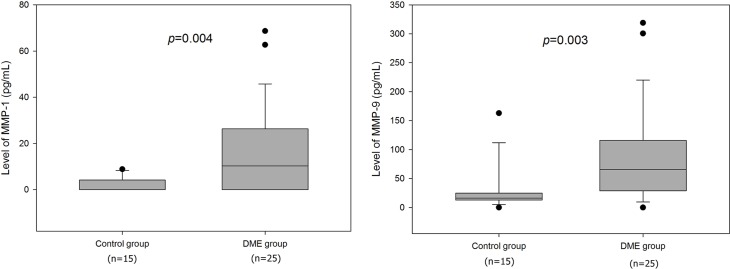
We found a significant difference between the average MMP-1 concentrations in the study group (16 ± 19 pg/mL) and those in the control group (2 ± 3 pg/mL, p = 0.005). The average concentration of MMP-9 also differed significantly between the study group (84 ± 80 pg/mL) and the control group (31± 41 pg/mL, p = 0.002). The p-values in the box plots where median values were compared are slightly different from p-values in the text where mean values were compared. However, statistical significances were not different (Fig 1).
Fig 1. The box and whisker plots for levels of metalloproteinases (MMPs).
The bottom and top of the box are the first and third quartiles with a line to show the median value. The whiskers show the 10th and 90th percentiles. Circles represent outliers. The levels of MMP-1 and MMP-9 are increased in diabetic macular edema (DME) group with significant differences.
DME subgroup analysis by the stage of DR
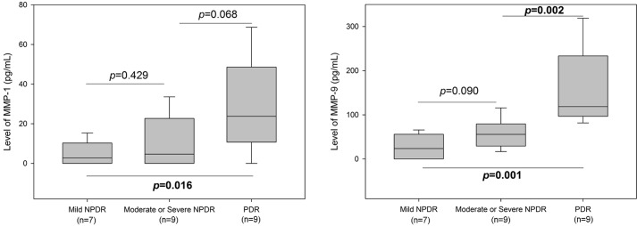
The concentrations of MMP-1 and MMP-9 by the stage of DR also differed significantly among groups (p = 0.03, p<0.001, respectively). Post hoc analysis showed a significant difference in MMP-1 concentrations between the mild NPDR group (n = 8, 5 ± 6 pg/mL) and the PDR group (n = 9, 29 ± 24 pg/mL, p = 0.012). We found no significant differences between the mild NPDR group and the moderate or severe NPDR group (n = 9, 11 ± 13 pg/mL) (p = 0.351), or between the moderate or severe NPDR group and the PDR group (p = 0.077).
MMP-9 concentrations differed significantly between the mild NPDR group (27 ± 24 pg/mL) and the PDR group (157 ± 90 pg/mL, p < 0.001); and between the moderate or severe NPDR group (56 ± 32 pg/mL) and the PDR group (p < 0.001). There was no significant difference between the mild NPDR and moderate or severe NPDR group (p = 0.091). The p-values were slightly different in box plots where the median values are compared instead of the mean values (Fig 2).
Fig 2. The concentrations of metalloproteinase (MMP)-1and MMP-9 by the stage of diabetic retinopathy differed significantly among groups.
Post hoc analysis showed a significant difference in MMP-1 concentrations between the mild NPDR (non-proliferative diabetic retinopathy) group and the PDR (proliferative diabetic retinopathy) group. MMP-9 concentrations differed significantly between the mild NPDR group and the PDR group; and between the moderate or severe NPDR group and the PDR group.
DME subgroup analysis by PRP-treatment
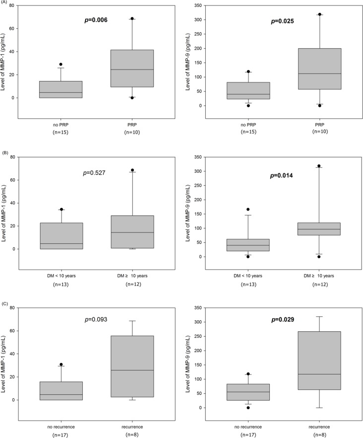
Both MMP-1 and MMP-9 concentrations were significantly higher in PRP-treated eyes (n = 10) compared to non- PRP-treated eyes (n = 15), (MMP-1: 8 ± 10 pg/mL versus 28 ± 23 pg/mL; MMP-9: 52 ± 37 pg/mL versus 133 ± 104 pg/mL, p = 0.005, p = 0.023, respectively) (Fig 3(A)).
Fig 3.
(A) Both metalloproteinase (MMP)-1 and MMP-9 concentrations were significantly higher in pan-retinal photocoagulation (PRP)-treated eyes. (B) MMP-9 concentration was significantly higher in DM eyes with diabetes ≥ 10 years. But there was no significant difference in level of MMP-1 between the two groups. (C) The eyes showed recurrence within 3 months of diabetic macular edema had significantly higher MMP-9 levels than non-recurrent eyes. But there was no statistical difference between the two groups in expression of MMP-1.
DME subgroup analysis by DM duration
The DM eyes were classified as those with diabetes<10 years (n = 13) and those with diabetes≥10 years (n = 12). There was no significant difference in MMP-1 between the two groups (12 ± 13 pg/mL versus 20 ± 23 pg/mL, p = 0.538), whereas MMP-9 differed significantly between the two groups (52 ± 45 pg/mL versus 120± 96, p = 0.011) (Fig 3(B)).
DME subgroup analysis by recurrence within 3 months
Recurrence within 3 months of DME occurred in eight eyes. These eyes had significantly higher MMP-9 levels than non-recurrent eyes (145 ± 113 versus 56 ± 37 pg/mL, p = 0.027), but no significant difference was evident in MMP-1 levels (10± 10 pg/mL versus 28 ± 26 pg/mL, p = 0.097) (Fig 3(C)).
The correlation analysis of concentration of MMPs with various factors
Spearman’s correlations were sought among age, axial length, and DM duration; between HbA1C and MMP levels; and between age and MMP levels. The MMP-1 level correlated significantly with age (r = -0.48, p = 0.01). The MMP-9 level correlated significantly with both axial length (r = -0.59, p < 0.01) and DM duration (r = 049, p = 0.01). The HbA1C level did not correlate with MMP levels (Table 2).
Table 2. The r-values (the rho values) derived by Spearman’s rank correlation (p-values, two-sided).
| Age | Axial length | DM duration | HbA1C level | MMP-1 level | MMP-9 level | |
|---|---|---|---|---|---|---|
| Age | -0.20 (0.35) | 0.04 (0.86) | 0.15 (0.58) | -0.48 (0.01) | -0.18 (0.38) | |
| Axial length | -0.20 (0.35) | -0.13 (0.56) | 0.36 (0.18) | -0.17 (0.44) | -0.59 (<0.01) | |
| DM duration | 0.04 (0.86) | -0.13 (0.56) | 0.52 (0.05) | 0.23 (0.26) | 0.49 (0.01) | |
| Hb1AC level | 0.15 (0.58) | 0.36 (0.18) | 0.52 (0.05) | 0.20 (0.48) | 0.25 (0.38) | |
| MMP-1 level | -0.48 (0.01) | -0.17 (0.44) | 0.23 (0.26) | 0.20 (0.48) | 0.63 (<0.01) | |
| MMP-9 level | -0.18 (0.38) | -0.59 (<0.01) | 0.49 (0.01) | 0.25 (0.38) | 0.63 (<0.01) |
MMP, matrix metalloproteinase; HbA1C, glycated hemoglobin
Discussion
Most studies have either measured vascular MMP levels in type 1 DM patients or been in vitro experiments[11–15, 32–34]. Although some studies have reported increased ocular MMP levels in diabetic patients, they did not report these levels in different types of patients classified according to DR stage, DM duration, PRP treatment, or degree of myopia [8, 16]. To the best of our knowledge, the present study is the first to characterize factors that may affect MMP-1 and MMP-9 levels in DME patients.
MMP-1 and MMP-9 concentrations in the anterior chamber of DR patients were increased, suggesting that ECM remodeling may be more active in DM patients than in patients without DM. This change was prominently found in patients with PDR and in proportion to the duration of DM.
Flaxel et al. reported that in cultured retinal pigment epithelial cells, MMP-2 and MMP-3 secretion increased to twice that of control values after PRP[35]. However, Sanchez et al. reported that proMMP-9 activity was significantly decreased in vitreous samples from patients with PDR after PRP[36]. In the present study, concentrations of MMPs in the aqueous humor were higher in patients with PRP. Although these results imply that there is increased ECM remodeling in PRP patients, the results may be confounded by other variables such as the stage of DR or the PRP status. We therefore analyzed PRP-treated patients according to the DR stage, to remove possible confounding variables, but the small sample size made it impossible to reach any significant conclusions. Additional studies involving larger cohorts with close follow-ups are necessary to confirm our results.
Axial myopia has a protective association with DR[37, 38]. There are several possible explanations for this relationship. First, decreased vascular diameters in myopic eyes may play a protective role in this association, because DR progression is associated with increased ocular blood flow[39–42]. Second, the dilution effects of cytokines such as transforming growth factor-beta may increase the ocular volume in axial myopia[7]. Third, posterior vitreous detachment (PVD) and vitreous syneresis, frequently present in myopia, may lower the risk of neovascularization progression[43–45]. This protective effect could be associated with the removal of vitreous scaffolds and improved oxygenation as a result of PVD[38, 46, 47] and MMP-9 has been associated with PVD[20]. Fourth, chorioretinal thinning in myopia may reduce oxygen demand and make it easier for oxygen to diffuse into the vitreous[38, 46]. We found that MMP-9 concentrations in the aqueous humor decreased with axial length elongation. In general, our results support these previous theories; however, we did not confirm the effects of PVD on MMP concentration in our study.
We found less significant differences in MMP-1 levels than in MMP-9 levels, although we did find significant differences in MMP-1 concentrations between the DME group and the control group and between the mild NPDR group and the PDR group. In animal model studies, MMP-9 has been shown to have important roles in DR progression to PDR, by facilitating apoptosis of the retinal capillary endothelium and altering tight junctions of the BRB[16, 17, 48]. Studies in humans have also shown significantly increased concentrations of MMP-9 in the aqueous humor[8]. MMP-1 and interstitial collagenase also have roles in extracellular matrix remodeling. They are found mainly in tissues with abundant collagens, such as the macrovascular structure, adipocytes, and the skin of animals and humans[49–52]. The results of the present study suggest that MMP-1 may have a less important role than MMP-9 in inducing DM complications in retinas with a microvascular structure and little connective tissue. A previous study also showed no significant differences in level of MMP-1 between DME and a control group[8].
Our study had several limitations. First, the small number of patients limited our results, and further studies enrolling a larger number of subjects are warranted in the future. Second, the retrospective cross-sectional design of our study made it difficult to identify causal relationships. A longitudinal study such as a randomized controlled trial is needed to confirm the causal relationships of MMP and DR.
In summary, compared to controls, the concentrations of MMP-9 and MMP-1 were increased in the eyes of DR patients, suggesting that MMP-9 and MMP-1 may be involved in the pathogenesis of DME. Furthermore, MMP-9 may have a more important role in the pathogenesis. Additional studies involving larger cohorts are necessary to confirm the results of this study and to show that suppression of MMP levels can reduce the complications of DME. Such studies should increase our understanding of the pathogenesis of MMPs in DR, and may provide a basis for therapeutic approaches to the treatment of DM patients.
Supporting Information
The raw data of this study.
(XLSX)
The standard curves of MMPs of this study.
(JPG)
Data Availability
All relevant data are within the paper and its Supporting Information files.
Funding Statement
The authors have no support or funding to report.
References
- 1.Klein BE. Overview of epidemiologic studies of diabetic retinopathy. Ophthalmic Epidemiol. 2007;14(4):179–83. Epub 2007/09/27. 10.1080/09286580701396720 . [DOI] [PubMed] [Google Scholar]
- 2.Varma R, Bressler NM, Doan QV, Gleeson M, Danese M, Bower JK, et al. Prevalence of and risk factors for diabetic macular edema in the United States. JAMA Ophthalmol. 2014;132(11):1334–40. Epub 2014/08/16. 10.1001/jamaophthalmol.2014.2854 . [DOI] [PMC free article] [PubMed] [Google Scholar]
- 3.Wong TY, Hyman L. Population-based studies in ophthalmology. American journal of ophthalmology. 2008;146(5):656–63. Epub 2008/11/06. 10.1016/j.ajo.2008.07.048 . [DOI] [PubMed] [Google Scholar]
- 4.Aiello LP, Avery RL, Arrigg PG, Keyt BA, Jampel HD, Shah ST, et al. Vascular endothelial growth factor in ocular fluid of patients with diabetic retinopathy and other retinal disorders. N Engl J Med. 1994;331(22):1480–7. Epub 1994/12/01. 10.1056/nejm199412013312203 . [DOI] [PubMed] [Google Scholar]
- 5.Wells JA, Glassman AR, Ayala AR, Jampol LM, Aiello LP, Antoszyk AN, et al. Aflibercept, bevacizumab, or ranibizumab for diabetic macular edema. N Engl J Med. 2015;372(13):1193–203. Epub 2015/02/19. 10.1056/NEJMoa1414264 ; PubMed Central PMCID: PMCPmc4422053. [DOI] [PMC free article] [PubMed] [Google Scholar]
- 6.Das A, McGuire PG, Rangasamy S. Diabetic Macular Edema: Pathophysiology and Novel Therapeutic Targets. Ophthalmology. 2015;122(7):1375–94. Epub 2015/05/04. 10.1016/j.ophtha.2015.03.024 . [DOI] [PubMed] [Google Scholar]
- 7.Yamamoto N, Itonaga K, Marunouchi T, Majima K. Concentration of transforming growth factor beta2 in aqueous humor. Ophthalmic Res. 2005;37(1):29–33. Epub 2005/01/08. 10.1159/000083019 . [DOI] [PubMed] [Google Scholar]
- 8.Jonas JB, Jonas RA, Neumaier M, Findeisen P. Cytokine concentration in aqueous humor of eyes with diabetic macular edema. Retina (Philadelphia, Pa). 2012;32(10):2150–7. Epub 2012/05/24. 10.1097/IAE.0b013e3182576d07 . [DOI] [PubMed] [Google Scholar]
- 9.Visse R, Nagase H. Matrix metalloproteinases and tissue inhibitors of metalloproteinases: structure, function, and biochemistry. Circ Res. 2003;92(8):827–39. Epub 2003/05/06. 10.1161/01.res.0000070112.80711.3d . [DOI] [PubMed] [Google Scholar]
- 10.Hu J, Van den Steen PE, Sang QX, Opdenakker G. Matrix metalloproteinase inhibitors as therapy for inflammatory and vascular diseases. Nature reviews Drug discovery. 2007;6(6):480–98. Epub 2007/06/02. 10.1038/nrd2308 . [DOI] [PubMed] [Google Scholar]
- 11.Thrailkill KM, Bunn RC, Moreau CS, Cockrell GE, Simpson PM, Coleman HN, et al. Matrix metalloproteinase-2 dysregulation in type 1 diabetes. Diabetes Care. 2007;30(9):2321–6. Epub 2007/06/15. 10.2337/dc07-0162 ; PubMed Central PMCID: PMCPmc2239013. [DOI] [PMC free article] [PubMed] [Google Scholar]
- 12.Toni M, Hermida J, Goni MJ, Fernandez P, Parks WC, Toledo E, et al. Matrix metalloproteinase-10 plays an active role in microvascular complications in type 1 diabetic patients. Diabetologia. 2013;56(12):2743–52. Epub 2013/10/01. 10.1007/s00125-013-3052-4 . [DOI] [PubMed] [Google Scholar]
- 13.Gharagozlian S, Svennevig K, Bangstad HJ, Winberg JO, Kolset SO. Matrix metalloproteinases in subjects with type 1 diabetes. BMC Clin Pathol. 2009;9:7 Epub 2009/09/18. 10.1186/1472-6890-9-7 ; PubMed Central PMCID: PMCPmc2753592. [DOI] [PMC free article] [PubMed] [Google Scholar]
- 14.Jacqueminet S, Ben Abdesselam O, Chapman MJ, Nicolay N, Foglietti MJ, Grimaldi A, et al. Elevated circulating levels of matrix metalloproteinase-9 in type 1 diabetic patients with and without retinopathy. Clin Chim Acta. 2006;367(1–2):103–7. Epub 2006/01/24. 10.1016/j.cca.2005.11.029 . [DOI] [PubMed] [Google Scholar]
- 15.Thrailkill KM, Moreau CS, Cockrell GE, Jo CH, Bunn RC, Morales-Pozzo AE, et al. Disease and gender-specific dysregulation of NGAL and MMP-9 in type 1 diabetes mellitus. Endocrine. 2010;37(2):336–43. Epub 2010/10/21. 10.1007/s12020-010-9308-6 ; PubMed Central PMCID: PMCPmc2959192. [DOI] [PMC free article] [PubMed] [Google Scholar]
- 16.Giebel SJ, Menicucci G, McGuire PG, Das A. Matrix metalloproteinases in early diabetic retinopathy and their role in alteration of the blood-retinal barrier. Laboratory investigation; a journal of technical methods and pathology. 2005;85(5):597–607. Epub 2005/02/16. 10.1038/labinvest.3700251 . [DOI] [PubMed] [Google Scholar]
- 17.Kowluru RA, Zhong Q, Santos JM. Matrix metalloproteinases in diabetic retinopathy: potential role of MMP-9. Expert opinion on investigational drugs. 2012;21(6):797–805. Epub 2012/04/24. 10.1517/13543784.2012.681043 ; PubMed Central PMCID: PMCPmc3802521. [DOI] [PMC free article] [PubMed] [Google Scholar]
- 18.Descamps FJ, Martens E, Kangave D, Struyf S, Geboes K, Van Damme J, et al. The activated form of gelatinase B/matrix metalloproteinase-9 is associated with diabetic vitreous hemorrhage. Experimental eye research. 2006;83(2):401–7. Epub 2006/04/29. 10.1016/j.exer.2006.01.017 . [DOI] [PubMed] [Google Scholar]
- 19.Noda K, Ishida S, Inoue M, Obata K, Oguchi Y, Okada Y, et al. Production and activation of matrix metalloproteinase-2 in proliferative diabetic retinopathy. Investigative ophthalmology & visual science. 2003;44(5):2163–70. Epub 2003/04/26. . [DOI] [PubMed] [Google Scholar]
- 20.Jin M, Kashiwagi K, Iizuka Y, Tanaka Y, Imai M, Tsukahara S. Matrix metalloproteinases in human diabetic and nondiabetic vitreous. Retina (Philadelphia, Pa). 2001;21(1):28–33. Epub 2001/02/24. . [DOI] [PubMed] [Google Scholar]
- 21.Kosano H, Okano T, Katsura Y, Noritake M, Kado S, Matsuoka T, et al. ProMMP-9 (92 kDa gelatinase) in vitreous fluid of patients with proliferative diabetic retinopathy. Life sciences. 1999;64(25):2307–15. Epub 1999/06/22. . [DOI] [PubMed] [Google Scholar]
- 22.Hawinkels LJ, Zuidwijk K, Verspaget HW, de Jonge-Muller ES, van Duijn W, Ferreira V, et al. VEGF release by MMP-9 mediated heparan sulphate cleavage induces colorectal cancer angiogenesis. European journal of cancer (Oxford, England: 1990). 2008;44(13):1904–13. Epub 2008/08/12. 10.1016/j.ejca.2008.06.031 . [DOI] [PubMed] [Google Scholar]
- 23.Ebrahem Q, Chaurasia SS, Vasanji A, Qi JH, Klenotic PA, Cutler A, et al. Cross-talk between vascular endothelial growth factor and matrix metalloproteinases in the induction of neovascularization in vivo. The American journal of pathology. 2010;176(1):496–503. Epub 2009/12/02. 10.2353/ajpath.2010.080642 ; PubMed Central PMCID: PMCPmc2797907. [DOI] [PMC free article] [PubMed] [Google Scholar]
- 24.Abu El-Asrar AM, Mohammad G, Nawaz MI, Siddiquei MM, Van den Eynde K, Mousa A, et al. Relationship between vitreous levels of matrix metalloproteinases and vascular endothelial growth factor in proliferative diabetic retinopathy. PloS one. 2013;8(12):e85857 Epub 2014/01/07. 10.1371/journal.pone.0085857 ; PubMed Central PMCID: PMCPmc3877391. [DOI] [PMC free article] [PubMed] [Google Scholar]
- 25.Yildirim N, Sahin A, Erol N, Kara S, Uslu S, Topbas S. The relationship between plasma MMP-9 and TIMP-2 levels and intraocular pressure elevation in diabetic patients after intravitreal triamcinolone injection. Journal of glaucoma. 2008;17(4):253–6. Epub 2008/06/17. 10.1097/IJG.0b013e31815c3a07 . [DOI] [PubMed] [Google Scholar]
- 26.Vandooren J, Born B, Solomonov I, Zajac E, Saldova R, Senske M, et al. Circular trimers of gelatinase B/matrix metalloproteinase-9 constitute a distinct population of functional enzyme molecules differentially regulated by tissue inhibitor of metalloproteinases-1. Biochem J. 2015;465(2):259–70. Epub 2014/11/02. 10.1042/bj20140418 ; PubMed Central PMCID: PMCPmc4399976. [DOI] [PMC free article] [PubMed] [Google Scholar]
- 27.Rohani MG, Pilcher BK, Chen P, Parks WC. Cdc42 inhibits ERK-mediated collagenase-1 (MMP-1) expression in collagen-activated human keratinocytes. J Invest Dermatol. 2014;134(5):1230–7. Epub 2013/12/20. 10.1038/jid.2013.499 ; PubMed Central PMCID: PMCPmc3989453. [DOI] [PMC free article] [PubMed] [Google Scholar]
- 28.Lin A, Salvador A, Carter JM. Multiplexed Microsphere Suspension Array-Based Immunoassays. Methods in molecular biology (Clifton, NJ). 2015;1318:107–18. Epub 2015/07/15. 10.1007/978-1-4939-2742-5_11 . [DOI] [PubMed] [Google Scholar]
- 29.Jia Y, Hu DN, Zhu D, Zhang L, Gu P, Fan X, et al. MMP-2, MMP-3, TIMP-1, TIMP-2, and TIMP-3 protein levels in human aqueous humor: relationship with axial length. Investigative ophthalmology & visual science. 2014;55(6):3922–8. Epub 2014/05/31. 10.1167/iovs.14-13983 . [DOI] [PubMed] [Google Scholar]
- 30.Kramer M, Hasanreisoglu M, Feldman A, Axer-Siegel R, Sonis P, Maharshak I, et al. Monocyte chemoattractant protein-1 in the aqueous humour of patients with age-related macular degeneration. Clinical & experimental ophthalmology. 2012;40(6):617–25. Epub 2011/12/17. 10.1111/j.1442-9071.2011.02747.x . [DOI] [PubMed] [Google Scholar]
- 31.Morgan E, Varro R, Sepulveda H, Ember JA, Apgar J, Wilson J, et al. Cytometric bead array: a multiplexed assay platform with applications in various areas of biology. Clinical immunology (Orlando, Fla). 2004;110(3):252–66. Epub 2004/03/30. 10.1016/j.clim.2003.11.017 . [DOI] [PubMed] [Google Scholar]
- 32.Peeters SA, Engelen L, Buijs J, Chaturvedi N, Fuller JH, Schalkwijk CG, et al. Plasma levels of matrix metalloproteinase-2, -3, -10, and tissue inhibitor of metalloproteinase-1 are associated with vascular complications in patients with type 1 diabetes: the EURODIAB Prospective Complications Study. Cardiovascular diabetology. 2015;14:31 Epub 2015/04/08. 10.1186/s12933-015-0195-2 ; PubMed Central PMCID: PMCPmc4355971. [DOI] [PMC free article] [PubMed] [Google Scholar]
- 33.Kowluru RA, Santos JM, Mishra M. Epigenetic modifications and diabetic retinopathy. BioMed research international. 2013;2013:635284 Epub 2013/11/29. 10.1155/2013/635284 ; PubMed Central PMCID: PMCPmc3826295. [DOI] [PMC free article] [PubMed] [Google Scholar]
- 34.Kowluru RA. Role of matrix metalloproteinase-9 in the development of diabetic retinopathy and its regulation by H-Ras. Investigative ophthalmology & visual science. 2010;51(8):4320–6. Epub 2010/03/12. 10.1167/iovs.09-4851 ; PubMed Central PMCID: PMCPmc2910650. [DOI] [PMC free article] [PubMed] [Google Scholar]
- 35.Flaxel C, Bradle J, Acott T, Samples JR. Retinal pigment epithelium produces matrix metalloproteinases after laser treatment. Retina (Philadelphia, Pa). 2007;27(5):629–34. Epub 2007/06/15. 10.1097/01.iae.0000249561.02567.fd . [DOI] [PubMed] [Google Scholar]
- 36.Sanchez MC, Luna JD, Barcelona PF, Gramajo AL, Juarez PC, Riera CM, et al. Effect of retinal laser photocoagulation on the activity of metalloproteinases and the alpha(2)-macroglobulin proteolytic state in the vitreous of eyes with proliferative diabetic retinopathy. Experimental eye research. 2007;85(5):644–50. Epub 2007/09/11. 10.1016/j.exer.2007.07.018 . [DOI] [PubMed] [Google Scholar]
- 37.Man RE, Sasongko MB, Wang JJ, Lamoureux EL. Association between myopia and diabetic retinopathy: a review of observational findings and potential mechanisms. Clinical & experimental ophthalmology. 2013;41(3):293–301. Epub 2012/09/11. 10.1111/j.1442-9071.2012.02872.x . [DOI] [PubMed] [Google Scholar]
- 38.Lim LS, Lamoureux E, Saw SM, Tay WT, Mitchell P, Wong TY. Are myopic eyes less likely to have diabetic retinopathy? Ophthalmology. 2010;117(3):524–30. Epub 2009/12/25. 10.1016/j.ophtha.2009.07.044 . [DOI] [PubMed] [Google Scholar]
- 39.Patel V, Rassam S, Newsom R, Wiek J, Kohner E. Retinal blood flow in diabetic retinopathy. Bmj. 1992;305(6855):678–83. Epub 1992/09/19. ; PubMed Central PMCID: PMCPmc1882919. [DOI] [PMC free article] [PubMed] [Google Scholar]
- 40.Lim LS, Cheung CY, Lin X, Mitchell P, Wong TY, Mei-Saw S. Influence of refractive error and axial length on retinal vessel geometric characteristics. Investigative ophthalmology & visual science. 2011;52(2):669–78. Epub 2010/09/18. 10.1167/iovs.10-6184 . [DOI] [PubMed] [Google Scholar]
- 41.Quigley M, Cohen S. A new pressure attenuation index to evaluate retinal circulation. A link to protective factors in diabetic retinopathy. Archives of ophthalmology (Chicago, Ill: 1960). 1999;117(1):84–9. Epub 1999/02/04. . [DOI] [PubMed] [Google Scholar]
- 42.Cunha-Vaz JG, Fonseca JR, de Abreu JR, Lima JJ. Studies on retinal blood flow. II. Diabetic retinopathy. Archives of ophthalmology (Chicago, Ill: 1960). 1978;96(5):809–11. Epub 1978/05/01. . [DOI] [PubMed] [Google Scholar]
- 43.Tagawa H, McMeel JW, Furukawa H, Quiroz H, Murakami K, Takahashi M, et al. Role of the vitreous in diabetic retinopathy. I. Vitreous changes in diabetic retinopathy and in physiologic aging. Ophthalmology. 1986;93(5):596–601. Epub 1986/05/01. . [DOI] [PubMed] [Google Scholar]
- 44.Tagawa H, McMeel JW, Trempe CL. Role of the vitreous in diabetic retinopathy. II. Active and inactive vitreous changes. Ophthalmology. 1986;93(9):1188–92. Epub 1986/09/01. . [DOI] [PubMed] [Google Scholar]
- 45.Akiba J, Arzabe CW, Trempe CL. Posterior vitreous detachment and neovascularization in diabetic retinopathy. Ophthalmology. 1990;97(7):889–91. Epub 1990/07/01. . [DOI] [PubMed] [Google Scholar]
- 46.Stefansson E. Ocular oxygenation and the treatment of diabetic retinopathy. Survey of ophthalmology. 2006;51(4):364–80. Epub 2006/07/05. 10.1016/j.survophthal.2006.04.005 . [DOI] [PubMed] [Google Scholar]
- 47.Hendrikse F, Yeo KT. [Role of the vitreous body in diabetic retinopathy]. Klinische Monatsblatter fur Augenheilkunde. 1993;203(5):319–23. Epub 1993/11/01. 10.1055/s-2008-1045684 . [DOI] [PubMed] [Google Scholar]
- 48.Mohammad G, Kowluru RA. Diabetic retinopathy and signaling mechanism for activation of matrix metalloproteinase-9. Journal of cellular physiology. 2012;227(3):1052–61. Epub 2011/05/14. 10.1002/jcp.22822 ; PubMed Central PMCID: PMCPmc3801274. [DOI] [PMC free article] [PubMed] [Google Scholar]
- 49.Galis ZS, Sukhova GK, Lark MW, Libby P. Increased expression of matrix metalloproteinases and matrix degrading activity in vulnerable regions of human atherosclerotic plaques. The Journal of clinical investigation. 1994;94(6):2493–503. Epub 1994/12/01. 10.1172/jci117619 ; PubMed Central PMCID: PMCPmc330083. [DOI] [PMC free article] [PubMed] [Google Scholar]
- 50.Nambi V, Morrison AC, Hoogeveen RC, Coresh J, Miles S, Rhodes CE, et al. Matrix metalloproteinase-1 and tissue inhibitors do not predict incident coronary artery disease in the atherosclerosis risk in communities (ARIC) study. Texas Heart Institute journal / from the Texas Heart Institute of St Luke's Episcopal Hospital, Texas Children's Hospital. 2008;35(4):388–94. Epub 2009/01/22. ; PubMed Central PMCID: PMCPmc2607088. [PMC free article] [PubMed] [Google Scholar]
- 51.Zeng W, Tahrani A, Shakher J, Varani J, Hughes S, Dubb K, et al. Effects of a synthetic retinoid on skin structure, matrix metalloproteinases, and procollagen in healthy and high-risk subjects with diabetes. Journal of diabetes and its complications. 2011;25(6):398–404. Epub 2011/11/08. 10.1016/j.jdiacomp.2011.10.002 ; PubMed Central PMCID: PMCPmc3240843. [DOI] [PMC free article] [PubMed] [Google Scholar]
- 52.Papazoglou D, Papatheodorou K, Papanas N, Papadopoulos T, Gioka T, Kabouromiti G, et al. Matrix metalloproteinase-1 and tissue inhibitor of metalloproteinases-1 levels in severely obese patients: what is the effect of weight loss? Experimental and clinical endocrinology & diabetes: official journal, German Society of Endocrinology [and] German Diabetes Association. 2010;118(10):730–4. Epub 2010/04/03. 10.1055/s-0030-1249671 . [DOI] [PubMed] [Google Scholar]
Associated Data
This section collects any data citations, data availability statements, or supplementary materials included in this article.
Supplementary Materials
The raw data of this study.
(XLSX)
The standard curves of MMPs of this study.
(JPG)
Data Availability Statement
All relevant data are within the paper and its Supporting Information files.


