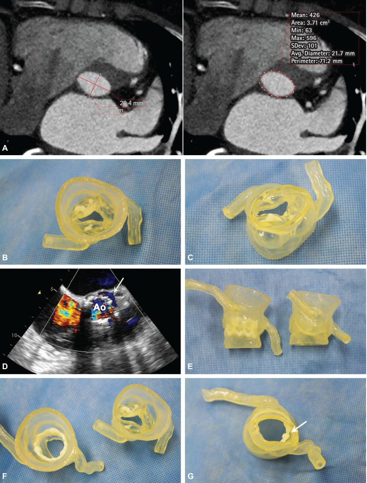
A 71-year-old woman had severe aortic stenosis (AS) (aortic valve area, 0.6 cm2; mean valve gradient, 47 mmHg; and peak velocity, 4.2 m/s) and cardiac symptoms. She had a history of stroke and paroxysmal atrial fibrillation. Our multidisciplinary heart team decided to perform transcatheter aortic valve implantation (TAVI). Before TAVI, computed tomography (CT) was performed, and the measurements of the aortic annulus were as follows: diameter, 25.4×19 mm; perimeter, 71.2 mm; area, 3.72 cm2; distance to the right coronary artery, 13.2 mm; and distance to the left coronary artery, 12.3 mm (Fig. 1A). Using the CT data, a 3D printing model was created (Materialise®, Luven, Belgium). 3D printing model provided the cardiologist with an intuitive perception of the cardiovascular anatomy and measurements in preparation for TAVI (Fig. 1B and C).
Fig. 1.
A 23-mm Edward-Sapien prosthetic valve (Edwards Lifesciences, Irvine, CA, USA) was selected. TAVI was successfully performed with mild paravalvular leakage of the left coronary sinus (Fig. 1D). A post-TAVI 3D model clearly showed the position of the replaced valve (Fig. 1E), change in the aortic annulus (i.e., from an oval to a round shape) (Fig. 1F), and the site of mild paravalvular leakage (Fig. 1G).
CT is essential tool for TAVI.1),2),3),4) Although CT measurements increase the success rate of TAVI, its success is highly dependent on the operator's skill and expertise. A 3D printing model may offer a greater possibility for simulating patient specific complex structural characterization procedures5) and overcome the limitation of non-invasive TAVI-poor direct visualization.
Footnotes
The authors have no financial conflicts of interest.
References
- 1.Makkar RR, Fontana GP, Jilaihawi H, et al. Transcatheter aortic-valve replacement for inoperable severe aortic stenosis. N Engl J Med. 2012;366:1696–1704. doi: 10.1056/NEJMoa1202277. [DOI] [PubMed] [Google Scholar]
- 2.Kodali SK, Williams MR, Smith CR, et al. Two-year outcomes after transcatheter or surgical aortic-valve replacement. N Engl J Med. 2012;366:1686–1695. doi: 10.1056/NEJMoa1200384. [DOI] [PubMed] [Google Scholar]
- 3.Cao C, Ang SC, Indraratna P, et al. Systematic review and meta-analysis of transcatheter aortic valve implantation versus surgical aortic valve replacement for severe aortic stenosis. Ann Cardiothorac Surg. 2013;2:10–23. doi: 10.3978/j.issn.2225-319X.2012.11.09. [DOI] [PMC free article] [PubMed] [Google Scholar]
- 4.Nguyen G, Leipsic J. Cardiac computed tomography and computed tomography angiography in the evaluation of patients prior to transcatheter aortic valve implantation. Curr Opin Cardiol. 2013;28:497–504. doi: 10.1097/HCO.0b013e32836245c1. [DOI] [PubMed] [Google Scholar]
- 5.Valverde I, Gomez G, Coserria JF, et al. 3D printed models for planning endovascular stenting in transverse aortic arch hypoplasia. Catheter Cardiovasc Interv. 2015;85:1006–1012. doi: 10.1002/ccd.25810. [DOI] [PubMed] [Google Scholar]


