

Abstract
A 37-year-old male presented with severe oral and genital mucosal ulcers, lichenoid eruption and twenty-nail dystrophy. Systemic examination was normal, except for anemia. On investigations, he was found to have persistently elevated peripheral eosinophilia, absolute eosinophil count >5000/mm3, bone marrow showing increased eosinophilic precursors, and infiltration by atypical cells. The serum vitamin B12 levels were grossly elevated, and Philadelphia chromosome study was negative. Thus, a diagnosis of chronic eosinophilic leukemia was made. The patient showed excellent response to imatinib mesylate. We are reporting a rare type of leukemia presenting with predominantly cutaneous manifestations.
Keywords: Chronic eosinophilic leukemia, lichen planus, twenty-nail dystrophy
Introduction
What was known?
Chronic eosinophilic leukemia (CEL) is rare leukemia considered to be a type of idiopathic hypereosinophilic syndrome
CEL is characterized by an increase in eosinophils in the peripheral smear, bone marrow, and tissues and consequently, the patient has severe systemic manifestations.
Chronic eosinophilic leukemia (CEL) is a chronic myeloproliferative disease of unknown etiology in which clonal proliferation of eosinophilic precursors results in a persistently elevated number of eosinophils in blood, bone marrow or peripheral tissues resulting in organ injury, and mortality.[1] CEL is considered to be a subtype of a myeloproliferative variant of hypereosinophilic syndrome (HES) by some authors, whereas others consider it as an independent entity. Under normal circumstances, CEL is characterized by severe systemic involvement with subtle cutaneous manifestations. CEL is by itself rare, and we are reporting a case of CEL where the cutaneous manifestations predominated the systemic manifestations.
Case Report
A 37-year-old male presented with recurrent ulcerations of lips, glans penis, hyperpigmented skin lesions and dystrophic nails of 2½ years duration. Lesions started as painless erosions on glans penis and later on lips and oral cavity. Later, he noticed nail changes along with skin lesions on the elbows and trunk. He did not give any history of drug intake before the development of the lesions. He did not have any systemic symptoms and denied extramarital or premarital sexual exposures.
Examination revealed pallor and generalized nontender lymphadenopathy. Multiple confluent erosions with adherent whitish plaques and yellowish crusting were present over lips, buccal mucosa, uvula, glans penis, scrotum, and perianal region. Multiple well-defined hyperpigmented plaques with scaling were present over elbows, back of the trunk, and right buttock. All twenty nails were dystrophic [Figure 1]. Systemic examination was within normal limits.
Figure 1.
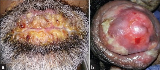
(a) Crusted plaques on the lips, (b) whitish plaques and erosions on the glans penis
Blood and peripheral smear examination showed normochromic anemia and severe eosinophilia (eosinophils – 30%). Absolute eosinophil count was elevated (5040 cells/cm3). Blood sugar, liver and renal function tests, and urine examination were normal, and stool examination for parasites was negative.
Serum B12 levels were elevated (>2000 IU/ml). Bone marrow aspirate showed severe eosinophilia and mild basophilia. Bone marrow trephine biopsy showed increase in eosinophilic precursors, eosinophils, and infiltration of bone marrow by atypical mononuclear cells suggestive of blast cells [Figure 2].
Figure 2.
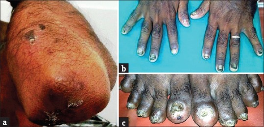
(a) Hyperpigmented plaques with scaling on the elbows, (b) dystrophic fingernails, (c) dystrophic toenails
Viral markers (human immunodeficiency virus, hepatitis B surface antigen, anti-hepatitis C virus, and herpes simplex virus 1 and 2) were negative. Antinuclear antibody profile, serum IgE levels (37.75 IU/ml), thyroid function test, serum electrophoresis, chest X-ray, electrocardiogram, echocardiogram, gastrointestinal endoscopy, Mantoux and pathergy test did not reveal any abnormalities. Fine needle aspiration and excision biopsy of lymph node showed reactive hyperplasia. Ultrasound abdomen showed mild hepatosplenomegaly. Computed tomography scan of the chest and abdomen did not reveal any malignancy.
Oral mucosal scraping and culture did not yield any fungal elements. Skin biopsies from the lesions on the trunk and elbows showed dermal edema, diffuse infiltration in the upper dermis and perivascular infiltration by neutrophils, and few eosinophils. However, direct immunofluorescence showed ragged fibrin in the basement membrane zone and colloid bodies staining with IgA, suggestive of lichen planus (LP) [Figure 3].
Figure 3.

Direct immunofluorescence showing ragged fibrin in the basement membrane zone (arrow) (direct immunofluorescence, ×400)
Genital biopsy showed features of a mild vasculitis with neutrophils and eosinophils. Philadelphia chromosome study was negative. F1P1-PDGFRA/ETV6-PDGFRB gene mutation studies and serum tryptase levels could not be done as these tests were not available.
The presentation with peripheral eosinophilia, absolute eosinophil count >5000/mm3, bone marrow showing increased eosinophilic precursors and infiltration by atypical cells suggestive of blast transformation, elevated Vitamin B12 levels, and negative Philadelphia chromosome enabled us to make a final diagnosis of CEL with cutaneous manifestations [Figure 4a–c].
Figure 4.

(a) Peripheral smear showing eosinophils (Leishman, ×1000), (b) bone marrow aspiration showing eosinophils (Leishman, ×1000), (c) bone marrow trephine biopsy showing eosinophils, eosinophil precursors, and blast cells (arrows) (Leishman, ×400)
The patient was started on imatinib mesylate 400 mg once daily and showed excellent response with the healing of the mucosal ulcers, disappearances of the LP lesions, and the eosinophil count became normal [Figure 5]. The patient is currently on follow-up.
Figure 5.

(a) Complete healing of oral lesion and (b) healing of genital lesions following therapy with imatinib mesylate 400 mg
Discussion
CEL is a rare entity, often under-diagnosed due to its overlapping features with HES with a male preponderance in the age group 25–55 years.[1,2,3] CEL is considered to be a subtype of the so-called idiopathic HES while others consider it to be a distinct entity of myeloproliferative disorder.[1,2,3] Allergic, parasitic, and other common causes of peripheral eosinophilia should be ruled out before diagnosing CEL. According to the classification of eosinophilic diseases by the WHO, CEL, not otherwise specified, is operationally defined by the absence of Philadelphia chromosome or rearrangement involving PDGFRA/B and FGFR1 gene and exclusion of other acute or chronic primary marrow neoplasms associated with eosinophilia. The FIP1L1-PDGRA fusion gene encodes an aberrant, constitutively activated tyrosine kinase that drives the eosinophil proliferation without the need for external growth stimuli.[2] CEL is histologically characterized by an increase in blasts in the bone marrow, and there is an evidence for clonality in the eosinophil lineage. If appropriate molecular analysis is not available, the diagnosis of CEL is made if there is a Philadelphia chromosome-negative myeloproliferative neoplasm with hematological features of CEL associated with splenomegaly, a marked elevation of serum Vitamin B12, and increased bone marrow eosinophil precursors.[4] Our patient satisfied the aforementioned criteria; therefore, we made a diagnosis of CEL with cutaneous manifestations.
The hyperproliferation of eosinophils and deposition in end organs including the skin is responsible for the clinical symptoms. The most common manifestations include weakness, fatigue, cough, dyspnea, myalgia, rash, and rhinitis.[5,6] All organ systems may be susceptible to effects of sustained eosinophilia. Progressive heart failure is a classical example of eosinophil-mediated organ injury and is the most common cause for mortality.[5,6] Other important manifestations include lung fibrosis, eosinophilic gastritis, hypertension, encephalopathy, ataxia, and thromboembolism. However, our case was unique, as the only systemic findings were anemia, lymphadenopathy, and hepatosplenomegaly without any systemic symptoms and it was the cutaneous manifestations which prompted the patient to consult the physician. Cutaneous manifestations are rare in CEL but include eczemas, dermographism, mucosal ulcers, angioedema, vasculitis, erythroderma, and erythema annulare centrifugam.[7] Mucosal ulceration has been reported in CEL, and the histopathology shows dermal infiltrate with eosinophils and perivascular neutrophilic and mononuclear infiltrate without true vasculitis as seen in our case.[4,7] The LP-like lesions and twenty-nail dystrophy seen in our patient have not been described in CEL previously to the best of our knowledge. LP has been described as a paraneoplastic manifestation rarely in thymoma and adult T-cell leukemia, but not in CEL.[8]
The main-stay of treatment of CEL is systemic steroids, hydroxyurea and the tyrosine kinase inhibitors imatinib, nilotinib, and sorafenib.[9] CEL patients require long-term follow-up as they can develop later acute myeloid leukemia and the presence of vital organ damage, carry a poor prognosis.[10]
We are reporting rare leukemia, CEL, presenting with an even rarer presentation of predominantly cutaneous manifestations rather than systemic manifestations and the LP with twenty-nail dystrophy seen in our patient has previously not been reported in literature. LP with twenty-nail dystrophy could be a new paraneoplastic syndrome.
Financial support and sponsorship
Nil.
Conflicts of interest
There are no conflicts of interest.
What is new?
Chronic eosinophilic leukemia (CEL) can present with predominant cutaneous manifestations, rather than systemic manifestations
Lichen planus (LP) with trachyonychia is a new cutaneous manifestation of CEL
LP with trachyonychia can be considered as a new paraneoplastic manifestation.
References
- 1.Kumar A, Sinha S, Tripathi AK. Chronic eosinophilic leukemia: A case report and review of literature. Indian J Hematol Blood Transfus. 2007;23:112–5. doi: 10.1007/s12288-008-0010-2. [DOI] [PMC free article] [PubMed] [Google Scholar]
- 2.Bain BJ, Fletcher SH. Chronic eosinophilic leukemias and the myeloproliferative variant of the hypereosinophilic syndrome. Immunol Allergy Clin North Am. 2007;27:377–88. doi: 10.1016/j.iac.2007.06.001. [DOI] [PubMed] [Google Scholar]
- 3.Gotlib J. World Health Organization-defined eosinophilic disorders: 2014 update on diagnosis, risk stratification, and management. Am J Hematol. 2014;89:325–37. doi: 10.1002/ajh.23664. [DOI] [PubMed] [Google Scholar]
- 4.Najera JE, Harn L. Chronic eosinophilic leukemia presenting with mouth and penile ulcers. Am J Med. 2012;125:e5–6. doi: 10.1016/j.amjmed.2011.11.017. [DOI] [PubMed] [Google Scholar]
- 5.Zachée P. Atypical myeloproliferative disorders in adults. Transfus Apher Sci. 2011;44:211–21. doi: 10.1016/j.transci.2011.01.017. [DOI] [PubMed] [Google Scholar]
- 6.Roufosse F, Weller PF. Practical approach to the patient with hypereosinophilia. J Allergy Clin Immunol. 2010;126:39–44. doi: 10.1016/j.jaci.2010.04.011. [DOI] [PMC free article] [PubMed] [Google Scholar]
- 7.Leiferman KM, Gleich GJ, Peters MS. Dermatologic manifestations of the hypereosinophilic syndromes. Immunol Allergy Clin North Am. 2007;27:415–41. doi: 10.1016/j.iac.2007.07.009. [DOI] [PubMed] [Google Scholar]
- 8.Sumida H, Sugaya M, Kamata M, Suga H, Miyagaki T, Ohmatsu H, et al. Lichen planus-like lesions as the first manifestation of adult T-cell leukaemia/lymphoma. Acta Derm Venereol. 2013;93:461–3. doi: 10.2340/00015555-1498. [DOI] [PubMed] [Google Scholar]
- 9.Butterfield JH, Weiler CR. Treatment of hypereosinophilic syndromes – The first 100 years. Semin Hematol. 2012;49:182–91. doi: 10.1053/j.seminhematol.2012.01.001. [DOI] [PubMed] [Google Scholar]
- 10.Podjasek JC, Butterfield JH. Mortality in hypereosinophilic syndrome: 19 years of experience at Mayo Clinic with a review of the literature. Leuk Res. 2013;37:392–5. doi: 10.1016/j.leukres.2012.12.016. [DOI] [PubMed] [Google Scholar]