Figure 1.
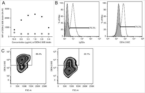
Staining method optimization using a dengue-specific hybridoma cell line, 4G2 (A) Titration of DEN-2–80E biotin for use in FACS staining. Final concentration of DEN-2–80E biotin and corresponding geomean fluorescent intensity (MFI) of the PE signal from the 4G2 pan dengue hybridoma (filled circles) and negative control Staphylococcus aureus antigen-specific hybridoma, UKNKC (open circles). (B) DEN-2–80E SA-PE staining identifies antibody secreting cells comparably to an IgG-specific stain. 4G2 hybridoma (transparent histogram), was stained with DEN-2–80E PE (right) or for the hybridoma subtype, IgG2a (left). For comparison, an IgG-1 type Staphylococcus aureus specific hybridoma (filled histogram) is overlayed, (right). (C) Effects of 100X concentration unlabeled DEN-2 80E pre-incubation on DEN-2–80E PE staining. 4G2 hybridomas were stained with 1.6 μg/mL of DEN-2–80E following pre-incubation with (right) or without (left) of 160 μg/mL of unlabeled DEN-2–80E.

