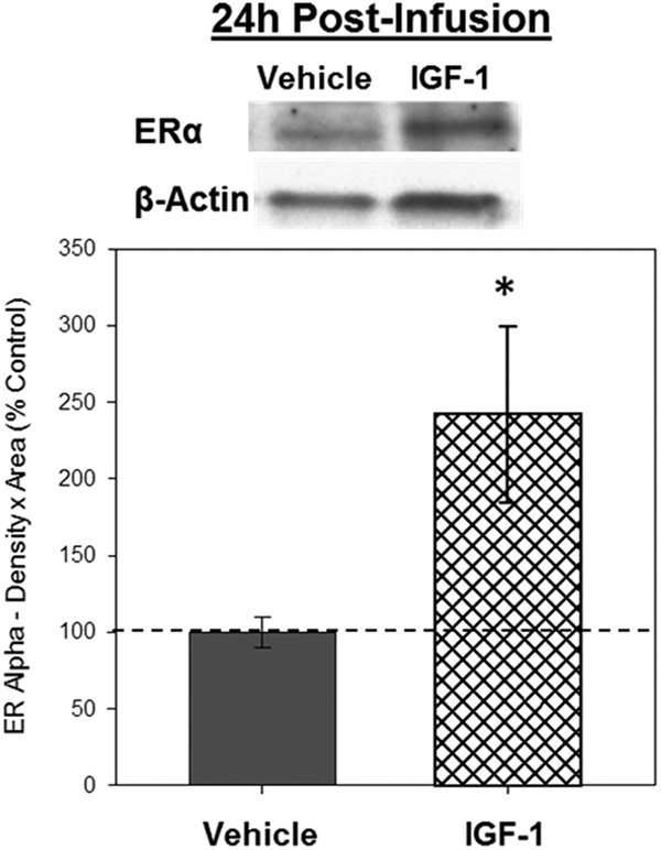
Figure 3.

Effects of IGF-1 infusion on total levels of ERα in the hippocampus 24 hours after infusion. Hippocampi were collected from ovariectomized rats 24 hours after an i.c.v. infusion of aCSF vehicle or IGF-1. Western blotting data showing effects of treatments on levels of ERα. Mean density × area (±SEM) relative to vehicle control group; *, P < .05. Representative blot images for ERα and the loading control β-actin are shown in insets above the graph.
