This study reveals that the simple behavior of fidgeting is sufficient to counteract the detrimental effects of prolonged sitting on leg endothelial function. Thus, we provide the first evidence that the unfavorable vascular effects of sitting are avoidable with small amounts of leg movement while seated for an extended period.
Keywords: physical inactivity, blood flow, endothelial function, leg movement, sitting
Abstract
Prolonged sitting impairs endothelial function in the leg vasculature, and this impairment is thought to be largely mediated by a sustained reduction in blood flow-induced shear stress. Indeed, preventing the marked reduction of shear stress during sitting with local heating abolishes the impairment in popliteal artery endothelial function. Herein, we tested the hypothesis that sitting-induced reductions in shear stress and ensuing endothelial dysfunction would be prevented by periodic leg movement, or “fidgeting.” In 11 young, healthy subjects, bilateral measurements of popliteal artery flow-mediated dilation (FMD) were performed before and after a 3-h sitting period during which one leg was subjected to intermittent fidgeting (1 min on/4 min off) while the contralateral leg remained still throughout and served as an internal control. Fidgeting produced a pronounced increase in popliteal artery blood flow and shear rate (prefidgeting, 33.7 ± 2.6 s−1 to immediately postfidgeting, 222.7 ± 28.3 s−1; mean ± SE; P < 0.001) that tapered off during the following 60 s. Fidgeting did not alter popliteal artery blood flow and shear rate of the contralateral leg, which was subjected to a reduction in blood flow and shear rate throughout the sitting period (presit, 71.7 ± 8.0 s−1 to 3-h sit, 20.2 ± 2.9 s−1; P < 0.001). Popliteal artery FMD was impaired after 3 h of sitting in the control leg (presit, 4.5 ± 0.3% to postsit: 1.6 ± 1.1%; P = 0.039) but improved in the fidgeting leg (presit, 3.7 ± 0.6% to postsit, 6.6 ± 1.2%; P = 0.014). Collectively, the present study provides evidence that prolonged sitting-induced leg endothelial dysfunction is preventable with small amounts of leg movement while sitting, likely through the intermittent increases in vascular shear stress.
NEW & NOTEWORTHY
This study reveals that the simple behavior of fidgeting is sufficient to counteract the detrimental effects of prolonged sitting on leg endothelial function. Thus, we provide the first evidence that the unfavorable vascular effects of sitting are avoidable with small amounts of leg movement while seated for an extended period.
alterations in local hemodynamic forces play a fundamental role in the etiology of atherosclerotic disease (7). As early as 1969, it was noted that atherosclerotic lesions preferentially develop in arterial regions characterized by low shear stress (3). Indeed, studies in cultured endothelial cells as well as in vivo studies in animals and humans demonstrate that low shear stress impairs endothelial function and instigates atherosclerosis (4, 9, 10, 16, 22, 24, 27). Notably, arteries of the lower limbs are exposed to marked reductions in blood flow and shear stress during sitting, and this in turn leads to transient endothelial dysfunction (15, 19, 20, 25). Consistent with the notion that sitting-induced leg endothelial dysfunction is mediated by a reduction in shear stress, we recently found that preventing the reduction of shear stress during prolonged sitting using local heating to increase blood flow abolished the impairment in popliteal artery endothelial function (20). Taken together, it is conceivable that the proatherogenic hemodynamic environment to which the vasculature of the lower limbs is exposed to during sitting contributes to the increased propensity of the leg vasculature to disease.
Herein we examined whether periodic leg muscle contraction, or “fidgeting,” during sitting would be effective at averting the marked reduction in leg blood flow and shear stress leading to preservation of leg endothelial function. Bilateral measurements of popliteal artery flow-mediated dilation (FMD) were performed before and after a 3-h sitting period during which one leg was subjected to intermittent fidgeting (1 min on/4 min off) while the contralateral leg remained still throughout and served as an internal control. We hypothesized that sitting-induced reductions in shear stress and ensuing endothelial dysfunction would be prevented by intermittent fidgeting.
METHODS
Eleven young healthy and recreationally active subjects (male, n = 7; and female, n = 4) recruited from the University of Missouri campus participated in this study (age, 26 ± 1 yr; height, 173.4 ± 3.3 cm; weight, 76.8 ± 6.1 kg; body mass index, 25.0 ± 1.1 kg/m2). All experimental procedures and measurements conformed to the Declaration of Helsinki and were approved by the University of Missouri Health Sciences Institutional Review Board. Before participating in the study, each subject provided written, informed consent. Subjects were recreationally active and nonsmokers with no history or symptoms of cardiovascular, pulmonary, metabolic, or neurological disease as determined from a medical health history questionnaire. No subjects were using prescribed or over-the-counter medications.
Experimental procedures.
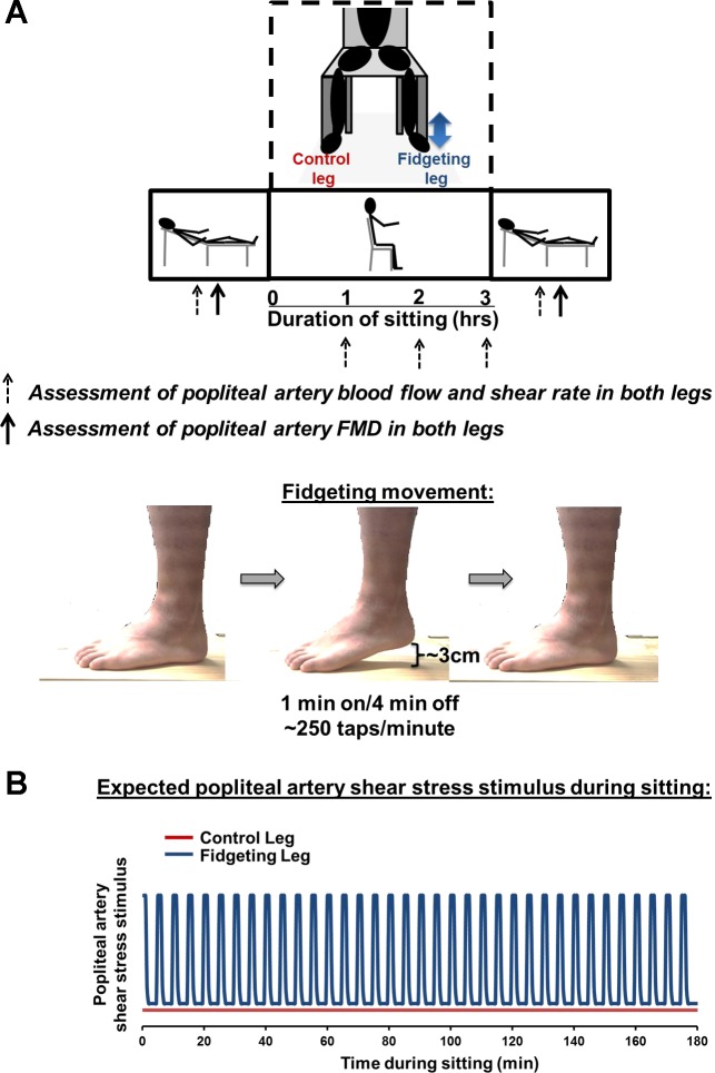
A schematic of the study design is presented in Fig. 1A, illustrating the sequence of events and various positions in which measurements were made. Subjects were instructed to eat a light meal 2 h or more before arriving to the laboratory. In addition, subjects were asked to refrain from caffeine and supplements for at least 12 h as well as from exercise for 24 h before the study visit. Ten subjects were tested at 8:00 am and one subject was tested at 2:00 pm. All study visits were performed in a temperature-controlled room kept at 22–23°C. Upon arrival to the laboratory, subjects were placed in a supine position with an automated sphygmomanometer (SphygmoCor XCEL, AtCor Medical, Itasca, IL) for periodic measurements of brachial artery blood pressure (BP), which were obtained after resting quietly for 10 min. Popliteal artery diameter and blood velocity were measured using duplex-Doppler ultrasound (Logiq P5; GE Medical Systems, Milwaukee, WI). An 11-MHz linear array transducer was placed over the popliteal artery just distal to the popliteal fossa. Simultaneous diameter and velocity signals were obtained in duplex mode at a pulsed frequency of 5 MHz and corrected with an insonation angle of 60°. Sample volume was adjusted to encompass the entire lumen of the vessel without extending beyond the walls, and the cursor was set at midvessel. Popliteal artery FMD was assessed in both legs in the supine position as previously described (2, 19, 20). Briefly, a rapid inflating cuff was placed on the lower leg. Two minutes of baseline hemodynamics were recorded and then the cuff was inflated to a pressure of 220 mmHg for 5 min. Continuous diameter and blood velocity measures were recorded for 3 min following cuff deflation. Recordings of all vascular variables were analyzed offline using specialized edge-detection software (Cardiovascular Suite, Quipu, Pisa, Italy).
Fig. 1.
Experimental design. A: schematic diagram of experimental protocol and positional changes over the course of the study. Measurements taken at the time points of 1, 2, and 3 h were made while the subject was in the seated position, whereas presit and postsit measurements were taken while subject was in the supine position. B: expected popliteal artery shear stress stimulus during sitting in the control and fidgeting legs. FMD, flow-mediated dilation.
Following baseline FMD measurements, subjects were positioned into a seated position for 3 h. Ankle circumference (taken at midpoint of malleolus lateralis) was measured on both legs as a crude estimate of venous congestion. One leg remained still throughout the sitting period (control leg), whereas the contralateral leg was subjected to intermittent fidgeting (fidgeting leg, 1 min on/4 min off). For the intermittent fidgeting intervention, subjects tapped the heel and bounced the knee at their own natural cadence (Fig. 1A). The number of heel taps was quantified during a fidgeting bout for each subject. Single leg fidgeting was designed to create intermittent increases in blood flow and shear stress in one leg while the other leg remained still (Fig. 1B). Throughout the entire protocol, a study representative monitored the subject to ensure no leg movements or muscle contractions occurred in the control leg and during the nonfidgeting time. In addition, monitoring of fidgeting was performed at each time interval. Right and left legs were randomly assigned the condition of control leg or fidgeting leg. Throughout the course of the sitting period, BP, popliteal artery diameter and blood velocity, and ankle circumference were measured on both legs every hour. It should be noted that vascular measurements in control and fidgeting legs were not performed simultaneously because only one Doppler ultrasound was available for this study. We used a chair that allows sufficient space for the ultrasound probe to access the popliteal artery during sitting. Because it was technically challenging to obtain Doppler ultrasound images during fidgeting, to quantify the blood flow and shear responses to fidgeting (in both legs), measures were initiated immediately (within 2 or 3 s) after the fidgeting bout. The basal measurements of popliteal artery blood flow and shear (in both legs) during sitting were performed after 2 min of rest following the 1-min fidgeting. At every hour, BP was measured after the measurements of blood flow.
Following the 3-h sitting period, subjects were manually lifted and placed back into the supine position to avoid any muscle activity of the legs. FMD assessments were then immediately (within 5 min) repeated. The order of FMD assessments was randomized between control and fidgeting legs within each subject. Once FMD was completed in one leg; the other leg was immediately set up for the FMD measurement (<5 min). After the FMD measurements, one last measurement of BP was performed while the subject rested supine.
Data analysis.
Blood flow was calculated from continuous diameter and mean blood velocity recordings at each of the experimental time points using the following equation: 3.14 × (diameter/2)2 × mean blood velocity × 60. Popliteal artery FMD percent change was calculated using the following equation: %FMD = (peak diameter − base diameter)/(base diameter) × 100. Shear rate was defined as 8 × mean blood velocity/diameter (17). Hyperemic shear rate area under the curve (AUC) up to peak diameter was calculated as stimulus for FMD, as previously described (2, 24).
Statistical analysis.
A two-way (time × leg) repeated-measures ANOVA using the Tukey HSD test for pairwise comparisons was performed on all dependent variables. FMD was also adjusted for hyperemic shear rate AUC via ANCOVA to statistically control for the influence of shear stimulus on the FMD response. ANCOVA and ANOVA test were performed using SPSS software (version 23). Significance was accepted at P < 0.05. Data are expressed as means ± SE.
RESULTS
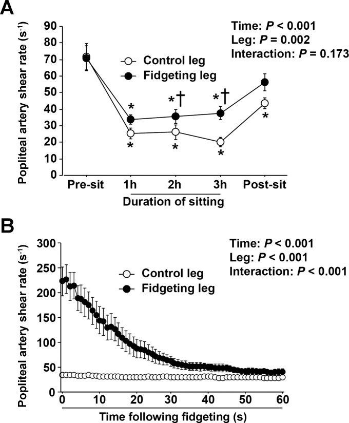
Over the course of the sitting period, popliteal artery blood flow (Table 1) and shear rate (Fig. 2A) were progressively reduced in both legs (P < 0.05). However, the fidgeting leg exhibited a significantly higher blood flow (Table 1, P < 0.05) and shear rate (Fig. 2A, P < 0.05) than the control leg during the sitting period. Notably, each bout of fidgeting produced a pronounced increase in popliteal artery blood flow and shear rate (Fig. 2B, P < 0.05) that tapered off during the following 60 s. Fidgeting did not alter popliteal artery blood flow and shear rate in the contralateral leg (Fig. 2B). During fidgeting, subjects were instructed to adhere to their own cadence of heel taps, which turned out to be fairly similar across individuals (average, 248 ± 6 taps/min; range, 220 to 290 taps/min).
Table 1.
Popliteal artery hemodynamics in control and fidgeting legs before, during, and after sitting for 3 h
| Duration of Sitting |
||||||
|---|---|---|---|---|---|---|
| Presit | 1 h | 2 h | 3 h | Postsit | ANOVA | |
| Basal diameter, cm | ||||||
| Control leg | 0.557 ± 0.02 | 0.585 ± 0.02 | 0.574 ± 0.02 | 0.589 ± 0.03 | 0.584 ± 0.03 | Time, P = 0.649 |
| Fidgeting leg | 0.571 ± 0.02 | 0.573 ± 0.02 | 0.573 ± 0.03 | 0.567 ± 0.02 | 0.562 ± 0.02 | Leg, P = 0.440 |
| Interaction, P = 0.183 | ||||||
| Blood flow, ml/min | ||||||
| Control leg | 71.75 ± 8.6 | 29.92 ± 4.5* | 27.26 ± 3.9* | 26.74 ± 3.5* | 50.75 ± 5.8* | Time, P = 0.027 |
| Fidgeting leg | 75.57 ± 8.5 | 38.00 ± 4.4* | 37.36 ± 3.4* | 38.39 ± 3.5*† | 61.19 ± 9.2 | Leg, P < 0.001 |
| Interaction, P = 0.855 | ||||||
| Hyperemic shear-rate AUC, arbitrary units | ||||||
| Control leg | 39,987 ± 5,135 | 21,679 ± 4,564* | Time, P = 0.026 | |||
| Fidgeting leg | 34,294 ± 7,182 | 24,785 ± 5,736* | Leg, P = 0.829 | |||
| Interaction, P = 0.579 | ||||||
| ANCOVA-corrected FMD, % | ||||||
| Control leg | 4.3 ± 0.9 | 1.8 ± 0.9 | Time, P = 0.635 | |||
| Fidgeting leg | 3.6 ± 0.9 | 6.8 ± 0.9† | Leg, P = 0.021 | |||
| Interaction, P = 0.004 | ||||||
| Mean arterial pressure, mmHg | 88 ± 2 | 94 ± 2* | 95 ± 2* | 94 ± 2* | 90 ± 1 | Time, P = 0.011 |
Values are means ± SE. Basal measurements of popliteal artery diameter and blood flow (in both legs) during sitting were performed after 2 min of rest following the 1-minute fidgeting. ANCOVA-corrected flow-mediated dilation (FMD) data are adjusted for hyperemic shear rate area under the curve (AUC).
P < 0.05 vs. presit;
P < 0.05, between legs.
Fig. 2.
Popliteal artery shear rate in the control and fidgeting legs. A: popliteal artery shear rate before, during, and after sitting for 3 h in the control and fidgeting legs. Basal measurements of popliteal artery shear rate (in both legs) during sitting were performed after 2 min of rest following the 1-min fidgeting. B: popliteal artery shear rate during sitting immediately after fidgeting in the control and fidgeting legs. Measurements of shear rate responses to fidgeting (in both legs) were initiated immediately (within 2 or 3 s) after the fidgeting bout. In the control leg, error bars are within symbols. Data are expressed as means ± SE. *P < 0.05 vs. presit; †P < 0.05, between legs.
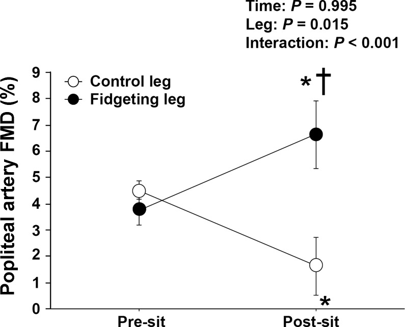
Importantly, 3 h of sitting caused a significant impairment in popliteal artery FMD in the control leg Fig. 3 (Cohen's d = 1.08; P = 0.039), whereas, in contrast, the fidgeting leg exhibited an increase in FMD (Fig. 3, Cohen's d = 0.92; P = 0.014). All 11 subjects decreased FMD in the control leg, and 9 out of 11 subjects increased FMD in the fidgeting leg. The improvement in FMD from fidgeting was not dependent on whether the fidgeting leg was assessed first or second following the sitting period (P = 0.858).
Fig. 3.
Popliteal artery FMD in the control and fidgeting legs before and after sitting for 3 h. FMD data are noncorrected for hyperemic shear rate area under the curve. Data are expressed as means ± SE. *P < 0.05 vs. presit; †P < 0.05, between legs.
Hyperemic shear rate AUC was similar between legs before sitting and significantly reduced in both legs after sitting (Table 1, P < 0.05). FMD corrected for hyperemic shear rate AUC by ANCOVA did not affect the interpretation of the main findings as FMD was reduced in the control leg and elevated in the fidgeting leg (Table 1). No changes were observed in popliteal artery diameter over time and no differences between legs were detected across time points (Table 1).
Mean arterial pressure (MAP) was significantly elevated during the sitting period but returned to presitting levels thereafter (Table 1). Ankle circumference was increased over time in the control leg (3-h sit, 2.58 ± 0.7%, relative to presit, P < 0.05), whereas no changes were observed in the fidgeting leg (3-h sit, 0.15 ± 0.1%, relative to presit, P > 0.05).
DISCUSSION
The novel finding of the present study is that leg endothelial dysfunction caused by prolonged sitting can be prevented by small amounts of leg movement or fidgeting. Indeed, we found that leg endothelial function was preserved in the leg subjected to a natural pattern of intermittent fidgeting but impaired in the leg that remained still. These findings support the notion that small amounts of leg movement during sitting are sufficient to prevent leg endothelial dysfunction in healthy young individuals, likely through the intermittent increases in blood flow-induced shear stress.
The prevalence of sedentary jobs in modern societies has augmented radically in the last few decades such that occupations involving some level of physical activity now represent a small fraction of the total workforce (5). Excessive sitting is not only confined to the workplace as our home and social environment is also full of incessant enticements to sit. While it is becoming more appreciated that sedentary behavior is correlated with an increased incidence of cardiovascular disease (5, 8), the mechanisms linking prolonged sitting time to increased cardiovascular disease risk still remain unclear. An understanding of the mechanisms by which too much sitting imposes cardiovascular risk becomes of paramount importance to create effective strategies that can mitigate the risks associated with this emerging “occupational” and “societal” hazard (14).
Along these lines, we (19, 20) and others (15, 25) have observed that during prolonged sitting, there is a marked reduction in blood flow and shear stress in the lower extremities that subsequently leads to leg endothelial dysfunction. We recently conducted a study designed to prevent the reduction in leg blood flow and thus shear stress during prolonged sitting with local heating of the foot (20). We found that leg endothelial dysfunction following sitting was abrogated by preventing the decrease in shear during sitting, supporting the hypothesis that sitting-induced leg endothelial dysfunction is indeed mediated by a reduction in shear stress (20).
While local heating of the foot was effective at sustaining leg blood flow and shear stress over the sitting period and consequently prevent the impairment in endothelial function (20), alternative more applicable strategies that could be used to offset the detrimental effects of sitting need investigation. As such, in the present study, we examined whether transient elevations in blood flow and shear stress with just fidgeting was sufficient to avert endothelial dysfunction caused by sitting. This was accomplished by subjecting one leg to intermittent fidgeting (1 min on/4 min off) during sitting while the contralateral leg remained still throughout and served as an internal control. Our model of fidgeting was chosen to reflect the natural behavior of fidgeting, and, as such, subjects were instructed to adhere to their own cadence of heel taps which turned out to be fairly similar across individuals (average, 248 ± 6 taps/min). Importantly, we found that this small amount of leg movement during sitting prevented the decline in leg endothelial function. Each single 1-min bout of fidgeting caused a marked and transient increase in blood flow and shear stress that remained slightly elevated postfidgeting. Of note, fidgeting did not alter blood flow and shear stress in the contralateral nonfidgeting leg. Given our previous work indicating that sustained reductions in flow-induced shear stress is a primary mechanism by which sitting induces leg endothelial dysfunction (20), it is likely that the beneficial vascular effects of fidgeting are mediated through the intermittent increases in shear stress throughout the prolonged sitting period; however, the involvement of other potential factors cannot be excluded.
Sitting-induced endothelial dysfunction in the lower extremities is of clinical relevance in light of evidence establishing that the leg vasculature is highly vulnerable to atherosclerosis, relative to other disease-resistant vasculatures such as the brachial artery (1, 12, 13, 21, 23). Our previous findings (19, 20) and findings from others (15, 26) that sitting causes endothelial dysfunction in the leg, but not the arm, fuel the idea that increased susceptibility of the leg vasculature to atherosclerosis may be, in part, attributable to the direct detrimental effects of prolonged sitting on that vasculature. Strategies such as fidgeting can offset the decay in leg blood flow and shear stress during sitting, thus preventing the consequent impairment of endothelial function and potentially providing important vascular benefits in the long term. However, more research is needed to examine the extent to which preventing leg endothelial dysfunction with sitting, through fidgeting or other approaches, leads to long-lasting effects. Furthermore, it remains unknown whether fidgeting with both legs would produce even greater beneficial vascular effects. It is also undetermined the extent to which the acute effects of fidgeting last and whether there are sex differences in the vascular responses to sitting as well as an influence of the menstrual cycle. Lastly, future work should determine if the present results can be extrapolated to clinical populations that are susceptible to peripheral artery disease such as patients with type 2 diabetes.
The mechanisms by which sitting increases leg vascular resistance and contributes to subsequent reductions in shear stress warrant consideration. It is possible that in the absence of muscle contraction, the increased hydrostatic pressure to which the legs are exposed during sitting lead to blood pooling and associated venous distension-induced arterial constriction and pressure-induced myogenic constriction (11). The increased ankle circumference observed during sitting in the control leg suggests that venous congestion occurred in the noncontracting lower limb. Venous distension has been shown to evoke sympathetic activation via stimulation of limb afferents (6), which may partially explain why muscle sympathetic nerve activity is greater in the upright position compared with supine (17). As a result, adrenergic vasoconstriction may also contribute to the increased leg vascular resistance. In this regard, we found a mild but significant increase in blood pressure during sitting.
In conclusion, the present study revealed that the simple behavior of fidgeting is sufficient to counteract the detrimental effects of prolonged sitting on leg endothelial function, likely through the intermittent increases in blood flow-induced shear stress. This study provides the first evidence that the detrimental vascular effects of sitting are preventable with small amounts of leg movement while seated for an extended period. Accordingly, people should be encouraged to consciously engage in leg movement when sitting for prolonged periods of time either at work or at home.
GRANTS
This work were supported by the National Institutes of Health Grants K01-HL-125503 and R21-DK-105368 (to J. Padilla) and a Japan Society for the Promotion of Science Grant-in-Aid for Scientific Research 14J09537 (to T. Morishima).
DISCLOSURES
No conflicts of interest, financial or otherwise, are declared by the author(s).
AUTHOR CONTRIBUTIONS
T.M., R.M.R., L.K.W., J.A.K., P.J.F., and J.P. conception and design of research; T.M., R.M.R., and J.P. performed experiments; T.M. analyzed data; T.M., R.M.R., L.K.W., J.A.K., P.J.F., and J.P. interpreted results of experiments; T.M. and J.P. prepared figures; T.M. and J.P. drafted manuscript; T.M., R.M.R., L.K.W., J.A.K., P.J.F., and J.P. edited and revised manuscript; T.M., R.M.R., L.K.W., J.A.K., P.J.F., and J.P. approved final version of manuscript.
ACKNOWLEDGMENTS
The authors appreciate the time and effort put in by all volunteer subjects.
REFERENCES
- 1.Aboyans V, McClelland RL, Allison MA, McDermott MM, Blumenthal RS, Macura K, Criqui MH. Lower extremity peripheral artery disease in the absence of traditional risk factors. The Multi-Ethnic Study of Atherosclerosis. Atherosclerosis 214: 169–173, 2011. [DOI] [PMC free article] [PubMed] [Google Scholar]
- 2.Boyle LJ, Credeur DP, Jenkins NT, Padilla J, Leidy HJ, Thyfault JP, Fadel PJ. Impact of reduced daily physical activity on conduit artery flow-mediated dilation and circulating endothelial microparticles. J Appl Physiol 115: 1519–1525, 2013. [DOI] [PMC free article] [PubMed] [Google Scholar]
- 3.Caro CG, Fitz-Gerald JM, Schroter RC. Arterial wall shear and distribution of early atheroma in man. Nature 223: 1159–1161. 1969. [DOI] [PubMed] [Google Scholar]
- 4.Cheng C, Tempel D, van Haperen R, van der Baan A, Grosveld F, Daemen MJ, Krams R, de Crom R. Atherosclerotic lesion size and vulnerability are determined by patterns of fluid shear stress. Circulation 113: 2744–2753, 2006. [DOI] [PubMed] [Google Scholar]
- 5.Church TS, Thomas DM, Tudor-Locke C, Katzmarzyk PT, Earnest CP, Rodarte RQ, Martin CK, Blair SN, Bouchard C. Trends over 5 decades in US occupation-related physical activity and their associations with obesity. PloS One 6: e19657, 2011. [DOI] [PMC free article] [PubMed] [Google Scholar]
- 6.Cui J, MacQuillan PM, Blaha C, Kunselman AR, Sinoway LI. Limb venous distension evokes sympathetic activation via stimulation of the limb afferents in humans. Am J Physiol Heart Circ Physiol 303: H457–H463, 2012. [DOI] [PMC free article] [PubMed] [Google Scholar]
- 7.Hahn C, Schwartz MA. Mechanotransduction in vascular physiology and atherogenesis. Nat Rev Mol Cell Biol 10: 53–62, 2009. [DOI] [PMC free article] [PubMed] [Google Scholar]
- 8.Hamilton MT, Hamilton DG, Zderic TW. Role of low energy expenditure and sitting in obesity, metabolic syndrome, type 2 diabetes, and cardiovascular disease. Diabetes 56: 2655–2667, 2007. [DOI] [PubMed] [Google Scholar]
- 9.Jenkins NT, Padilla J, Boyle LJ, Credeur DP, Laughlin MH, Fadel PJ. Disturbed blood flow acutely induces activation and apoptosis of the human vascular endothelium. Hypertension 61: 615–621, 2013. [DOI] [PMC free article] [PubMed] [Google Scholar]
- 10.Johnson BD, Mather KJ, Newcomer SC, Mickleborough TD, Wallace JP. Vitamin C prevents the acute decline of flow-mediated dilation after altered shear rate patterns. Appl Physiol Nutr Metab 38: 268–274, 2013. [DOI] [PubMed] [Google Scholar]
- 11.Kitano A, Shoemaker JK, Ichinose M, Wada H, Nishiyasu T. Comparison of cardiovascular responses between lower body negative pressure and head-up tilt. J Appl Physiol 98: 2081–2086, 2005. [DOI] [PubMed] [Google Scholar]
- 12.Kroger K, Kucharczik A, Hirche H, Rudofsky G. Atherosclerotic lesions are more frequent in femoral arteries than in carotid arteries independent of increasing number of risk factors. Angiology 50: 649–654, 1999. [DOI] [PubMed] [Google Scholar]
- 13.Li MF, Ren Y, Zhao CC, Zhang R, Li LX, Liu F, Lu JX, Tu YF, Zhao WJ, Bao YQ, Jia WP. Prevalence and clinical characteristics of lower limb atherosclerotic lesions in newly diagnosed patients with ketosis-onset diabetes: a cross-sectional study. Diabetol Metab Syndr 6: 71, 2014. [DOI] [PMC free article] [PubMed] [Google Scholar]
- 14.Martens CR. Taking steps in the workplace to improve vascular function. Exp Physiol 100: 774–775, 2015. [DOI] [PubMed] [Google Scholar]
- 15.McManus AM, Ainslie PN, Green DJ, Simair RG, Smith K, Lewis N. Impact of prolonged sitting on vascular function in young girls. Exp Physiol 100: 1379–1387, 2015. [DOI] [PubMed] [Google Scholar]
- 16.Nam D, Ni CW, Rezvan A, Suo J, Budzyn K, Llanos A, Harrison D, Giddens D, Jo H. Partial carotid ligation is a model of acutely induced disturbed flow, leading to rapid endothelial dysfunction and atherosclerosis. Am J Physiol Heart Circ Physiol 297: H1535–H1543, 2009. [DOI] [PMC free article] [PubMed] [Google Scholar]
- 17.Parker BA, Trehearn TL, Meendering JR. Pick your Poiseuille: normalizing the shear stimulus in studies of flow-mediated dilation. J Appl Physiol 107: 1357–1359, 2009. [DOI] [PubMed] [Google Scholar]
- 18.Ray CA, Rea RF, Clary MP, Mark AL. Muscle sympathetic nerve responses to dynamic one-legged exercise: effect of body posture. Am J Physiol Heart Circ Physiol 264: H1–H7, 1993. [DOI] [PubMed] [Google Scholar]
- 19.Restaino RM, Holwerda SW, Credeur DP, Fadel PJ, Padilla J. Impact of prolonged sitting on lower and upper limb micro- and macrovascular dilator function. Exp Physiol 100: 829–838, 2015. [DOI] [PMC free article] [PubMed] [Google Scholar]
- 20.Restaino RM, Walsh LK, Morishima T, Vranish JR, Martinez-Lemus LA, Fadel PJ, Padilla J. Endothelial dysfunction following prolonged sitting is mediated by a reduction in shear stress. Am J Physiol Heart Circ Physiol 310: H648–H653, 2016. [DOI] [PMC free article] [PubMed] [Google Scholar]
- 21.Ross R, Wight TN, Strandness E, Thiele B. Human atherosclerosis I. Cell constitution and characteristics of advanced lesions of the superficial femoral artery. Am J Pathol 114: 79–93, 1984. [PMC free article] [PubMed] [Google Scholar]
- 22.Schreuder TH, Green DJ, Hopman MT, Thijssen DH. Acute impact of retrograde shear rate on brachial and superficial femoral artery flow-mediated dilation in humans. Physiol Rep 2: e00193, 2014. [DOI] [PMC free article] [PubMed] [Google Scholar]
- 23.Stary HC, Chandler AB, Dinsmore RE, Fuster V, Glagov S, Insull W Jr, Rosenfeld ME, Schwartz CJ, Wagner WD, Wissler RW. A definition of advanced types of atherosclerotic lesions and a histological classification of atherosclerosis A report from the Committee on Vascular Lesions of the Council on Arteriosclerosis, American Heart Association. Circulation 92: 1355–1374, 1995. [DOI] [PubMed] [Google Scholar]
- 24.Thijssen DH, Dawson EA, Tinken TM, Cable NT, Green DJ. Retrograde flow and shear rate acutely impair endothelial function in humans. Hypertension 53: 986–992, 2009. [DOI] [PubMed] [Google Scholar]
- 25.Thosar SS, Bielko SL, Mather KJ, Johnston JD, Wallace JP. Effect of prolonged sitting and breaks in sitting time on endothelial function. Med Sci Sports Exerc 47: 843–849, 2015. [DOI] [PubMed] [Google Scholar]
- 26.Thosar SS, Bielko SL, Wiggins CC, Wallace JP. Differences in brachial and femoral artery responses to prolonged sitting. Cardiovasc Ultrasound 15: 12–50, 2014. [DOI] [PMC free article] [PubMed] [Google Scholar]
- 27.Totosy de Zepetnek JO, Jermey TL, MacDonald MJ. Superficial femoral artery endothelial responses to a short-term altered shear rate intervention in healthy men. PloS One 9: e113407, 2014. [DOI] [PMC free article] [PubMed] [Google Scholar]


