

Abstract
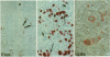
An immunohistological study of paraffin wax embedded tissue from three cases of plasmacytoid monocyte neoplasms, using a panel of antibodies which react with fixation resistant leucocyte markers, is reported. This neoplasm was found to have a distinctive antigenic profile, being negative for CD3 and elastase, but positive for CD43 and CD68. This immunological phenotype, coupled with its characteristic morphological features, should facilitate the recognition of this rare neoplasm in routinely processed tissue. Furthermore, the term "plasmacytoid monocyte sarcoma" is proposed to designate it because it is inappropriate to refer to it as a lymphoma. As all cases have been associated with a myeloproliferative disorder (usually an acute or chronic myeloid leukaemia), these tumours probably represent the accumulation in lymphoid tissue of neoplastic cells which have differentiated along the plasmacytoid monocyte pathway.
Full text
PDF


Images in this article


Selected References
These references are in PubMed. This may not be the complete list of references from this article.
- Beiske K., Langholm R., Godal T., Marton P. F. T-zone lymphoma with predominance of 'plasmacytoid T-cells' associated with myelomonocytic leukaemia--a distinct clinicopathological entity. J Pathol. 1986 Dec;150(4):247–255. doi: 10.1002/path.1711500404. [DOI] [PubMed] [Google Scholar]
- Beiske K., Munthe-Kaas A., Davies C. D., Marton P. F., Godal T. Single cell studies on the immunological marker profile of plasmacytoid T-zone cells. Lab Invest. 1987 Apr;56(4):381–393. [PubMed] [Google Scholar]
- Cordell J. L., Falini B., Erber W. N., Ghosh A. K., Abdulaziz Z., MacDonald S., Pulford K. A., Stein H., Mason D. Y. Immunoenzymatic labeling of monoclonal antibodies using immune complexes of alkaline phosphatase and monoclonal anti-alkaline phosphatase (APAAP complexes). J Histochem Cytochem. 1984 Feb;32(2):219–229. doi: 10.1177/32.2.6198355. [DOI] [PubMed] [Google Scholar]
- Facchetti F., De Wolf-Peeters C., Kennes C., Rossi G., De Vos R., van den Oord J. J., Desmet V. J. Leukemia-associated lymph node infiltrates of plasmacytoid monocytes (so-called plasmacytoid T-cells). Evidence for two distinct histological and immunophenotypical patterns. Am J Surg Pathol. 1990 Feb;14(2):101–112. doi: 10.1097/00000478-199002000-00001. [DOI] [PubMed] [Google Scholar]
- Facchetti F., De Wolf-Peeters C., van den Oord J. J., De vos R., Desmet V. J. Plasmacytoid T cells: a cell population normally present in the reactive lymph node. An immunohistochemical and electronmicroscopic study. Hum Pathol. 1988 Sep;19(9):1085–1092. doi: 10.1016/s0046-8177(88)80091-1. [DOI] [PubMed] [Google Scholar]
- Facchetti F., de Wolf-Peeters C., Mason D. Y., Pulford K., van den Oord J. J., Desmet V. J. Plasmacytoid T cells. Immunohistochemical evidence for their monocyte/macrophage origin. Am J Pathol. 1988 Oct;133(1):15–21. [PMC free article] [PubMed] [Google Scholar]
- Facchetti F., de Wolf-Peeters C., van den Oord J. J., de Vos R., Desmet V. J. Plasmacytoid monocytes (so-called plasmacytoid T-cells) in Kikuchi's lymphadenitis. An immunohistologic study. Am J Clin Pathol. 1989 Jul;92(1):42–50. doi: 10.1093/ajcp/92.1.42. [DOI] [PubMed] [Google Scholar]
- Facchetti F., de Wolfe-Peeters C., van den Oord J. J., Desmet V. J. Immunohistochemical visualization of plasmacytoid T cells in paraffin sections. Hum Pathol. 1987 Dec;18(12):1300–1300. doi: 10.1016/s0046-8177(87)80419-7. [DOI] [PubMed] [Google Scholar]
- Feller A. C., Lennert K., Stein H., Bruhn H. D., Wuthe H. H. Immunohistology and aetiology of histiocytic necrotizing lymphadenitis. Report of three instructive cases. Histopathology. 1983 Nov;7(6):825–839. doi: 10.1111/j.1365-2559.1983.tb02299.x. [DOI] [PubMed] [Google Scholar]
- Flavell D. J., Jones D. B., Wright D. H. Identification of tissue histiocytes on paraffin sections by a new monoclonal antibody. J Histochem Cytochem. 1987 Nov;35(11):1217–1226. doi: 10.1177/35.11.3309045. [DOI] [PubMed] [Google Scholar]
- Harris N. L., Bhan A. K. "Plasmacytoid T cells" in Castleman's disease. Immunohistologic phenotype. Am J Surg Pathol. 1987 Feb;11(2):109–113. doi: 10.1097/00000478-198702000-00004. [DOI] [PubMed] [Google Scholar]
- Horny H. P., Feller A. C., Horst H. A., Lennert K. Immunocytology of plasmacytoid T cells: marker analysis indicates a unique phenotype of this enigmatic cell. Hum Pathol. 1987 Jan;18(1):28–32. doi: 10.1016/s0046-8177(87)80189-2. [DOI] [PubMed] [Google Scholar]
- Koo C. H., Mason D. Y., Miller R., Ben-Ezra J., Sheibani K., Rappaport H. Additional evidence that "plasmacytoid T-cell lymphoma" associated with chronic myeloproliferative disorders is of macrophage/monocyte origin. Am J Clin Pathol. 1990 Jun;93(6):822–827. doi: 10.1093/ajcp/93.6.822. [DOI] [PubMed] [Google Scholar]
- LENNERT K., REMMELE W. Karyometrische Untersuchungen an Lymphknotenzellen des Menschen. I. Germinoblasten, Lymphoblasten und Lymphozyten. Acta Haematol. 1958 Feb;19(2):99–113. doi: 10.1159/000205419. [DOI] [PubMed] [Google Scholar]
- Mason D. Y., Cordell J., Brown M., Pallesen G., Ralfkiaer E., Rothbard J., Crumpton M., Gatter K. C. Detection of T cells in paraffin wax embedded tissue using antibodies against a peptide sequence from the CD3 antigen. J Clin Pathol. 1989 Nov;42(11):1194–1200. doi: 10.1136/jcp.42.11.1194. [DOI] [PMC free article] [PubMed] [Google Scholar]
- Müller-Hermelink H. K., Kaiserling E., Lennert K. Pseudofollikuläre Nester von Plasmazellen (eines besonderen Typs?) in der paracorticalen Pulpa menschlicher Lymphknoten. Virchows Arch B Cell Pathol. 1973;14(1):47–56. [PubMed] [Google Scholar]
- Müller-Hermelink H. K., Stein H., Steinmann G., Lennert K. Malignant lymphoma of plasmacytoid T-cells. Morphologic and immunologic studies characterizing a special type of T-cell. Am J Surg Pathol. 1983 Dec;7(8):849–862. [PubMed] [Google Scholar]
- Poppema S., Hollema H., Visser L., Vos H. Monoclonal antibodies (MT1, MT2, MB1, MB2, MB3) reactive with leukocyte subsets in paraffin-embedded tissue sections. Am J Pathol. 1987 Jun;127(3):418–429. [PMC free article] [PubMed] [Google Scholar]
- Prasthofer E. F., Prchal J. T., Grizzle W. E., Grossi C. E. Plasmacytoid T-cell lymphoma associated with chronic myeloproliferative disorder. Am J Surg Pathol. 1985 May;9(5):380–387. doi: 10.1097/00000478-198505000-00009. [DOI] [PubMed] [Google Scholar]
- Pulford K. A., Erber W. N., Crick J. A., Olsson I., Micklem K. J., Gatter K. C., Mason D. Y. Use of monoclonal antibody against human neutrophil elastase in normal and leukaemic myeloid cells. J Clin Pathol. 1988 Aug;41(8):853–860. doi: 10.1136/jcp.41.8.853. [DOI] [PMC free article] [PubMed] [Google Scholar]
- Pulford K. A., Rigney E. M., Micklem K. J., Jones M., Stross W. P., Gatter K. C., Mason D. Y. KP1: a new monoclonal antibody that detects a monocyte/macrophage associated antigen in routinely processed tissue sections. J Clin Pathol. 1989 Apr;42(4):414–421. doi: 10.1136/jcp.42.4.414. [DOI] [PMC free article] [PubMed] [Google Scholar]
- Rivano M. T., Falini B., Stein H., Canino S., Ciani C., Gerdes J., Ribacchi R., Gobbi M., Pileri S. Histiocytic necrotizing lymphadenitis without granulocytic infiltration (Kikuchi's lymphadenitis). Morphological and immunohistochemical study of eight cases. Histopathology. 1987 Oct;11(10):1013–1027. doi: 10.1111/j.1365-2559.1987.tb01842.x. [DOI] [PubMed] [Google Scholar]
- Sheibani K., Fritz R. M., Winberg C. D., Burke J. S., Rappaport H. "Monocytoid" cells in reactive follicular hyperplasia with and without multifocal histiocytic reactions: an immunohistochemical study of 21 cases including suspected cases of toxoplasmic lymphadenitis. Am J Clin Pathol. 1984 Apr;81(4):453–458. doi: 10.1093/ajcp/81.4.453. [DOI] [PubMed] [Google Scholar]
- Stross W. P., Warnke R. A., Flavell D. J., Flavell S. U., Simmons D., Gatter K. C., Mason D. Y. Molecule detected in formalin fixed tissue by antibodies MT1, DF-T1, and L60 (Leu-22) corresponds to CD43 antigen. J Clin Pathol. 1989 Sep;42(9):953–961. doi: 10.1136/jcp.42.9.953. [DOI] [PMC free article] [PubMed] [Google Scholar]
- Vollenweider R., Lennert K. Plasmacytoid T-cell clusters in non-specific lymphadenitis. Virchows Arch B Cell Pathol Incl Mol Pathol. 1983;44(1):1–14. doi: 10.1007/BF02890155. [DOI] [PubMed] [Google Scholar]
- Wood G. S., Warner N. L., Warnke R. A. Anti-Leu-3/T4 antibodies react with cells of monocyte/macrophage and Langerhans lineage. J Immunol. 1983 Jul;131(1):212–216. [PubMed] [Google Scholar]