Abstract
Aging is associated with a fall in maximal aerobic capacity as well as with a decline in lean body mass. The purpose of the study was to investigate the influence of aging on the relationship between aerobic capacity and lean body mass in subjects that chronically train both their upper and lower bodies. Eleven older rowers (58±5 yrs) and 11 younger rowers (27±4 yrs) participated in the study. Prior to the VO2max testing, subjects underwent a dual energy X-ray absorptiometry scan to estimate total lean body mass. Subsequently, VO2max was quantified during a maximal exercise test on a rowing ergometer as well as a semi-recumbent cycle ergometer. The test protocol included a pre-exercise stage followed by incremental exercise until VO2max was reached. The order of exercise modes was randomized and there was a wash-out period between the two tests. Oxygen uptake was obtained via a breath-by-breath metabolic cart (Vmax™ Encore, San Diego, CA). Rowing VO2max was higher than cycling VO2max in both groups (p<0.05). Older subjects had less of an increase in VO2max from cycling to rowing (p<0.05). There was a significant relationship between muscle mass and VO2max for both groups (p<0.05). After correcting for muscle mass, the difference in cycling VO2max between groups disappeared (p>0.05), however, older subjects still demonstrated a lower rowing VO2max relative to younger subjects (p<0.05). Muscle mass is associated with the VO2max obtained, however, it appears that VO2max in older subjects may be less influenced by muscle mass than in younger subjects.
Introduction
Maximal oxygen consumption (VO2max) is the maximum capacity to transport and utilize oxygen and is often used as a measure of an individual’s aerobic capacity. Generally, VO2max decreases gradually with advancing age, and the rate of decline is approximately 10% per decade after the age of 25 years, and more specifically was suggested to be 15% between the ages of 50 and 75 [1, 2, 3]. In addition, a previous meta-analysis illustrated that age-related declines in VO2max were approximately 0.40, 0.39 and 0.46 ml/kg/min per year for sedentary, active and trained males respectively and 0.35, 0.44, 0.62 ml/kg/min per year for sedentary, active and trained female respectively [4, 5, 6]. Age-related decline in VO2max results from multiple factors: decreased maximal heart rate and stroke volume, reductions in blood volume due to pooling from less effective muscle pump action of the valves in the extremities, and stiffening of both the heart muscle fibers and thickening and stiffening of the arterial walls, reduced peripheral oxygen extraction and maximal A-V O2 difference [7, 8, 9, 10]. Age-related muscle loss (sarcopenia) also appears to play an important role in the decline in VO2max; by the age of 50 about 10% of muscle area is lost and this rate only increases in the decades that follow. In concert with sarcopenia, muscle strength declines by 15% per decade in the 60s and 70s and doubles to 30% after 80 [11]. A previous study by Fleg and Lakatta (1988) demonstrated that whole body lean mass contributed to a decline in VO2max with advancing age in sedentary individuals [12]. Similar results were observed in active individuals [13, 14]. However, active individuals demonstrated a slower rate of decline than sedentary individuals [3, 15]. Thus the influence of muscle mass on VO2max can be influenced by training and possibly type of training (e.g., upper vs lower extremity exercise) as well as other factors, such as how muscle mass is quantified (e.g., only active muscles vs total lean body or skeletal muscle mass) [13].
Rowing is a demanding sport requiring a high aerobic capacity and the ability to sprint; yet it is a common sport amongst older individuals. It is an activity requiring large muscle mass and the ability to distribute blood flow to both upper and lower extremity skeletal muscles simultaneously. Accordingly, rowers exhibit both upper and lower body muscle adaptations, and thus offer a unique population to better understand the effect of muscle mass on VO2max.
The purpose of study was to investigate the relationship between active muscle mass and VO2max, and how this relationship might be influenced by age. Two separate exercise modes–recumbent cycling (targeting leg muscles only) and rowing (targeting leg and upper body muscles)–were utilized to differentiate active muscle mass. A mode specific VO2max was quantified based on active muscle for a group of younger and older rowers.
Methods
For the present study, 22 subjects were recruited from rowing teams in Augusta, Georgia (GA). Eleven older subjects (age = 58±5 yr, 6 males and 5 females) and 11 younger subjects (age = 27±4 yr, 6 males and 5 females) who had at least a one year of competitive rowing experience, a current baseline level of training that included rowing and typical synergistic activities–running, swimming, resistive training and etc. They performed for a minimum of one hr four times a week, and were planning to participate in seasonal rowing competitions–regattas. Subjects who had a body mass index (BMI) >30, a history of cardiovascular or pulmonary disease, an inability to perform repeated volitional level of exercise or females who were pregnant were excluded from the study. The study was approved by Mayo Clinic Institutional Review Board and Joseph M. Still Research Foundation, Inc. (on behalf of Doctors Hospital). Prior to participating, all subjects provided their written and informed consent. The baseline pulmonary function and body composition metrics for the subjects is provided in Table 1. The study protocol included two maximal oxygen consumption tests (VO2max) one on a semi-recumbent bike and a second on a rowing ergometer. These exercise modes are thought to recruit a different active muscle mass with cycling being regional (leg muscle only) whereas rowing is more of a whole body muscle mass. BothVO2max tests were on the same day, with a 90min rest period given between tests. Subjects were randomized and counterbalanced to a test order such that half the subjects completed the cycling test first and the other half completed the rowing test first. Whole body muscle mass and regional muscle mass were estimated via dual energy X-ray absorptiometry (DEXA) scan. Quantification of specific regional muscle masses allowed for a logical estimation of active muscle mass following exercise modes. For the cycling test the representative active muscle mass was leg muscle mass (LMM), and for the rowing test whole body lean muscle mass (LBM) was used.
Table 1. Lung function and body composition.
| Older subjects | Younger subjects | |
|---|---|---|
| Lung function and diffusing capacity | ||
| Total lung capacity (L) | 5.9±1.4 | 7.0±1.2 |
| Vital capacity (L) | 3.9±1.1* | 5.1±1.2* |
| Residual volume (L) | 2.1±0.4 | 2.0±0.3 |
| Expiratory reserve volume (L) | 1.3±0.4* | 1.8±0.4* |
| Inspiratory capacity (L) | 2.75±0.7* | 3.46±0.6* |
| RV/TLC (%) | 36.8±1.8* | 27.6±2.2* |
| Forced vital capacity (FVC) (%pre) | 111±16 | 107±16 |
| FEV 1(%pre) | 102±17 | 103±17 |
| FEV25-75 (%pre) | 78±22 | 93±22 |
| Diffusing capacity for CO (%pre) | 119±21 | 116±17 |
| Alveolar gas volume (L) | 6.1±1.5 | 7.0±1.3 |
| Body composition | ||
| Total mass (kg) | 74.4±16 | 78.6±11 |
| Body mass index | 25.4±4 | 24.1±3 |
| Percent body fat (%) | 29.4±13 | 21.1±1 |
| LBM (kg) | 50.3±13 | 58.9±10 |
| Fat free mass (kg) | 53.2±13 | 62.7±10 |
| LM (kg) | 16.3±4 | 19.4±3 |
| Arms lean mass (kg) | 5.9±2 | 6.8±2 |
* indicates a significant difference between older and younger rowers (p<0.05).
Respiratory exchange ratio (no exertion ~ maximal exertion: 6 ~ 20).
Dyspnea (no breathlessness at all ~ maximum: 0 ~ 10).
All experimental trials and DEXA scans were conducted in Doctors Hospital (Augusta, GA). Subjects reported to the laboratory either on the same day of testing or prior to testing for review of the study protocols and signing consent form. In addition, they completed simple questionnaires of health (PAR-Q: physical activity readiness questionnaire) and activity levels (IPAQ SF: short from of international physical activity questionnaire), and female subjects competed a urine pregnancy test. Next subjects underwent a DEXA scan for body composition and a basic spirometry including single breath diffusion capacity for carbon monoxide (Vmax™ Encore, San Diego, CA). This was then followed by the VO2max tests on a rowing ergometer (Concept 2, Inc., Morrisville, VT) and semi-recumbent cycling ergometer (Lode B. V., Groningen, Netherlands).
The VO2max testing protocol included a warming-up stage (3min resting and 2min warm-up with 25W) followed by incremental exercise to volitional exhaustion. The exercise intensity was increased every 2min, with the first three stages consistent for all subjects (50W, 75W and 100W respectively). To ensure all subjects had a similar time to exhaustion, power output during the subsequent stages was increased by 20W for old females, 30W for old males and young females and 40W for young males until a termination of exercise.
Subjects were asked to maintain a pedal rate of 60–80 rpm for cycling and a stroke rate of 20–30 for rowing with exhaustion classified as the inability to maintain this rate despite verbal encouragement. During cycling on a recumbent cycle ergometer, the subjects were asked not to hold the handle to minimize the involvement of upper body muscle groups. Initial mode of testing were randomized and counter-balanced. Oxygen uptake and gas exchange was measured via breath by breath metabolic cart (Vmax™ Encore, San Diego, CA). The VO2max was determined as the highest 60sec average VO2 observed within the last 2min of incremental exercise. Heart rate was monitored via electrocardiography (ECG) and peripheral oxygen saturation via forehead probe pulse-oximeter. Perceived exertion (RPE) was obtained every stage using the Borg Scale and dyspnea by Borg’s Breathlessness Scale. A true VO2max was verified to have been reached when two of the following three were observed: peak heart rate (HR)> 90% predicted max (220-age), a rating of perceived exertion (RPE) 18 or greater out of 20, or a respiratory exchange ratio (RER) greater than or equal to 1.15.
To compare baseline pulmonary function and body composition between older and younger subjects, independent t-tests were conducted. To estimate the differences in VO2max between groups and exercise modes, repeated measures analysis of variance was conducted and subsequently paired-sample t-tests and independent t-tests were conducted. Furthermore, the relationships between VO2max (l/min) and active muscle mass for both exercise modes and the relationship between age and exercise modes for VO2max (corrected by LBM or LMM) were analyzed. For cycling, absolute VO2max (l/min) was corrected for leg muscle mass (LMM) and illustrated with ml/kg-LMM/min, while for rowing it was corrected with whole body muscle mass (LBM) and illustrated with ml/kg-LBM/min. The significance level was set at 0.05.
Results
Peak heart rate (HR), respiratory exchange ratio (RER), rate of perceived exertion (RPE) and dyspnea obtained during cycling and rowing were illustrated in Table 2. While older subjects demonstrated no significant difference in peak HR during cycling and rowing (p>0.05), younger subjects showed a significant difference (p<0.05). In addition, both older and younger groups demonstrated no significant difference in peak HR during cycling (p>0.05), however, older subjects reached significant lower peak HR during rowing than younger subjects (p<0.05). Both older and younger subjects demonstrated a higher RER at peak during cycling than rowing (p<0.05 and p<0.05 respectively). However, no significant differences in RPE were observed between cycling and rowing in either older (p>0.05) or younger (p>0.05) subjects. Furthermore, older subjects showed no significant difference in dyspnea score at peak exercise between exercise modes (p>0.05), however, younger subjects reported higher scores during rowing (p<0.05).
Table 2. The assessment of exertion at peak level.
| Older subjects | Younger subjects | |
|---|---|---|
| Cycling vs. Rowing | Cycling vs. Rowing | |
| Peak heart rate (beat/min) | 164±10 vs. 167±7 | 173±6 vs. 180±8* |
| Respiratory exchange ratio (RER) | 1.26±0.10 vs. 1.18±0.08* | 1.17±0.09 vs. 1.15±0.08* |
| Rating of perceived exertion (RPE) | 18.5±1.1 vs. 18.6±1.1 | 18.3±0.8 vs. 18.7±1.3 |
| Dyspnea | 7.9±1.6 vs. 7.9±1.6 | 7.5±1.7 vs. 8.5±1.3* |
| SpO2 (%) | 98.4±1.5 vs. 97.6±1.7 | 98.2±1.2 vs. 96.4±1.3* |
| Peak power (watts) | 186.4±61.9 vs. 186.4±61.9 | 251.8±41.9 vs. 261.1±47.8 |
* indicates a significant difference between cycling and rowing (p<0.05.)
Respiratory exchange ratio (no exertion ~ maximal exertion: 6 ~ 20).
Dyspnea (no breathlessness at all ~ maximum: 0 ~ 10).
Oxygen saturation (SpO2)
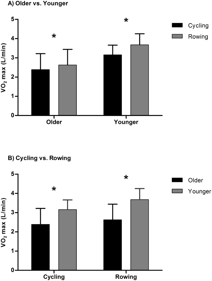
Both older and younger subjects had higher VO2max (l/min) values with rowing than cycling, however, the effect of mode of exercise, difference in VO2max (l/min) between cycling and rowing was significantly less in older subjects relative to the difference noted in younger subjects (p<0.05). Older subjects increased VO2max (l/min) by 10% from cycling to rowing (Fig 1A, p<0.05), while younger rowers improved VO2max (l/min) by 16.7% (Fig 1A, p<0.05). In addition, VO2max (l/min) of younger subjects was 29.8% higher during cycling (Fig 1B, p<0.05) and 37.6% higher during rowing (Fig 1B, p<0.05) when compared to older rowers.
Fig 1. (A) The differences in VO2max between cycling and rowing in older and younger. (B) The differences in VO2max between older and younger subjects in cycle and rowing.
* denotes a significant difference between exercise modes (A) and groups (B).
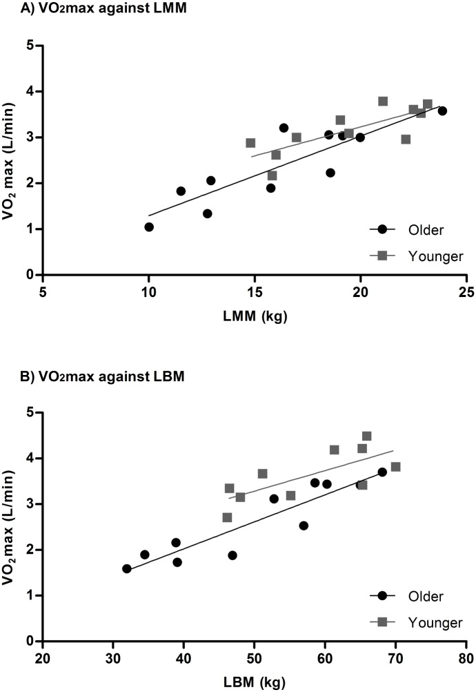
The relationships between cycling VO2max (l/min) and LMM in older (r2 = 0.764, p<0.05) and younger subjects (r2 = 0.613, p<0.05) are illustrated Fig 2A. In addition, the relationship between rowing VO2max and LBM in older (r2 = 0.851, p<0.05) and younger subjects (r2 = 0.531, p<0.05) are illustrated in Fig 2B.
Fig 2. Individual VO2max against muscle mass.
The slope of relationship between LMM and cycling VO2max was not significant different between older (y = 0.1737x-0.4418) and younger (y = 0.1263x+0.7057) subjects (p>0.05). The slope of relationship between LBM and rowing VO2max was not significant different between older (y = 0.05907x-0.3382) and younger (y = 0.04480x+1.047) subjects (p>0.05).
There was a modest relationship between cycling VO2max (ml/kg-LMM/min) and age (r2 = 0.218, p<0.05) while a strong relationship between rowing VO2max (ml/kg-LBM/min) and age (r2 = 0.521, p<0.01) was observed. In addition, rowing VO2max (ml/kg-LBM/min) in older subjects remained lower than younger subjects (p<0.05). However, there was no longer a difference in cycling VO2max (ml/kg-LMM/min) between groups (p>0.05).
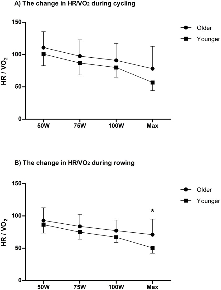
Furthermore, older subjects demonstrated higher HR/VO2 ratio, which may indicate a higher HR at a given VO2 than younger subjects during rowing (p<0.05). However, there was no significant difference in HR/VO2 ratio between groups observed during cycling (p>0.05). Fig 3A and 3B illustrates HR/VO2 at a given workload during cycling (3A) and rowing (3B).
Fig 3. (A) VO2 at a given HR.
The changes in VO2/HR at each workload (50W, 75W, 100W and Max) during cycling (3A) and rowing (3B). * denotes a significant difference between older and younger subjects.
Discussion
Aerobic capacity (VO2max) is strongly associated with muscle mass, and rowing VO2max was higher than cycling VO2max in old and younger athletes. Furthermore, when comparing to older subjects, younger subjects demonstrated a larger change in VO2max with rowing compared to cycling. After correcting for the respective active muscle mass being recruited for the specific mode of exercise, older subjects showed no significant difference in VO2max during cycling from younger subjects where less muscle mass is being recruited, however, VO2max during rowing remained lower in older subjects compared to younger subjects where more muscle mass was being recruited. These findings suggest that VO2max is influenced by the total active muscle mass being recruited during exercise, where the more muscle mass involved during exercise, the higher the VO2max. However, these data also suggest that VO2max in older subjects may be less influenced by muscle mass than younger subjects. In addition, older subjects required higher cardiac effort (higher HR) at a given VO2.
Aerobic capacity generally declines with the aging process. In the present experiment, we observed that older subjects demonstrated lower VO2max than younger subjects regardless of exercise modes. Generally, VO2max declines about 10% per decade [1, 2, 3]. The rate of decline in VO2max appears to be greater in athletic or active population than sedentary population [16, 17, 18]. This may result from a higher initial VO2max [17]. However, this hypotheses is still not fully tested as results can be inconsistent due to study design (i.e., cross-sectional and longitudinal) and different non-linearity of changes in VO2max in athletic/active and sedentary subjects [3, 19].
Over the years, a number of studies have investigated the mechanisms of age-related decline in aerobic capacity. As described by the Fick equation, VO2max is determined by maximal cardiac output and arterio-venous O2 difference. Age-related declines in VO2max are the consequence of decreases in maximal heart rate and stroke volume [20, 21, 22] and maximal arterio-venous O2 difference [23]. However, the mechanism for this is not simply explained since there are several confounding factors: diffusion capacity of the lungs, vascular conductance, skeletal muscle mitochondria density and enzyme activity, skeletal muscle mass and training volume.
Skeletal muscle mass loss reaches 10% by age 50 yrs. and decreases further to 30% by age 80 yrs [24, 25]. The natural age-related decline in muscle mass (sarcopenia) and the additional changes in body composition that tend to result from a reduction in physical activity in older subjects both further provoke the age-related declines in VO2max [26]. Earlier work by Flegg and Lakatta (1988) assessed 24 hour urinary creatinine excretion to measure muscle mass and reported that skeletal muscle mass was an important determinant for aerobic capacity [12]. In addition, Proctor and Joyner (1997) utilized DEXA scan to assess upper and lower appendicular muscle mass (logical active muscle mass during exercise on treadmill) and found a significant relationship between active muscle mass and VO2max [13]. Alternatively, to be more specific to the active muscles involved during exercise in our experiment, we compared the relationship between VO2max and LBM for rowing exercise and LMM for cycling exercise. Consistent with previous studies, we observed a strong relationship between muscle mass and VO2max in both groups. Further as demonstrated in this study, Secher et al. (1974) observed that subjects showed greater oxygen uptake during combined arm and leg exercise relative to arm or leg exercise alone [27]. Moreover, VO2max improved after strength training [26, 28]. Consequently, skeletal muscle mass and VO2max are closely related and this phenomenon contributes to the age-related decline in VO2max. Nevertheless, we observed that older subjects demonstrated a smaller change in VO2max from cycling to rowing than younger subjects even though a similar proportion of muscle mass was added and during rowing correcting for LBM did not account for all the age-related differences in VO2max between old and young individuals. This result may suggest that VO2max in older subjects is less influenced by skeletal muscle mass than younger subjects. However, further studies are required to truly test this hypothesis.
Interestingly, we observed that during the cycling test (where LMM was significantly different between young and older subjects), there was no difference in VO2 max corrected for LMM; whereas during the rowing test where there was no difference in LBM between age groups, there was a significant difference in VO2max corrected for LBM. From these results, it appears that the difference in VO2max with aging is likely due to other factors such as an age related decline in max HR and consequently cardiac output rather than specific to muscle mass.
Age-related declines in maximal HR and stroke volume in healthy and trained old cohorts are relatively well documented [20, 21, 29]. In addition, decreased cardiac output and reduced blood supply contribute to the age-related decline in VO2max [30, 31]. In the present experiment, older subjects had a significantly higher HR at a given VO2 during rowing, where they demonstrated a lower VO2max, than younger subjects. However, this relationship was not significantly different between groups during cycling as no significant difference in VO2max was observed between groups. Therefore, HR seems to be a limiting factor for VO2 for older subjects as they may be unable to increase HR to the same extent as such limits the peak VO2 they can attain. This hypothesis is consistent with previous finding by Hagberg et al. (1985) reporting that the reduced maximal HR is mainly account for hemodynamic difference between older athletes and younger runners [21].
Although we did not intend to determine the sex differences in this study, subsequent analysis revealed no difference in VO2max (L/min) between male and female subjects during cycling and rowing when the data was corrected for LMM and LBM, respectively (p>0.05). Furthermore, the changes in VO2max (L/min) from cycling to rowing were not significantly different between groups (p>0.05). These results are in accordance with previous findings by Proctor and Joyner (1997) who reported similar aerobic capacity per muscle mass between highly trained male and female subjects [13].
The present study applied a unique protocol to assess age related differences in VO2max, and its’ relationship with active muscle mass. The study provides a cross sectional perspective of the specific population with a practical approach. Nevertheless, a longitudinal study design would provide additional information beyond that which can be extrapolated from cross sectional studies. In this study, rowing VO2max was corrected by LBM and cycling VO2max by LMM; direct measurement of active muscle mass being recruited was not assessed but rather we theoretically took the approach that rowing exercise would utilize nearly all the lean muscle mass, whereas cycling would recruit primarily leg muscles only. Although a feasible assumption, other muscle groups (e.g., respiratory muscles) will also be recruited during cycling, and this muscle mass was not included in the cycling correction. However, after subsequent analysis which corrected cycling VO2max with LBM, there was still no significant difference in VO2max between older and younger subjects. Thus, we presumed that this limitation did not influence the outcome. However, exercise-induced adaptations in rowers might influence muscle recruitment (e.g., force production related to concentric versus eccentric contractions) during exercise, and this was not assessed in this study. Although body posture or position might be expected to influence the study outcomes, the impact of posture in this study should be minimal because body posture was similar between exercise modes.
One other potential limitation is the lack of representation of subjects with less muscle mass in the younger group. This may have influenced the relationship between muscle mass and VO2max in the younger group.
Conclusion
Active muscle mass involved during exercise is highly associated with VO2max and this relationship may explain partially age-related decline in VO2max. However, the influence of muscle mass on aerobic capacity in elderly athletes may be less, compared to younger athletes. Furthermore, central factors, such as the loss of HR and maximal cardiac output with aging clearly contribute to the reductions in aerobic capacity.
Acknowledgments
We would like to thank the subjects for their dedicated participation in the study, Angie Holland and Lynda Lance for their help on data collection and subject recruitment and Doctors Hospital for financial supports and collaboration. The present study does not constitute endorsement by ACSM.
Data Availability
Participants did not consent to sharing data publicly and the institutions involved feel that it may be possible for individuals to uniquely identify subjects based on some of the physiological and anthropometric data if provided in a raw format. Thus the only clean way to do this is through averaged values as presented in the manuscript.
Funding Statement
Doctors Hospital, Augusta, GA, funded the present study.
References
- 1.Robinson S. Experimental studies of physical fitness in relation to age. Arbeitsphysiologie. 1938; 10: 251–323. [Google Scholar]
- 2.Astrand I. Aerobic work capacity in men and women with special reference to age. Acta Physiol Scand Suppl. 1960; 49: 1–92. [PubMed] [Google Scholar]
- 3.Hawkins S, Wiswell R. Rate and mechanism of maximal oxygen consumption decline with aging: implications for exercise training. Sports Med. 2003; 33: 877–888. [DOI] [PubMed] [Google Scholar]
- 4.Fitzgerald MD, Tanaka H, Tran ZV, Seals DR. Age-related declines in maximal aerobic capacity in regularly exercising vs. sedentary women: a meta analysis. J Appl Physiol. 1997; 83: 160–165. [DOI] [PubMed] [Google Scholar]
- 5.Brown SJ, Ryan HJ, Brown JA. Age-associated changes in VO2 and power output-A cross-sectional study of endurance trained New Zealand cyclists. J Sports Sci Med. 2007; 6: 477–483. [PMC free article] [PubMed] [Google Scholar]
- 6.Wilson TM, Tanaka H. Meta-analysis of the age-associated decline in maximal aerobic capacity in men: relation to training status. Am J Physiol Heart Circ Physiol. 2000; 278: H829–H834. [DOI] [PubMed] [Google Scholar]
- 7.Lakatta EG, Levy D. Arterial and cardiac aging: major shareholders in cardiovascular disease enterprises: Part I: aging arteries: a "set up" for vascular disease. Circulation. 2003; 107: 139–146. [DOI] [PubMed] [Google Scholar]
- 8.Lakatta EG, Levy D. Arterial and cardiac aging: major shareholders in cardiovascular disease enterprises: Part II: the aging heart in health: links to heart disease. Circulation. 2003; 107: 346–354. [DOI] [PubMed] [Google Scholar]
- 9.Tanaka H, Seals DR. Endurance exercise performance in Masters athletes: age-associated changes and underlying physiological mechanisms. J Physiol. 2008; 586: 55–63. [DOI] [PMC free article] [PubMed] [Google Scholar]
- 10.North BJ, Sinclair DA. The intersection between aging and cardiovascular disease. Circ Res. 2012; 110: 1097–1108. doi: 10.1161/CIRCRESAHA.111.246876 [DOI] [PMC free article] [PubMed] [Google Scholar]
- 11.Mazzeo RS. Exercise and the older adult. ACSM Current Comment [Internet] 2014 [cited 2015 May 9]. Available from: https://www.acsm.org/docs/current-comments/exerciseandtheolderadult.pdf.
- 12.Fleg JL, Lakatta EG. Role of Muscle Loss in the Age-Associated Reduction in VO2max. J Appl Physiol. 1988; 65: 1147–1151. [DOI] [PubMed] [Google Scholar]
- 13.Proctor DN, Joyner MJ. Skeletal muscle mass and the reduction of VO2max in trained older subjects. J Appl Physiol. 1997; 82: 1411–1415. [DOI] [PubMed] [Google Scholar]
- 14.Cosgrove MJ, Wilson J, Watt D, Grant SF. The relationship between selected physiological variables of rowers and rowing performance as determined by a 2000 m ergometer test. J Sport Sci. 1999; 17: 845–852. [DOI] [PubMed] [Google Scholar]
- 15.Rogers MA, Hagberg JM, Martin WH 3rd, Ehsani AA, Holloszy JO. Decline in Vo2max with Aging in Master Athletes and Sedentary Men. J Appl Physiol. 1990; 68: 2195–2199. [DOI] [PubMed] [Google Scholar]
- 16.Eskurza I, Donato AJ, Moreau KL, Seals DR, Tanaka H. Changes in maximal aerobic capacity with age in endurance-trained women: 7-yr follow-up. J Appl Physiol. 2002; 92: 2303–2308. [DOI] [PubMed] [Google Scholar]
- 17.Pimentel AE, Gentile CL, Tanaka H, Seals DR, Gates PE. Greater rate of decline in maximal aerobic capacity with age in endurance-trained than in sedentary men. J Appl Physiol. 2003; 94: 2406–2413. [DOI] [PubMed] [Google Scholar]
- 18.Ladyga M, Faff J, Burkhard-Jagodzinska K. Age-Related Decrease of the Indices of Aerobic Capacity in the Former Elite Rowers and Kayakers. Biol Sport. 2008; 25: 245–261. [Google Scholar]
- 19.Betik AC, Hepple RT. Determinants of VO2 max decline with aging: an integrated perspective. Appl Physiol Nutr Metab. 2008; 33: 130–140. doi: 10.1139/H07-174 [DOI] [PubMed] [Google Scholar]
- 20.Heath GW, Hagberg JM, Ehsani AA, Holloszy JO. A physiological comparison of young and older endurance athletes. J Appl Physiol Respir Environ Exerc Physiol. 1981; 51: 634–640. [DOI] [PubMed] [Google Scholar]
- 21.Hagberg JM, Allen WK, Seals DR, Hurley BF, Ehsani AA, Holoszy JO. A hemodynamic comparison of young and older endurance athletes during exercise. J Appl Physiol. 1985; 58: 2041–2046. [DOI] [PubMed] [Google Scholar]
- 22.Fuchi T, Iwaoka K, Higuchi M, Kobayashi S. Cardiovascular changes associated with decreased aerobic capacity and aging in long-distance runners. Eur J Appl Physiol Occup Physiol. 1989; 58: 884–889. [DOI] [PubMed] [Google Scholar]
- 23.Seals DR, Hagberg JM, Hurley BF, Ehsani AA, Holloszy JO. Endurance Training in Older Men and Women .1. Cardiovascular-Responses to Exercise. J Appl Physiol. 1984; 57: 1024–1029. [DOI] [PubMed] [Google Scholar]
- 24.Lexell J, Taylor CC, Sjostrom M. What is the cause of the ageing atrophy? Total number, size and proportion of different fiber types studied in whole vastus lateralis muscle from 15- to 83-year-old men. J Neurol Sci. 1988; 84: 275–294. [DOI] [PubMed] [Google Scholar]
- 25.Deschenes MR. Effects of aging on muscle fibre type and size. Sports Med. 2004; 34: 809–824. [DOI] [PubMed] [Google Scholar]
- 26.Evans WJ, Campbell WW. Sarcopenia and age-related changes in body composition and functional capacity. The Journal of nutrition. 1993; 123: 465–468. [DOI] [PubMed] [Google Scholar]
- 27.Secher NH, Ruberg-Larsen N, Binkhorst RA, Bonde-Petersen F. Maximal oxygen uptake during arm cranking and combined arm plus leg exercise. J Appl Physiol. 1974; 36: 515–518. [DOI] [PubMed] [Google Scholar]
- 28.Frontera WR, Meredith CN, O'Reilly KP, Evans WJ. Strength training and determinants of VO2max in older men. J Appl Physiol. 1990; 68: 329–333. [DOI] [PubMed] [Google Scholar]
- 29.McElvaney GN, Blackie SP, Morrison NJ, Fairbarn MS, Wilcox PG, Pardy RL. Cardiac output at rest and in exercise in elderly subjects. Med Sci Sport Exer. 1989; 21: 293–298. [PubMed] [Google Scholar]
- 30.Rivera AM, Pels AE 3rd, Sady SP, Sady MA, Cullinane EM, Thompson PD. Physiological factors associated with the lower maximal oxygen consumption of master runners. J Appl Physiol. 1989; 66: 949–954. [DOI] [PubMed] [Google Scholar]
- 31.Ogawa T, Spina RJ, Martin WH 3rd, Kohrt WM, Schechtman KB, Holloszy JO et al. Effects of aging, sex, and physical training on cardiovascular responses to exercise. Circulation. 1992; 86: 494–503. [DOI] [PubMed] [Google Scholar]
Associated Data
This section collects any data citations, data availability statements, or supplementary materials included in this article.
Data Availability Statement
Participants did not consent to sharing data publicly and the institutions involved feel that it may be possible for individuals to uniquely identify subjects based on some of the physiological and anthropometric data if provided in a raw format. Thus the only clean way to do this is through averaged values as presented in the manuscript.


