Abstract
Mycobacterium leprae is the causative agent of leprosy and also known to possess unique features such as inability to proliferate in vitro. Among the cellular components of M. leprae, various glycolipids present on the cell envelope are well characterized and some of them are identified to be pathogenic factors responsible for intracellular survival in host cells, while other intracellular metabolites, assumed to be associated with basic physiological feature, remain largely unknown. In the present study, to elucidate the comprehensive profile of intracellular metabolites, we performed the capillary electrophoresis-mass spectrometry (CE-MS) analysis on M. leprae and compared to that of M. bovis BCG. Interestingly, comparison of these two profiles showed that, in M. leprae, amino acids and their derivatives are significantly accumulated, but most of intermediates related to central carbon metabolism markedly decreased, implying that M. leprae possess unique metabolic features. The present study is the first report demonstrating the unique profiles of M. leprae metabolites and these insights might contribute to understanding undefined metabolism of M. leprae as well as pathogenic characteristics related to the manifestation of the disease.
Author Summary
Mycobacterium leprae, the causative agent of leprosy, has unique physiological features including being uncultivable in artificial media. This fact raises the possibility that M. leprae possesses specific metabolism that are different from other cultivable mycobacteria. Among the components of M. leprae, the glycolipids are known to be involved in pathogenicity, while the dynamics of intracellular metabolites such as organic acids, amino acids and nucleic acids remain unclear. Aiming to understand the metabolism of M. leprae, we characterized the profile of intracellular metabolites. Unexpectedly, we found that amino acid species are significantly accumulated, while most of intermediates related to central carbon metabolism markedly decreased in the metabolite fraction of M. leprae, as compared with that of other mycobacteria. These specific metabolic features of M. leprae was presented for the first time and these insights may contribute to understanding the mechanism of physiology including obligate growth in vivo, which is one of the key characteristics of leprosy.
Introduction
Mycobacterium leprae, the causative agent of leprosy, is an obligate intracellular pathogen having unique features. The doubling time of M. leprae is about 14 days as compared to approximately 24 hours of M. tuberculosis or the vaccine strain M. bovis BCG. The inability to cultivate M. leprae in vitro may be due to its intrinsic characteristics of inert nature, probably resulting from the reduced coding capacity of M. leprae genome. Comparative genomics have shown that there are numerous pseudogenes (1,116 pseudogenes versus 1,604 functional genes) in M. leprae [1]. So far key studies including comparative genomics and proteomics have been performed, and its unique characteristics have been elucidated [1–4]. However, the interaction of the changes in genomic structures or protein expressions leading to the uncultivable nature of M. leprae is still unclear. On the other hand, the cell envelope components known to be highly involved in the differentiation of mycobacterial species are becoming clearer. Generally, various glycolipids and lipids, which are abundantly present in the outer layer of mycobacterial cell envelope, play an important role in achieving the pathogenicity including resistance to immune response and entrance into host cells [5]. M. leprae also has most of such glycolipids in common [6], while the phenolic glycolipid-I (PGL-I) is shown to appear specifically in M. leprae as a one of major cellular components involved in pathogenicity [7–8].
Focusing on the cytosol of mycobacteria, various metabolites such as free amino acids or organic acid are present, which could be the valuable metabolic fuels, for subsequent intermediary metabolism. Therefore, understanding of the intricate balance of these metabolites, which play a role in driving and maintaining the cellular metabolism, is important. In cultivable organisms like M. tuberculosis, their dynamics has been investigated under different growth conditions and its alterations have been shown to be associated with the physiological feature as well as unique life cycle [9–12]. However, in the case of M. leprae, there have been no studies focusing on the fate of intracellular metabolites, which are assumed to be involved in the cellular metabolism. In some studies, the detection of intermediates was performed by labeling with isotopes or in other studies, enzymatic activity was measured by biochemical means [13–16]. Wheeler et al. showed that in M. leprae carbon from glycerol could be incorporated into the glycerol moiety of acylglycerols but not into the fatty acid moieties [16]. Unfortunately, most of other pathways and metabolism remain unknown probably due to the difficulties in culturing the bacilli. In addition, although the genes involved in basic metabolism are conserved without large deletions, sporadic distribution of pseudogenes was observed in M. leprae genome, [1], which make the speculation of the metabolism from the genomic analyses of M. leprae difficult. These facts suggested that genomic analysis alone is not sufficient for elucidating whole metabolisms associated with its unique physiology.
Recently, metabolomics approach using sera of the leprosy patients was undertaken. Significant increase of certain polyunsaturated fatty acids and phospholipids in high bacterial index patients were observed [17]. Also urinary metabolites could discriminate endemic controls from untreated patients, as well as leprosy patients with reversal reactions [18].
In this study, we focused on characterizing the quantitative and qualitative profile of intracellular metabolites in M. leprae by capillary electrophoresis-mass spectrometry (CE-MS) analysis, and compared them with those in Mycobacterium bovis BCG, which would consequently lead to the elucidation of pathogenic mechanisms of leprosy.
Methods
Mycobacterial culture and metabolite preparation
M. leprae Thai-53 strain was infected into the footpads of each nude mouse (BALB/c nu/nu) [19]. To propagate M. leprae, infected nude mice were maintained for 12 months in an isolated chamber. In order to exclude the possibility of getting wrong results, from the mouse-derived metabolites in M. leprae extract, we also prepared the uninfected nude mice which were maintained for the same period as M. leprae-infected nude mouse. Briefly, footpads dissected from M. leprae-infected and uninfected nude mouse were mechanically homogenized in Hanks' balanced salt solution (HBSS) as previously described [20, 21]. An aliquot containing 2.5×10¹⁰ bacilli was taken from footpad homogenate of M. leprae-infected nude mouse and suspended in 5 ml of HBSS. In case of footpad homogenate from uninfected nude mouse, the volume equivalent to an average of above 2.5×10¹⁰ bacilli-contained aliquot was also taken and suspended in 5 ml of HBSS. For comparative study of the metabolites of M. leprae, we used M. bovis BCG Tokyo strain as control mycobacteria. 2.5×10¹⁰ bacilli were harvested from 1 week-culture in Middlebrook 7H9 broth supplemented with 10% ADC enrichment and suspended in 5 ml of HBSS. Above 5ml-HBSS suspensions containing 2.5×10¹⁰ mycobacterial cells and uninfected footpad homogenate were then incubated with 0.05% trypsin at 37°C for 1 hour. According to the metabolite extraction procedures [22–24], trypsin-treated samples were collected by suction filtration using the Isopore Membrane Filter (HTTP04700) (Millipore, Massachusetts, USA). The collected samples were washed with Milli-Q water, and then exposed to methanol with Internal Standard Solution 1 (Human Metabolome Technologies, Yamagata, Japan) to obtain crude intracellular extracts. These were further treated with chloroform to remove the lipid components, and then filtrated with 5-kDa cut-off filter (UFC3LCCNB) (Human Metabolome Technologies, Yamagata, Japan) to yield the intracellular metabolite extract suitable for CE-MS analysis. In three groups, all procedures were independently performed in triplicate.
CE-MS and statistical analysis
For metabolite identification and quantification, Agilent CE-TOFMS System was basically performed on above prepared extracts, according to the conditions previously reported [22–24]. Capillary electrophoresis was carried out with a fused silica capillary whose diameter and length are 50 μm and 80 cm, respectively. Cationic and anionic metabolites were ionized in the positive ion mode (4 kV) and negative ion mode (3.5 kV), respectively. The data were processed using MasterHands ver.2.16.0.15 (Keio University) for retrieving the m/z value, migration time and peak area. Each metabolite was identified from its m/z value and migration time by searching against metabolite database (Human Metabolome Technologies, Yamagata, Japan). Relative quantity of each metabolite was estimated by comparison of peak area with that of a standard compound in Internal Standard Solution 1 and resultant values were determined as relative peak area. In data processing of relative peak area, only the metabolites detected in triplicate were used for calculation of means, standard deviations and other analyses. By Welch’s t test, we assessed whether relative quantity of each metabolite was statistically different between two groups. Principal component analysis (PCA) was performed by using the MetaboAnalyst 3.0 (University of Alberta and McGill University) [25].
Ethics statement
Animal experiments were carried out in strict accordance to "Act on Welfare and Management of Animals" enacted in 1973. The protocol was approved by the Experimental Animal Committee of the National Institute of Infectious Diseases, Tokyo (Permit Number: 214001), whose guidelines are established by the Ministry of Health, Labour and Welfare, Japan (MHLW).
Results and Discussion
Since M. leprae cannot be cultivated in vitro, the propagation in experimental animals such as nude mice and armadillo is currently the only way to obtain sufficient M. leprae for biochemical experiments. In this study, we propagated M. leprae in footpads of nude mice for 12 months. Footpads from M. leprae-infected and uninfected nude mice were processed in similar manner as described in the Methods, and the resultant extracts were analyzed.
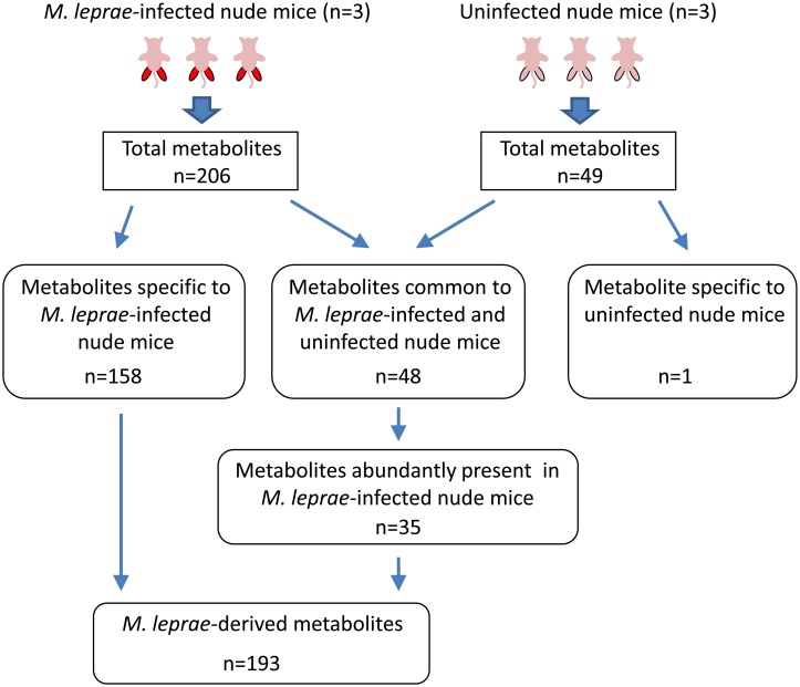
As shown in Fig 1, CE-MS analysis showed that 206 and 49 metabolites were present in detectable amount in the extracts of footpads dissected from M. leprae-infected (S1 Table) and uninfected nude mice (S2 Table), respectively (n = 3). Among 206 metabolites, 158 metabolites were specifically observed in M. leprae-infected nude mice, while only one compound was detected in uninfected nude mice (Fig 1). These observations mean that more than 75% of the detected metabolites in M. leprae-infected nude mice are derived from M. leprae itself. On the other hand, 48 metabolites from M. leprae-infected nude mice were also found in the extracts of uninfected nude mice. Quantitative comparison of 48 compounds between M. leprae-infected and uninfected nude mice revealed that most of relative peak areas from uninfected nude mice are much lower than those from M. leprae-infected nude mice (S1 Fig). To acquire the authentic value of the metabolites derived from M. leprae, we used the background subtraction method. Mean value of each metabolite obtained from uninfected nude mice was subtracted from all triplicate value of M. leprae-infected nude mice and thereby retrieved 35 metabolites which were abundantly present in M. leprae-infected nude mice (Fig 1). Eventually, 193 metabolites were totally determined to be specifically present in the M. leprae extract (S3 Table) (Fig 1).
Fig 1. Schematic representation for retrieving the M. leprae-derived metabolites identified by CE-MS analysis.
In each of common 48 metabolites, the mean of triplicate values (relative peak areas) of uninfected nude mice was subtracted from all triplicate value of M. leprae-infected nude mice and resultant 35 metabolites were retrieved as abundantly present in M. leprae-infected nude mice.
Although it became apparent that M. leprae possessed 193 specific intracellular metabolites, it is unknown how their qualitative and quantitative profiles differ from other mycobacteria. M. leprae is known to have unique properties such as long-term obligate growth in vivo, which is quite different from other mycobacteria, indicating that it is difficult to select the control mycobacteria for comparison. In this study, for comparative profiling of the M. leprae metabolites, we chose in vitro-cultured M. bovis BCG. Unlike M. leprae, M. bovis BCG does not remain in the footpad where inoculated but get disseminated all over the body and sufficient bacilli cannot be retrieved for analyses. [26]. By performing the extraction procedures of in vitro-cultured M. bovis BCG under the same conditions as M. leprae, we obtained the metabolite extracts of M. bovis BCG for comparative study.
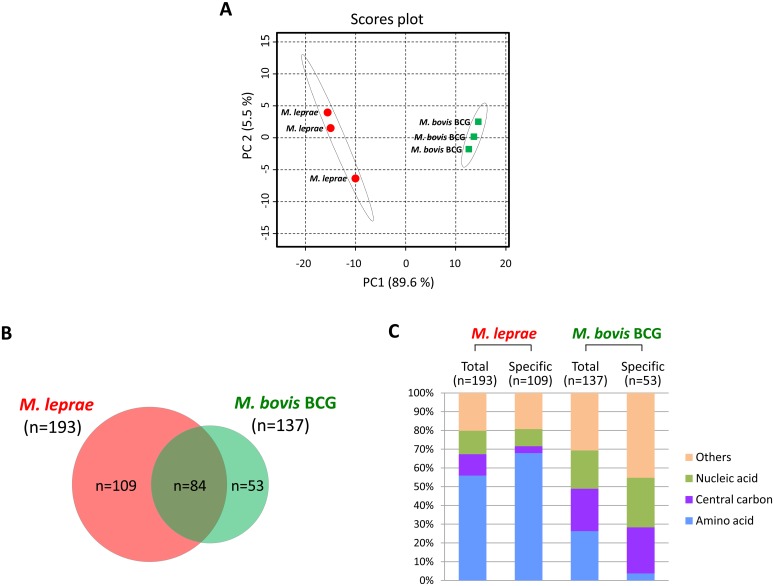
CE-MS analysis of M. bovis BCG indicated that 137 compounds could be detected (S4 Table). To obtain a comprehensive profile of metabolites, we performed the principal component analysis (PCA) on relative peak area of metabolites detected from three independent groups of M. leprae and M. bovis BCG (Fig 2A). The results showed that the groups cluster within each species but their clusters are clearly separated, suggesting that the sort and quantity of detected metabolites from M. leprae are close within three analyzed groups, but they are quite distinguishable from those of M. bovis BCG groups whose profiles were also similar to each other (Fig 2A). Therefore, this implied that metabolic profile of M. leprae and M. bovis BCG are functionally distinct. Comparison of detected metabolites between two species indicated that 84 metabolites are in common, while 109 and 53 metabolites are specifically observed in M. leprae and M. bovis BCG, respectively (Fig 2B). Additionally, we assessed and classified the metabolites in terms of their function in each category of metabolism (Fig 2C). As a consequence, we found that 56% of the total metabolites of M. leprae were categorized under the amino acid metabolism group, while in M. bovis BCG only 26% of the total metabolites belonged to the amino acid metabolism (Fig 2C). On the other hand, as for the metabolites associated with central carbon and nucleic acid metabolism, it was observed that their proportion in M. bovis BCG was almost twice as much as the metabolites of M. leprae (central carbon: 23% vs. 11%; nucleic acid: 20% vs. 12% in M. bovis BCG vs M. leprae respectively)(Fig 2C). These differences were more pronounced when the population of specifically detected metabolites from M. leprae and M. bovis BCG were considered. The amino acid-related compounds constitute 68% of 109 metabolites, which were specifically observed in M. leprae, but its proportion in 53 M. bovis BCG-specific metabolites was only 4% (Fig 2C). On the contrary, the percentage of the compounds related to the central carbon and nucleic acid metabolism was shown to be quite high in M. bovis BCG-specific metabolites, compared to those in M. leprae-specific metabolites (central carbon: 25% vs. 4%; nucleic acid: 26% vs. 9% in M. bovis BCG vs M. leprae respectively)(Fig 2C). These results indicated that, in M. leprae, amino acid-related compounds constituted a larger proportion of detected metabolites, while detection of metabolites associated with central carbon and nucleic acids were relatively small, compared to those of M. bovis BCG.
Fig 2. Comparative profiling of metabolites detected from M. leprae and M. bovis BCG.
A) Principal component analysis (PCA) on relative peak area of metabolites detected from three independent groups of M. leprae and M. bovis BCG. Total relative peak area for analysis was derived from 193 metabolites of M. leprae and 137 metabolites of M. bovis BCG. B) Venn diagram of identified metabolites. C) Proportion of the classified metabolites based on their putative functions in each metabolism. Each of mycobacterial metabolites was derived from 2.5x10¹⁰ cells.
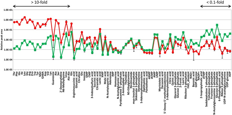
To better explore the mechanisms governing the fate of M. leprae, we focused on the quantitative evaluation of 84 metabolites, which were common to both M. leprae and M. bovis BCG (S5 Table). When the mean ratio of each relative peak area (M. leprae/M. bovis BCG) was arranged in decreasing order, it was found that 26% (22/84) of the metabolites significantly showed large differences (>10-fold), while 14% (12/84) showed <0.1-fold differences (Fig 3). Additionally, functional classification of the metabolites having 10-fold difference in the mean ratio revealed that most of the compounds listed are involved in amino acid metabolism (Table 1). This is supported by the fact that M. leprae-specific metabolites were dominated by those related to amino acid metabolism (Fig 2C), suggesting that amino acid and its derivatives abundantly accumulated as intracellular metabolites in M. leprae, when compared to those metabolites of M. bovis BCG. These results raise two possibilities regarding the metabolic aspects of M. leprae: (1) M. leprae itself has the capacity to produce high amount of amino acids, (2) M. leprae activates the uptake of amino acids from host, in order to maintain and control the metabolism suited for long-term, obligate growth in vivo. At present, it is not clear which mechanism could better explain the cause of amino acid accumulation, because no direct evidence was obtained from the M. leprae genomic analyses of the regions that could be involved. Probably there is yet unknown mechanism which causes the amino acid accumulation. On the contrary, Table 2 show that the metabolites in the cluster showing less than 0.1-fold differences in their mean ratio of relative peak area (M. leprae/M. bovis BCG) dominantly belonged to the intermediates related to the central carbon metabolism, for example metabolites such as glucose-6-phosphate, fructose-6-phosphate, sedoheptulose 7-phosphate, 6-phosphogluconic acid and ribose 5-phosphate. These metabolites play a critical role in cellular pathways of energy metabolism as well as other basic cellular processes. The same tendency is also observed when metabolites were classified according to their function (Fig 2C), suggesting that the central carbon metabolism in M. leprae is strikingly declined or repressed compared to those in M. bovis BCG. These results are exemplified by the M. leprae genomics demonstrating that around half of genes related to energy metabolism tuned out to be the pseudogenes, which might lead to the functional defect [1]. It is generally hypothesized that lack of energy generation machinery is the main reason for the inability of the bacilli to proliferate in vitro, while there are only genomics analysis available to prove the hypothesis. Thus, phenotypically, our study of comparative metabolomics for the first time supported this hypothesis.
Fig 3. Quantitative comparison of 84 metabolites common to M. leprae and M. bovis BCG.
Relative peak areas are expressed as means± S.D. of triplicate. These values were retrieved by CE-MS analysis performing on methanol extract from 2.5x10¹⁰ cells of each mycobacteria collected by filtration. As for those of M. leprae, nude mice-derived values were subtracted. Red circle and green square represent the mean value of each metabolite detected from M. leprae and M. bovis BCG, respectively. 84 metabolites are arranged in descending order of their mean ratio (M. leprae/M. bovis BCG). Of these metabolites, their mean ratio showing over 10-fold and less than 0.1-fold are indicated by arrows.
Table 1. Functional classification of the metabolites having over 10-fold difference in the mean ratio of relative peak areas [M. leprae (n = 3)/M. bovis BCG (n = 3)].
| Name | Class | Difference of relative peak area | |
|---|---|---|---|
| Mean ratio | P-value | ||
| Thr | Amino acid | 432.366 | 0.007 |
| Phe | Amino acid | 289.962 | 0.005 |
| His | Amino acid | 289.694 | 0.008 |
| Ile | Amino acid | 224.924 | 0.005 |
| Leu | Amino acid | 214.813 | 0.004 |
| Gly | Amino acid | 176.775 | 0.01 |
| Arg | Amino acid | 163.532 | 0.005 |
| Pro | Amino acid | 159.291 | 0.003 |
| Trp | Amino acid | 152.172 | 0.003 |
| Tyr | Amino acid | 133.469 | 0.006 |
| Met | Amino acid | 116.309 | 0.016 |
| GABA | Amino acid | 92.422 | 0.002 |
| Asp | Amino acid | 85.865 | 0.038 |
| Val | Amino acid | 68.687 | 0.005 |
| Lys | Amino acid | 53.537 | 0.009 |
| Guanosine | Nucleic acid | 47.326 | 0.178 |
| Ala | Amino acid | 38.597 | 0.006 |
| Ser | Amino acid | 33.751 | 0.013 |
| 2'-Deoxycytidine | Nucleic acid | 28.377 | 0.049 |
| N6-Methyllysine | Amino acid | 20.319 | 0.002 |
| Urea | Amino acid | 15.24 | 0.06 |
| Ala-Ala | Amino acid | 10.622 | 0.001 |
Metabolites are arranged in descending order of the mean ratio of each relative peak area.
Table 2. Functional classification of the metabolites having less than 0.1-fold difference in the mean ratio of relative peak areas [M. leprae (n = 3)/M. bovis BCG (n = 3)].
| Name | Class | Difference of relative peak area | |
|---|---|---|---|
| Mean ratio | P-value | ||
| 2-Isopropylmalic acid | Amino acid | 0.095 | 0.0003 |
| dTMP | Nucleic acid | 0.094 | 0.019 |
| Sedoheptulose 7-phosphate | Central carbon | 0.079 | 0.003 |
| Trehalose 6-phosphate | Other | 0.071 | 0.002 |
| Fructose 6-phosphate | Central carbon | 0.066 | 0.007 |
| N-Acetylglucosamine 1-phosphate | Central carbon | 0.064 | 0.017 |
| Glucose 6-phosphate | Central carbon | 0.061 | 0.006 |
| 6-Phosphogluconic acid | Central carbon | 0.053 | 0.006 |
| Ribose 5-phosphate | Central carbon | 0.052 | 0.011 |
| UDP-N-acetylglucosamine | Central carbon | 0.043 | 0.002 |
| UDP-glucose | Central carbon | 0.037 | 0.0001 |
| ADP | Central carbon | 0.017 | 0.006 |
Metabolites are arranged in descending order of the mean ratio of each relative peak area.
M. tuberculosis is known to possess the ability to degrade and use cholesterol as an energy source and for the biosynthesis of mycobacterial lipids. In M. leprae, the presence of host-derived cholesterol plays important role in the process of intracellular survival [27]. However, M. leprae lost essentially all the genes associated with cholesterol catabolism but retained only the ability to oxidize cholesterol to cholestenone, indicating that cholesterol metabolism was not coupled to central carbon metabolism in M. leprae [28]. Lipids in the foamy macrophages and Schwann cells were shown to be derived from the host lipids, favoring bacterial survival [29, 30]. In these contexts, M. leprae has its own unique metabolic pathways to sustain its growth and multiplication, which has to be further elucidated.
Focusing on minute detail of the M. leprae genome, numerous pseudogenes are distributed in the genomic region of each metabolic pathway, suggesting that such unique features of the genome might be one of the factors influencing the characteristic profiles of intracellular metabolites. However, at present, it is difficult to identify the pseudogenes causing M. leprae-specific profiles because each pathway is interrelated and is not thoroughly investigated, which leads to complications in deciphering the metabolism in M. leprae. Therefore, generation of mutants having mutations that mimic the pseudogenes of M. leprae in cultivable mycobacteria might partly elucidate the relationship between uniqueness in genomic organization and the metabolic profile obtained by CE-MS analysis.
Present study demonstrated that the dynamics of intracellular metabolites in M. leprae is quite different from those in M. bovis BCG. Although it is necessary to perform the metabolite profiling on other mycobacteria for more precise evaluation of M. leprae metabolism, the result retrieved from comparison with M. bovis BCG would partly contribute to the uncovering the M. leprae physiology associated with onset of leprosy. In M. tuberculosis, metabolomics analysis has been performed on in vitro-grown cells under conditions such as hypoxia and nutrient starvation which stimulate in vivo growth, while no study performing the metabolomics on actually in vivo-grown cells was reported [9–12]. Thus, our findings regarding in vivo-grown M. leprae might provide insights into the understanding of not only its physiology but also the metabolic behavior of mycobacteria in host, which remains unresolved.
Supporting Information
Red and blue bar represent the relative peak areas of each metabolite detected from M. leprae-infected and uninfected nude mouse, respectively. Relative peak areas are expressed as means ± S.D. of triplicate.
(PPTX)
(XLSX)
(XLSX)
Compounds also present in uninfected nude mouse (n = 35) and specifically detected from M. leprae-infected nude mouse (n = 158) were shown in shaded and plain column, respectively.
(XLSX)
(XLSX)
Metabolites are arranged in descending order of their mean ratio (M. leprae/M. bovis BCG).
(XLSX)
Data Availability
All relevant data are within the paper and its Supporting Information files.
Funding Statement
This work was supported by JSPS KAKENHI Grant Number 25871158 http://www.jsps.go.jp/english/e-grants/index.html Japan Society for the Promotion of Science to YMi; and Grant Number H24-Shinko-Ippan-009 http://www.mhlw.go.jp/english/index.html Ministry of Health, Labour and Welfare, Japan to TM. The funders had no role in study design, data collection and analysis, decision to publish, or preparation of the manuscript.
References
- 1.Cole ST, Eiglmeier K, Parkhill J, James KD, Thomson NR, et al. (2001) Massive gene decay in the leprosy bacillus. Nature 409: 1007–1011. [DOI] [PubMed] [Google Scholar]
- 2.Marques MA, Neves-Ferreira AG, da Silveira EK, Valente RH, Chapeaurouge A, et al. (2008) Deciphering the proteomic profile of Mycobacterium leprae cell envelope. Proteomics 8: 2477–2491. 10.1002/pmic.200700971 [DOI] [PubMed] [Google Scholar]
- 3.Parkash O, Singh BP (2012) Advances in Proteomics of Mycobacterium leprae. Scand J Immunol 75: 369–378. 10.1111/j.1365-3083.2012.02677.x [DOI] [PubMed] [Google Scholar]
- 4.Singh P, Cole ST (2011) Mycobacterium leprae: genes, pseudogenes and genetic diversity. Future Microbiol 6: 57–71. 10.2217/fmb.10.153 [DOI] [PMC free article] [PubMed] [Google Scholar]
- 5.Guenin-Macé L, Siméone R, Demangel C (2009) Lipids of pathogenic Mycobacteria: contributions to virulence and host immune suppression. Transbound Emerg Dis 56: 255–268. 10.1111/j.1865-1682.2009.01072.x [DOI] [PubMed] [Google Scholar]
- 6.Kai M, Fujita Y, Maeda Y, Nakata N, Izumi S, Yano I, Makino M (2007) Identification of trehalose dimycolate (cord factor) in Mycobacterium leprae. FEBS Lett 581: 3345–3350. [DOI] [PubMed] [Google Scholar]
- 7.Hunter SW, Fujiwara T, Brennan PJ (1982) Structure and antigenicity of the major specific glycolipid antigen of Mycobacterium leprae. J Biol Chem 257: 15072–15078. [PubMed] [Google Scholar]
- 8.Ng V, Zanazzi G, Timpl R, Talts JF, Salzer JL, Brennan PJ, Rambukkana A (2000) Role of the cell wall phenolic glycolipid-1 in the peripheral nerve predilection of Mycobacterium leprae. Cell 103: 511–524. [DOI] [PubMed] [Google Scholar]
- 9.Nandakumar M, Prosser GA, de Carvalho LP, Rhee K (2015) Metabolomics of Mycobacterium tuberculosis. Methods Mol Biol 1285: 105–115. 10.1007/978-1-4939-2450-9_6 [DOI] [PubMed] [Google Scholar]
- 10.de Carvalho LP, Fischer SM, Marrero J, Nathan C, Ehrt S, Rhee KY (2010) Metabolomics of Mycobacterium tuberculosis reveals compartmentalized co-catabolism of carbon substrates. Chem Biol 17: 1122–1131. 10.1016/j.chembiol.2010.08.009 [DOI] [PubMed] [Google Scholar]
- 11.Eoh H, Rhee KY (2014) Methylcitrate cycle defines the bactericidal essentiality of isocitrate lyase for survival of Mycobacterium tuberculosis on fatty acids. Proc Natl Acad Sci U S A 111: 4976–4981. 10.1073/pnas.1400390111 [DOI] [PMC free article] [PubMed] [Google Scholar]
- 12.Eoh H, Rhee KY (2013) Multifunctional essentiality of succinate metabolism in adaptation to hypoxia in Mycobacterium tuberculosis. Proc Natl Acad Sci U S A 110: 6554–6559. 10.1073/pnas.1219375110 [DOI] [PMC free article] [PubMed] [Google Scholar]
- 13.Prabhakaran K, Braganca BM (1962) Glutamic acid decarboxylase activity of Mycobacterium leprae and occurrence of gamma-amino butyric acid in skin lesions of leprosy. Nature 196: 589–590. [DOI] [PubMed] [Google Scholar]
- 14.Wheeler PR (1983) Catabolic pathways for glucose, glycerol and 6-phosphogluconate in Mycobacterium leprae grown in armadillo tissues. J Gen Microbiol 129: 1481–1495. [DOI] [PubMed] [Google Scholar]
- 15.Wheeler PR (1989) Pyrimidine biosynthesis de novo in M. leprae. FEMS Microbiol Lett 48: 185–189. [DOI] [PubMed] [Google Scholar]
- 16.Wheeler PR, Ratledge C (1988) Use of carbon sources for lipid biosynthesis in Mycobacterium leprae: a comparison with other pathogenic mycobacteria. J Gen Microbiol 134: 2111–2121. [DOI] [PubMed] [Google Scholar]
- 17.Al-Mubarak R, Vander Heiden J, Broeckling CD, Balagon M, Brennan PJ, Vissa VD (2011) Serum metabolomics reveals higher levels of polyunsaturated fatty acids in lepromatous leprosy: potential markers for susceptibility and pathogenesis. PLoS Negl Trop Dis 5: e1303 10.1371/journal.pntd.0001303 [DOI] [PMC free article] [PubMed] [Google Scholar]
- 18.Mayboroda OA, van Hooij A, Derks R, van den Eeden SJ, Dijkman K, et al. (2016) Exploratory Urinary Metabolomics of Type 1 Leprosy Reactions. Int J Infect Dis 45: 46–52. 10.1016/j.ijid.2016.02.012 [DOI] [PubMed] [Google Scholar]
- 19.Matsuoka M (2010) The history of Mycobacterium leprae Thai-53 strain. Lepr Rev 81: 137 [PubMed] [Google Scholar]
- 20.Kohsaka K, Matsuoka M, Hirata T, Nakamura M (1993) Preservation of Mycobacterium leprae in vitro for four years by lyophilization. Int J Lepr Other Mycobact Dis 61: 415–420. [PubMed] [Google Scholar]
- 21.Shepard CC, McRae DH (1968) A method for counting acid-fast bacteria. Int J Lepr Other Mycobact Dis 36: 78–82. [PubMed] [Google Scholar]
- 22.Soga T, Heiger DN (2000) Amino acid analysis by capillary electrophoresis electrospray ionization mass spectrometry. Anal Chem 72: 1236–1241. [DOI] [PubMed] [Google Scholar]
- 23.Soga T, Ueno Y, Naraoka H, Ohashi Y, Tomita M, Nishioka T (2002) Simultaneous determination of anionic intermediates for Bacillus subtilis metabolic pathways by capillary electrophoresis electrospray ionization mass spectrometry. Anal Chem 74: 2233–2239. [DOI] [PubMed] [Google Scholar]
- 24.Soga T, Ohashi Y, Ueno Y, Naraoka H, Tomita M, Nishioka T (2003) Quantitative metabolome analysis using capillary electrophoresis mass spectrometry. J Proteome Res 5: 488–94. [DOI] [PubMed] [Google Scholar]
- 25.Xia J, Sinelnikov IV, Han B, Wishart DS (2015) MetaboAnalyst 3.0-making metabolomics more meaningful. Nucleic Acids Res 43: W251–W257. 10.1093/nar/gkv380 [DOI] [PMC free article] [PubMed] [Google Scholar]
- 26.Sher NA, Chaparas SD, Greenberg LE, Merchant EB, Vickers JH. (1975) Response of congenitally athymic (nude) mice to infection with Mycobacterium bovis (strain BCG). J Natl Cancer Inst 54: 1419–1426. [DOI] [PubMed] [Google Scholar]
- 27.Lobato LS, Rosa PS, Ferreira Jda S, Neumann Ada S, da Silva MG, et al. (2014) Statins increase rifampin mycobactericidal effect. Antimicrob Agents Chemother 58: 5766–5774. 10.1128/AAC.01826-13 [DOI] [PMC free article] [PubMed] [Google Scholar]
- 28.Marques MA, Berrêdo-Pinho M, Rosa TL, Pujari V, Lemes RM, et al. (2015) The essential role of cholesterol metabolism in the intracellular survival of Mycobacterium leprae is not coupled to central carbon metabolism and energy production. J Bacteriol 197: 3698–3707. 10.1128/JB.00625-15 [DOI] [PMC free article] [PubMed] [Google Scholar]
- 29.Mattos KA, Oliveira VG, D'Avila H, Rodrigues LS, Pinheiro RO, et al. (2011) TLR6-driven lipid droplets in Mycobacterium leprae-infected Schwann cells: immunoinflammatory platforms associated with bacterial persistence. J Immunol 187: 2548–2558. 10.4049/jimmunol.1101344 [DOI] [PubMed] [Google Scholar]
- 30.Mattos KA, Sarno EN, Pessolani MCV, Bozza PT (2012) Deciphering the contribution of lipid droplets in leprosy: multifunctional organelles with roles in Mycobacterium leprae pathogenesis. Mem Inst Oswaldo Cruz 107: 156–166. [DOI] [PubMed] [Google Scholar]
Associated Data
This section collects any data citations, data availability statements, or supplementary materials included in this article.
Supplementary Materials
Red and blue bar represent the relative peak areas of each metabolite detected from M. leprae-infected and uninfected nude mouse, respectively. Relative peak areas are expressed as means ± S.D. of triplicate.
(PPTX)
(XLSX)
(XLSX)
Compounds also present in uninfected nude mouse (n = 35) and specifically detected from M. leprae-infected nude mouse (n = 158) were shown in shaded and plain column, respectively.
(XLSX)
(XLSX)
Metabolites are arranged in descending order of their mean ratio (M. leprae/M. bovis BCG).
(XLSX)
Data Availability Statement
All relevant data are within the paper and its Supporting Information files.


