Abstract
Objectives
Risk of knee osteoarthritis (OA) is much higher in women than in men. Previous studies have shown that bone shape is a risk factor for knee OA. However, few studies have examined whether knee bone shape differs between men and women. The purpose of the present study was to determine whether there are differences between men and women in knee bone shape.
Methods
We used information from the NIH-funded Osteoarthritis Initiative (OAI), a cohort of persons aged 45-79 at baseline who either had symptomatic knee OA or were at high risk of it. Among participants aged between 45 and 60 years, we randomly sampled 340 knees without radiographic OA (i.e., Kellgren/Lawrence grade of 0 in central readings on baseline radiograph). We characterized distal femur and proximal tibia shape of these selected radiographs using statistical shape modeling (SSM). We performed linear regression analysis to examine the association between sex and each knee shape mode (proximal tibia and distal femur), adjusting for age, race, body mass index (BMI) and clinic site.
Results
The mean age was 52.7 years (±4.3 SD) for both men and women. There were 192 female and 147 male knees for the distal femur analysis. Thirteen modes were derived for femoral shape, accounting for 95.5% of the total variance. Distal femur Mode 1 had the greatest difference in standardized score of knee shape between females and males (1.04, p<0.01); Modes 3, 5, 6, 8 and 12 were also significantly associated with sex. For tibial shape, 191 female knees and 149 male knees were used for the analysis. Ten modes explained 95.5% of shape variance. Of the significantly associated modes in the proximal tibia, Mode 2 had the greatest difference in standardized score of bone shape between males and females (−0.30, p=0.01); Modes 3 and 4 were also significantly associated.
Conclusion
The shapes of the distal femur and proximal tibia that form the knee joint differ by sex. Additional analyses are warranted to assess whether the difference in risk of OA between the sexes arises from bone shape differences.
Keywords: sex, knee, femur, gender, osteoarthritis, shape
INTRODUCTION
Risk of either radiographic or symptomatic osteoarthritis (OA) is much higher in women than men (1); however, the underlying causes for this sex difference in OA remain unknown. Several potential explanations have been proposed for the sex difference in OA, including differences in estrogen level, physical activity, and laxity or alignment (2-4), but each has only moderate supporting evidence, and none fully explains the observed sex differences.
Recently, several investigators have proposed that knee bone shape, assessed by anthropometric measures, cross-sectional findings, or statistical shape modeling, is associated with an increased risk of OA and with severity of OA (5-8). We have reported that several specific proximal femoral shapes, assessed by statistical shape modeling (SSM), are associated with incident radiographic hip OA in elderly women (9) and that proximal femoral shapes are associated with compartment-specific knee OA (10). Others have also reported that SSM-based knee shape is associated with severity of radiographic knee OA (11). If knee bone shape is an important mediator for the differences in risk of knee OA between men and women, then one would expect knee bone shape to be different between men and women. We measured bone shape of the distal femur and proximal tibia using SSM among knees without radiographic OA and compared bone shape difference between men and women among participants in Osteoarthritis Initiative.
METHODS
Study Subjects and Population
We selected our subjects from the Osteoarthritis Initiative (OAI) cohort, a database funded by the NIH-Foundation. Four clinical centers, with a coordinating center at University of California, San Francisco, enrolled 4796 subjects with or at high risk of knee osteoarthritis at baseline. More information is available online at http://www.oai.ucsf.edu/. Approval for the OAI project was given by the institutional review boards at each OAI Center, and for this project at the IRB at University of California, Davis.
Selection of Study Subjects
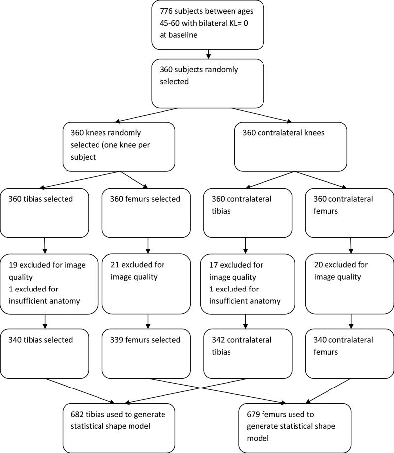
Two groups were selected for this study, a female group and a male group. A sub-population of 776 subjects ages 45 to 60 years old with a Kellgren/Lawrence (K/L) grade of 0 and with joint space narrowing (JSN) equal to 0 for both knees was selected from the OAI database. From the sub-population, 360 patients were randomly selected in the sex distribution of the total OAI. Some knees were ineligible due to poor knee film quality or if parts of the knee extended past the film window. All 679 femurs and 682 tibias remaining were used to generate the statistical shape models. Then one selected knee from each subject was used for the regression analysis. (See Figure 1)
Figure 1.
Flow diagram of subject selection.
Radiography
In plexiglass fixed-frame positioning, bilateral fixed-flexion posterior-anterior radiographs were taken at baseline and centrally read by two experienced readers with musculoskeletal training. Any disagreements were adjudicated between the readers. From the radiographs, Kellgren/Lawrence grade (K/L grade, scale from 0 to 4) and joint space narrowing (JSN, scale of 0-3) were provided. Only knees with a K/L grade of 0 and JSN of 0 were selected. Cross-sectional K/L grade scores had a kappa of 0.7. JSN scores had a kappa of 0.75.
Statistical Shape Modeling
The SSM methods used in this study are similar to methods described in previous publications (10). Radiographs where the edge of the knee extended beyond the border of the radiograph or where penetration was so poor that the edge of the knee could not be identified were eliminated because a complete shape could not be defined. One reader (LK) traced the shape of the distal femoral head and proximal tibial head on digitized radiographs using a standardized algorithm. The reader guided the placement of 41 landmark points outlining the shape of the distal femur and 40 landmark points outlining the proximal tibia. A total of 679 femurs and 682 tibias from all analyzed participants were compiled into composite distal femur and proximal tibia shapes to generate reference models representing a composite distal femur and proximal tibial shapes which were then used as reference shapes for measuring modes of variation from these references. Measurements of knee shape were acquired for each group using principal components analysis which gathered enough modes of variation in knee shape to explain 95% of total shape variance in this population, and by definition each mode of variation was independent of the other modes of variation. Mode scores for each knee were recorded as the number of standard deviations of a particular mode the given knee was away from the mean value of that bone shape, hereafter referred to as the “standardized score of bone shape”.
Reliability
Intra-rater reliability was evaluated by repeating measurements for 20% of the total sample and observing the point placement within 2 mm and within 3 mm of the prior hip shape points. Intra-rater reliability for the distal femur and proximal tibia were 97% and 92%, respectively, for point placement within 2 mm and 99% and 97% for point placement within 3 mm. These results are similar to reliability results reported in the literature for this type of SSM assessment (9).
Adjustment Variables
At the baseline visit for the OAI study, information was obtained for age, race, and clinic site by questionnaire; and body mass index (BMI) by measurement of the height and weight and then applying the appropriate equation to obtain kg/m2.
Statistical Methods
We compared the baseline characteristics between men and women using Chi-square tests for dichotomous variables and t-test for continuous variable. To test the association of sex differences and knee bone shape, we performed 10 independent linear regression models for the tibia and 13 for the femur, one for each knee shape mode as the outcome and sex as the predictor. Beta coefficients and 95% confidence intervals (CI) for the difference in mean of standardized score of each bone shape for women compared with men were reported. Models were adjusted for age, race, clinic site and BMI. SAS v9.2 (SAS, Inc, Cary, North Carolina) was used to complete statistical analyses.
RESULTS
We analyzed a total of 192 femurs from women and 147 from men and 191 tibias from women and 149 from men. The mean age of subjects contributing tibias in the analysis was 52.7 years (±4.3 SD) for both men and women. Mean BMI for women was 26.5 (±5.2) kg/m2 and was 27.7 (±4.2) kg/m2 for men. 76.4% of women were Caucasian and 85.2% of men were Caucasian in this sample. (See Table 1). For the distal femur, Modes 1, 3, 5, 6, 8, and 12 were significantly different by sex. For the proximal tibia, Modes 2, 3, and 4 differed by sex.
Table 1.
Characteristics of participants by sex.
| 1a. Characteristics of subjects with non-missing Femur shape modes | ||||
|---|---|---|---|---|
| Baseline Characteristics | Men | Women | Overall | P-value |
| (N= 147) | (N= 192) | (N= 339) | ||
| Age of enrollment | 52.7 +/− 4.3 | 52.8 +/− 4.4 | 52.7 +/− 4.3 | 0.84 |
| White | 126 (85.7) | 148 (77.1) | 274 (80.8) | 0.05 |
| BMI | 27.7 +/− 4.2 | 26.5 +/− 5.2 | 27.0 +/− 4.8 | 0.02 |
| Knee injury that caused immobility for 2 days | 61 (41.8) | 69 (36.3) | 130 (38.7) | 0.31 |
| College education or higher | 109 (75.2) | 124 (65.3) | 233 (69.6) | 0.05 |
| Modified Charlson Comorbidity score | 0.19 +/− 0.66 | 0.22 +/− 0.53 | 0.2 +/− 0.59 | 0.14 |
| 1b. Characteristics of subjects with non-missing Tibia shape modes | ||||
|---|---|---|---|---|
| Baseline Characteristics | Men | Women | Overall | P-value |
| (N= 149) | (N= 191) | (N= 340) | ||
| Age of enrollment | 52.7 +/− 4.3 | 52.7 +/− 4.3 | 52.7 +/− 4.3 | 0.88 |
| White | 127 (85.2) | 146 (76.4) | 273 (80.3) | 0.04 |
| BMI | 27.7 +/− 4.2 | 26.5 +/− 5.2 | 27.0 +/− 4.8 | 0.02 |
| Knee injury that caused immobility for 2 days | 62 (41.9) | 69 (36.5) | 131 (38.9) | 0.31 |
| College education or higher | 110 (74.8) | 124 (65.6) | 234 (69.6) | 0.07 |
| Modified Charlson Comorbidity score | 0.18 +/− 0.7 | 0.2 +/− 0.5 | 0.21 +/− 0.59 | 0.58 |
Statistical Shape Modeling
Thirteen modes were derived for femoral shape and accounted for 95.3% of the total variance of femoral shape in the population sampled (see Table 2). Ten modes described 95.5% of the total variance in proximal tibia shape in the population sampled (see Table 3).
Table 2.
Distal femur shape with sex.
| Mode | Variance Explained (%) | Difference in standardized score of knee shape mode between men and women (95%CI) | P-value for difference |
|---|---|---|---|
| 1 | 43.1 | 1.04 (0.85 to 1.23) | <0.01 |
| 2 | 16.6 | 0.02 (−0.18 to 0.23) | 0.83 |
| 3 | 11.5 | 0.23 (0.03 to 0.43) | 0.02 |
| 4 | 5.8 | 0.18 (−0.04 to 0.39) | 0.11 |
| 5 | 4.9 | −0.26 (−0.47 to −0.05) | 0.02 |
| 6 | 3.1 | −0.31 (−0.52 to −0.09) | <0.01 |
| 7 | 2.9 | −0.07 (−0.29 to 0.15) | 0.55 |
| 8 | 2.0 | −0.49 (−0.7 to −0.27) | <0.01 |
| 9 | 1.5 | 0.004 (−0.21 to 0.22) | 0.97 |
| 10 | 1.2 | −0.16 (−0.38 to 0.06) | 0.16 |
| 11 | 1.0 | 0.14 (−0.08 to 0.36) | 0.20 |
| 12 | 1.0 | 0.31 (0.1 to 0.54) | <0.01 |
| 13 | 0.6 | −0.20 (−0.42 to 0.01) | 0.07 |
Table 3.
Proximal tibial shape with sex.
| Mode | Variance Explained (%) | Difference in standardized score of knee shape mode between men and women (95%CI) | P-value for difference |
|---|---|---|---|
| 1 | 55.0 | −0.19 (−0.41 to 0.04) | 0.10 |
| 2 | 11.9 | −0.30 (−0.51 to −0.08) | 0.01 |
| 3 | 9.7 | −0.22 (−0.43 to −0.01) | 0.04 |
| 4 | 5.5 | −0.25 (−0.46 to −0.04) | 0.02 |
| 5 | 4.2 | −0.01 (−0.23 to 0.2) | 0.92 |
| 6 | 3.2 | 0.15 (−0.07 to 0.37) | 0.19 |
| 7 | 1.8 | 0.16 (−0.05 to 0.37) | 0.13 |
| 8 | 1.7 | 0.13 (−0.07 to 0.34) | 0.20 |
| 9 | 1.5 | 0.04 (−0.18 to 0.25) | 0.74 |
| 10 | 0.9 | −0.18 (−0.39 to 0.03) | 0.10 |
Femoral Modes
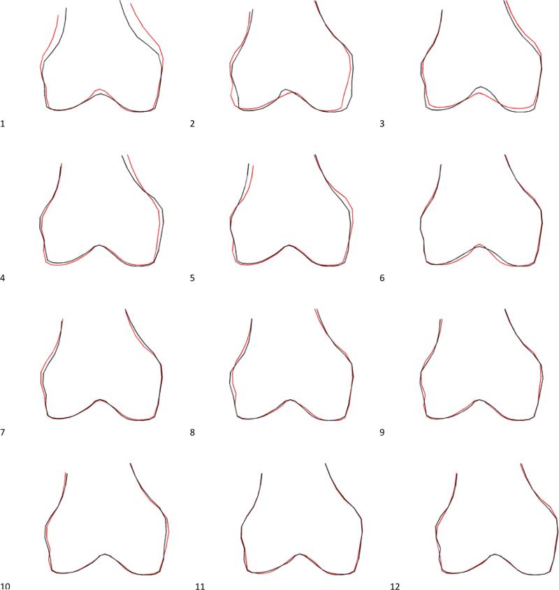
Several femoral modes significantly differed between men and women (p<0.01). The largest difference in means by sex for this association was in mode 1, where higher values of the mode were characterized by an increased shaft width relative to epicondylar width, along with a deepening of the intercondylar fossa. This mode explained 43.1% of the total variance in shape of the distal femur, and women had higher standardized scores for mode 1 than men (difference in means of standardized score for mode 1=1.04; 95%CI 0.85, 1.23). (See Figure 2)
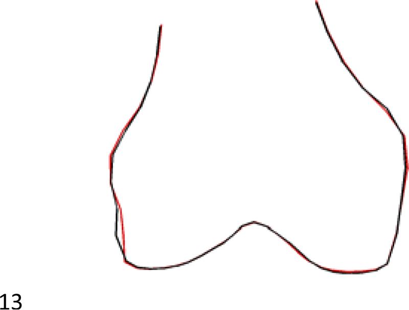
Figure 2.
All generated femoral modes. Red outline represents +2 SD and black outline represents −2 SD of each mode.
Mode 3 is characterized by a decreased inferior projection of the medial and lateral condylar heads with respect to the patellar groove with higher values of this mode (p=0.02). 11.5% of total variance in shape was explained by this mode. Women had higher standardized scores than men for mode 3 (difference in standardized scores for mode 3 =0.23; 95%CI 0.03, 0.43).
Higher values in mode 5 are characterized primarily by an increased excursion of the medial epicondyle, along with a narrowing of the femoral shaft. Higher values in this mode were also associated with a deepening of the groove between the lateral epicondyle and the condyle. Women had lower standardized scores than men for mode 5 (difference in standardized scores for mode 5 =−0.26; 95%CI −0.47, −0.05).
Higher values in mode 6 of the femur are characterized by an increased depth and acuity of angulation of the walls of the intercondylar fossa. Women had lower standardized scores than men for mode 6 (difference in standardized scores of mode 6 =−0.31; 95%CI −0.52, −0.09).
Positive values of femoral mode 8 displayed a decreased extension of the lateral epicondyle. Women had lower standardized scores than men for mode 8 (difference in standardized score of mode 8 =−0.49; 95%CI −0.7, −0.27).
Higher values of mode 12 are characterized by a decreased extension of the lateral epicondyle with increased extension of the medial epicondyle, and women had higher standardized scores than men for mode 12 (difference in standardized score of mode 12=0.32; 95%CI 0.1, 0.54).
Tibial Modes
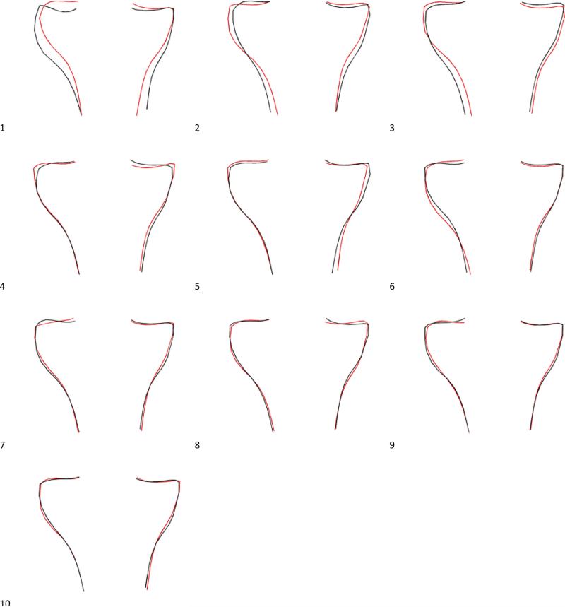
Tibial mode 2 had the highest significance for association with sex (p=0.01). Positive values of mode 2 represent an increased head width relative to the shaft width, along with a lateral shift of the tibial head with respect to the shaft, and a slightly more concave lateral tibial plateau and slightly more convex medial tibial plateaus. Women had lower standardized scores than men for mode 2 (difference in standardized score of mode 2 =−0.30; 95%CI −0.51, −0.08). (See figure 3.)
Figure 3.
All generated tibial modes. Red outline represents +2 SD and black outline represents −2 SD of each mode.
Tibial mode 3 was also significantly associated with sex (p=0.04). This mode displays slightly increased tibial width and depression of medial plateau and elevation of lateral plateau with increases in SD weighting in the mode. Women had lower standardized scores than men for mode 3 (difference in standardized score of mode 3=−0.22; 95%CI −0.43, −0.01). (See figure 3.)
Tibial mode 4 was significantly associated with sex at the p=0.02 level, and increases in SD weighting manifested as an increased concavity of the medial compartment and elevation of the medial lip, with a broadening of the lateral plateau. Women had lower standardized scores for mode 4 than men (difference in means of standardized score for mode 4 =−0.25; 95%CI −0.46, −0.04)(See figure 3.)
DISCUSSION
Our results confirm prior observations that the shape of the knee differs by sex in both the tibia and the femur among persons without knee OA and who by risk factor assessment were at the lowest risk of knee OA in the OAI cohort. The present study also extends understanding of shape differences by sex in the knee.
Our findings confirm and extend findings reported in previous studies. Yang et al. (12), Yan (13), and Yue (14), and Bellemans (15) each reported that women had significantly narrower distal femoral condyle width than men in a variety of different populations and using different radiographic approaches. In a study measuring knee dimensions intraoperatively rather than radiographically, mediolateral dimension of the distal femur was reported to be significantly larger in men (16). The results of these prior studies are similar in this respect and are in agreement with our study results. Specifically, we identified mode one for the distal femur as being an important mode of shape difference between women and men. This mode, which represents a different relative size of the distal shaft width to the condyle width can be interpreted to represent a similar shaft size for both men and women but with a smaller intercondylar width, essentially a similar finding to the prior reports referenced. All of these prior reports were based on data generated by measuring pre-designated morphological characteristics using a variety of different imaging or surgical approaches, so the fact that we identified a similar component of sex difference in shape lends substantial face validity to our approach and to our other findings.
Bellemans (15) also examined elements of morphometric geometry in the proximal tibia, and found that only 2% of variability in tibial geometry was explained by gender. Yue (14) found no significant gender differences in proximal tibial geometry. The present study utilized a different approach to the question of variability of shape by using active shape modeling and principal components analysis, and perhaps it is for this reason that we were able to identify differences in shape between the sexes in the proximal tibia where others didn't. However, we also found a smaller number of tibial shape modes to be significantly different between men and women than were femoral modes, and the effect sizes in the proximal tibia were overall smaller, with no modes having as large an effect size as mode 1 for the distal femur. These various findings together suggest that there is less sex based shape differentiation in the proximal tibia as compared overall with the distal femur.
The present study has a number of strengths, particularly that we used the Osteoarthritis Initiative, which has excellent standardization of radiograph acquisition and reading, as well as reliable collection of clinical and demographic characteristics. The OAI encompasses a diverse and representative population of persons both whites and African Americans and so in this sense this study is fairly generalizable to persons at risk of knee OA in the US population.
The present study also has limitations. First, study subject positioning at the time of the radiographs may influence the active shape modeling findings leading to a misclassification bias, despite every effort having been made to standardize the positioning, beam angle, and other factors related to acquisition of the radiographs. We also attempted to reduce the effect of poor positioning by critically scrutinizing the radiographs and eliminating those that were inadequate for ideal shape modeling. There is also a component of human error in the SSM reading which may have unknown effects on the shapes derived. Finally, there can be early OA in the joint prior to radiographically observable changes, which might have biased our findings; we addressed this by excluding knees with early radiographic evidence of knee OA, but some residual confounding may remain.
In summary, distal femoral and proximal tibial knee shapes differ by sex. Further work is warranted to understand how these shape modes may be associated with the development of knee OA and whether they can explain differences in prevalence of OA by sex.
ACKNOWLEDGMENTS
The authors thank the participants in the Osteoarthritis Initiative.
This work was supported by the following funding sources: the Center for Musculoskeletal Health at University of California, Davis School of Medicine; Nancy Lane support by NIH K24 AR048841; Barton Wise and Nancy Lane support by NIH P50 AR060752; NIH P50 AR063043; and the Endowed Chair for Aging at University of California, Davis School of Medicine. The Osteoarthritis Initiative (OAI) is a public–private partnership comprised of 5 contracts (N01-AR-2-2258, N01-AR-2-2259, N01-AR-2-2260, N01-AR-2-2261, and N01-AR-2-2262) funded by the NIH, a branch of the Department of Health and Human Services, and conducted by the OAI Study Investigators. Private funding partners include Pfizer, Novartis Pharmaceuticals, Merck Research Laboratories, and Glaxo-SmithKline. Private sector funding for the OAI is managed by the Foundation for the NIH.
Footnotes
Publisher's Disclaimer: This is a PDF file of an unedited manuscript that has been accepted for publication. As a service to our customers we are providing this early version of the manuscript. The manuscript will undergo copyediting, typesetting, and review of the resulting proof before it is published in its final citable form. Please note that during the production process errors may be discovered which could affect the content, and all legal disclaimers that apply to the journal pertain.
AUTHOR CONTRIBUTIONS
Conception and design: BLW, JAL, YZ, NEL
Analysis in interpretation of data: BLW, FL, JAL, NP, YZ, NEL
Drafting of the article: BLW, NP, YZ, NEL
Critical revision of the article for important intellectual content: BLW, FL, LK, JAL, NP, YZ, NEL
Final approval of the article: BLW, FL, LK, JAL, NP, YZ, NEL
Collection and assembly of data: BLW, FL, LK, JAL
Obtaining funding: BLW, NEL
REFERENCES
- 1.Lawrence RC, Felson DT, Helmick CG, Arnold LM, Choi H, Deyo RA, Gabriel S, Hirsch R, Hochberg MC, Hunder GG, Jordan JM, Katz JN, Kremers HM, Wolfe F. Estimates of the prevalence of arthritis and other rheumatic conditions in the United States. Part II. Arthritis and rheumatism. 2008;58(1):26–35. doi: 10.1002/art.23176. Epub 2008/01/01. doi: 10.1002/art.23176. PubMed PMID: 18163497. [DOI] [PMC free article] [PubMed] [Google Scholar]
- 2.Felson DT, Lawrence RC, Dieppe PA, Hirsch R, Helmick CG, Jordan JM, Kington RS, Lane NE, Nevitt MC, Zhang Y, Sowers M, McAlindon T, Spector TD, Poole AR, Yanovski SZ, Ateshian G, Sharma L, Buckwalter JA, Brandt KD, Fries JF. Osteoarthritis: new insights. Part 1: the disease and its risk factors. Annals of internal medicine. 2000;133(8):635–46. doi: 10.7326/0003-4819-133-8-200010170-00016. PubMed PMID: 11033593. [DOI] [PubMed] [Google Scholar]
- 3.Sharma L, Lou C, Cahue S, Dunlop DD. The mechanism of the effect of obesity in knee osteoarthritis: the mediating role of malalignment. Arthritis and rheumatism. 2000;43(3):568–75. doi: 10.1002/1529-0131(200003)43:3<568::AID-ANR13>3.0.CO;2-E. PubMed PMID: 10728750. [DOI] [PubMed] [Google Scholar]
- 4.Sharma L, Song J, Felson DT, Cahue S, Shamiyeh E, Dunlop DD. The role of knee alignment in disease progression and functional decline in knee osteoarthritis. Jama. 2001;286(2):188–95. doi: 10.1001/jama.286.2.188. PubMed PMID: 11448282. [DOI] [PubMed] [Google Scholar]
- 5.Bredbenner TL, Eliason TD, Potter RS, Mason RL, Havill LM, Nicolella DP. Statistical shape modeling describes variation in tibia and femur surface geometry between Control and Incidence groups from the Osteoarthritis Initiative database. Journal of biomechanics. doi: 10.1016/j.jbiomech.2010.02.015. PubMed PMID: 20227696. [DOI] [PMC free article] [PubMed] [Google Scholar]
- 6.Haverkamp DJ, Schiphof D, Bierma-Zeinstra SM, Weinans H, Waarsing JH. Variation in joint shape of osteoarthritic knees. Arthritis and rheumatism. 2011 doi: 10.1002/art.30575. Epub 2011/08/04. doi: 10.1002/art.30575. PubMed PMID: 21811994. [DOI] [PubMed] [Google Scholar]
- 7.Matsuda S, Miura H, Nagamine R, Mawatari T, Tokunaga M, Nabeyama R, Iwamoto Y. Anatomical analysis of the femoral condyle in normal and osteoarthritic knees. J Orthop Res. 2004;22(1):104–9. doi: 10.1016/S0736-0266(03)00134-7. Epub 2003/12/06. doi: S0736026603001347 [pii] 10.1016/S0736-0266(03)00134-7. PubMed PMID: 14656667. [DOI] [PubMed] [Google Scholar]
- 8.Miura H, Noguchi Y, Mitsuyasu H, Nagamine R, Urabe K, Matsuda S, Iwamoto Y. Clinical features of multiple epiphyseal dysplasia expressed in the knee. Clinical orthopaedics and related research. 2000(380):184–90. doi: 10.1097/00003086-200011000-00025. Epub 2000/11/07. PubMed PMID: 11064990. [DOI] [PubMed] [Google Scholar]
- 9.Lynch JA, Parimi N, Chaganti RK, Nevitt MC, Lane NE. The association of proximal femoral shape and incident radiographic hip OA in elderly women. Osteoarthritis and cartilage / OARS, Osteoarthritis Research Society. 2009;17(10):1313–8. doi: 10.1016/j.joca.2009.04.011. PubMed PMID: 19427402; PubMed Central PMCID: PMC2846640. [DOI] [PMC free article] [PubMed] [Google Scholar]
- 10.Wise BL, Kritikos L, Lynch JA, Liu F, Parimi N, Tileston KL, Nevitt MC, Lane NE. Proximal femur shape differs between subjects with lateral and medial knee osteoarthritis and controls: the Osteoarthritis Initiative. Osteoarthritis and cartilage / OARS, Osteoarthritis Research Society. 2014;22(12):2067–73. doi: 10.1016/j.joca.2014.08.013. doi: 10.1016/j.joca.2014.08.013. PubMed PMID: 25194496; PubMed Central PMCID: PMC4252863. [DOI] [PMC free article] [PubMed] [Google Scholar]
- 11.Barr RJGJ, Yoshida K, Alesci S, Reid DM, Aspden R. Knee joint shape assessed by a active shape modelling of plain radiographs is related to osteoarthritis severity. Osteoarthritis and Cartilage. 2009;17(Supplement):S200–1. [Google Scholar]
- 12.Yang B, Yu JK, Zheng ZZ, Lu ZH, Zhang JY. Comparative study of sex differences in distal femur morphology in osteoarthritic knees in a Chinese population. PLoS One. 2014;9(2):e89394. doi: 10.1371/journal.pone.0089394. doi: 10.1371/journal.pone.0089394. PubMed PMID: 24586746; PubMed Central PMCID: PMC3929686. [DOI] [PMC free article] [PubMed] [Google Scholar]
- 13.Yan M, Wang J, Wang Y, Zhang J, Yue B, Zeng Y. Gender-based differences in the dimensions of the femoral trochlea and condyles in the Chinese population: correlation to the risk of femoral component overhang. The Knee. 2014;21(1):252–6. doi: 10.1016/j.knee.2012.11.005. doi: 10.1016/j.knee.2012.11.005. PubMed PMID: 23245733. [DOI] [PubMed] [Google Scholar]
- 14.Yue B, Varadarajan KM, Ai S, Tang T, Rubash HE, Li G. Gender differences in the knees of Chinese population. Knee surgery, sports traumatology, arthroscopy : official journal of the ESSKA. 2011;19(1):80–8. doi: 10.1007/s00167-010-1139-8. doi: 10.1007/s00167-010-1139-8. PubMed PMID: 20407755. [DOI] [PubMed] [Google Scholar]
- 15.Bellemans J, Carpentier K, Vandenneucker H, Vanlauwe J, Victor J. The John Insall Award: Both morphotype and gender influence the shape of the knee in patients undergoing TKA. Clinical orthopaedics and related research. 2010;468(1):29–36. doi: 10.1007/s11999-009-1016-2. doi: 10.1007/s11999-009-1016-2. PubMed PMID: 19669385; PubMed Central PMCID: PMC2795809. [DOI] [PMC free article] [PubMed] [Google Scholar]
- 16.Lonner JH, Jasko JG, Thomas BS. Anthropomorphic differences between the distal femora of men and women. Clinical orthopaedics and related research. 2008;466(11):2724–9. doi: 10.1007/s11999-008-0415-0. doi: 10.1007/s11999-008-0415-0. PubMed PMID: 18719975; PubMed Central PMCID: PMC2565021. [DOI] [PMC free article] [PubMed] [Google Scholar]


