Erratum to: Human Cell (2016) 29:37–45 DOI 10.1007/s13577-015-0125-3
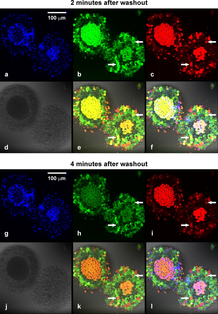
In the original publication of the article, Fig. 4k was published incorrectly. The correct figure is given in this erratum. The authors regret their errors.
Fig. 4.
Confocal microscopic images of 12 DIV MIN6 spheroids subjected to 100 μM of 2-NBDLG (green) and 20 μM of 2-TRLG (red) mixture for 3 min followed by washout. a Nuclear staining with DAPI in live cell condition. The central core region of spheroids appears to be necrotic (see also d). b and c Fluorescence images taken at 2 min after starting washout of the tracers in the green (b, 500–580 nm) and the red (c, 580–740 nm) channel, reflecting entrance of 2-NBDLG and 2-TRLG, respectively. d Differential interference contrast (DIC) image. e Overlay of the green, red, and DIC images. f Overlay of (a) and (e). Cellular heterogeneity is clearly seen by a combination of the two fluorescent colors. Cells indicated by arrows exhibited yellow color at 2 min (e), turned red at 4 min (k). This is because green 2-NBDLG was lost (b, h), while red 2-TRLG remained (c, i). If one saw a single 2-NBDLG image (b), cells indicated by arrows would have been misinterpreted to be similar to cells nearby. g–l Similar to a–f but images taken at 4 min after starting washout. Numbers of green cells with no red fluorescence, seen in the area surrounding the central core, preserved their color for at least up to 30 min (h, i, k). Also noted are dark cells in the area just surrounding the central core (b, e, h, k). Bars are common to all panels
Footnotes
The online version of the original article can be found under doi:10.1007/s13577-015-0125-3.


