Abstract
Objective:
To determine and compare the accuracy of pulp/tooth ratio method in mandibular first and second molar teeth in forensic age estimation.
Materials and Methods:
A total 300 panoramic radiographs of the Gujarati population (187 males and 113 females) were studied. The measurements of Pulp Chamber Height (PCH) and Crown Root Trunk Height (CRTH) were performed on the mandibular first and second molar teeth. The acquired data was subjected to correlation and regression.
Results:
The pulp chamber crown root trunk height ratios (PCTHR) of both the first (r = −0.609) and second molars (r = −0.422) were significantly correlated with the age of the individual. Individual regression formulae were derived for both the teeth which were then used separately to calculate the age. The standard errors estimate (SEE) for the first and second molars were 8.84 years and 10.11 years, respectively. There was no statistically significant difference between chronological and calculated age by both the teeth (P = 1.000).
Conclusion:
The mandibular first and second molar is a potential tool for age estimation in forensic dentistry. The pulp/tooth ratio of both the teeth is a useful method for forensic age prediction with reasonable accuracy in the Gujarati population.
Keywords: Adult, age estimation, molar, panoramic radiograph pulp/tooth ratio
Introduction
Age determination is an important and vital aspect in forensic sciences.[1] Teeth are the most durable and reliable tool for forensic age estimation.[2] Various methods can be utilized for age estimation in children as well as in adults by the use of a tooth.[3] The measurement of secondary dentin deposition by dental radiographs is a simple and nondestructive method for age estimation. This method is useful for both living as well as deceased persons.
Several studies have been performed to determine age by this method using canine,[2,4,5] first premolar, and second premolar either on intraoral periapical or panoramic radiographs.[2] Mathew et al.[6] developed an individual procedure of adult forensic age estimation by using mandibular first molar radiographs. But, there is no study performed by using second molar and/or third molar for age estimation by this method. Third molars are often congenitally missing, extracted, malformed or malpositioned making age estimation difficult by the said method. Conversely, first and second molars may be valuable sources for age estimation and many studies have accurately utilized these teeth for age estimation by other methods.[7] So, the present study was aimed to determine and compare the accuracy of age estimation using mandibular first and second molars.
Materials and Methods
Subject selection
A total of 300 digital panoramic radiographs obtained using KODAK 8000C panoramic radiograph machine were selected from the archives of the department based on the inclusion and exclusion criteria. The digital panoramic radiographs of the Gujarati patients between 20 to 59 years of age with good contrast and resolution and showing good image of mandibular first and second molars were selected for the present study. The digital panoramic radiographs showing image distortion, pathology in mandible, missing mandibular right first and/or second molars, and the molars with caries, periapical pathosis, radiopaque fillings and prosthetic crowns were excluded from the study.
Radiograph measurements
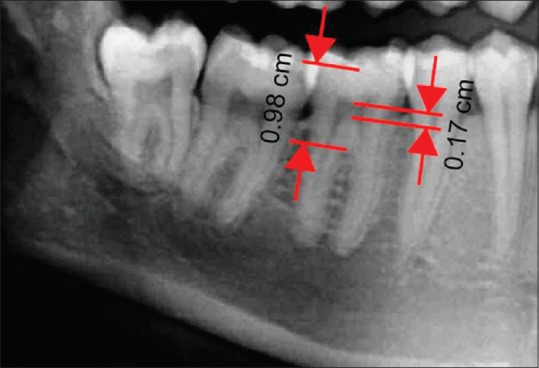
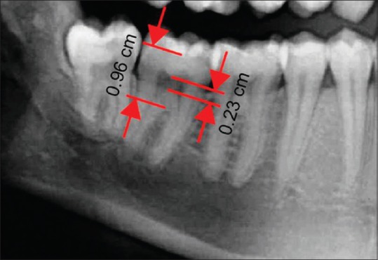
All 300 radiographs were subjected to measurements. The radiographs were exported into JPEG image format by using trophy Digital Imaging and Communications in Medicine (DICOM) software. The measurements were performed on these JPEG images by using Adobe Acrobat 8.0 Professional software. The measurements were performed according to the method described by Mathew et al.[4] on right mandibular first and second molar teeth. The distance between the central fossa and the highest point on the root furcation was measured as Crown Root Trunk Height (CRTH). The distance between the roof and the floor of the pulp chamber in the same axis was measured as the Pulp Chamber Height (PCH). A ratio of PCH and CRTH was derived as Pulp Chamber Crown Root Trunk Height Ratio (PCTHR).
(PCTHR = PCH/CRTH) [Figures 1 and 2].
Figure 1.

Cropped panoramic image showing measurements on the mandibular right first molar
Figure 2.

Cropped panoramic image showing measurements on the mandibular right second molar
Statistical analysis
The collected data was analyzed by a statistical software package It can be written as International Business Machines Corporation, Statistical Package for the Social Sciences version 19.0 (IBM SPSS v. 19.0). Pearson correlation was done between chronological age and PCTHR. Linear regression analysis was done on PCTHR of both first and second molars and two separate formulae were derived to estimate the age. To test the accuracy in age prediction by this method, the regression equation was applied for PCTHR obtained from all the 300 radiographic measurements. The standard error of estimate (SEE) was calculated to predict the deviation of the estimated age from the chronological age. The difference between the chronological age and the estimated age was recorded as error. Independent-samples t-test was performed between chronological age and estimated age and between estimated age by first and second molars. The level of significance was set at P ≤ 0.05.
Results and Observations
Digital orthopantomograms of total 300 subjects were analyzed in the present study of which there were 187 males and 113 females [Table 1].
Table 1.
Age and Sex distribution of study subjects

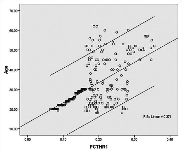
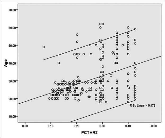
There was statistically significant negative correlation between chronological age and PCTHR of both mandibular first (r = −0.609) and second (r = −0.422) molars.
The regression formulae derived were:
For mandibular first molar:
Estimated age = 13.016 + (102.19 × PCTHR) years
For mandibular second molar:
Estimated age = 18.242 + (48.561 × PCHTR) years
The R2 values for regression equation were 0.371 and 0.178 for the first molar and second molar, respectively.
The SEE for the first molar was 8.84 years and for the second molar it was 10.11 years.
There was no statistically significant difference between actual age and calculated age for both first (P = 1.000) and second (P = 1.000) molars, when the age was calculated by applying the regression equations. In addition, there was no statistically significant difference found between the calculated age by first and second molars (P = 1.000) [Table 2].
Table 2.
Difference between chronological age and estimated age

Figures 3 and 4 shows scatterplot distribution between chronological age and the PCTHR of mandibular first and second molars, respectively.
Figure 3.

Scatterplot distribution of PCTHR of the first molar when compared to the subject's chronological age
Figure 4.

Scatterplot distribution of PCTHR of the second molar when compared to the subject's chronological age
Discussion
After root completion, formation of secondary dentin begins and it continues throughout one's life, reducing the pulp chamber dimension. Formation of secondary dentin is principally associated with advancement of age and is least affected by other environmental factors.[6] In 1925, Bodecker et al.[8] established that the apposition of the secondary dentin is correlated with age. Gustafson[9] introduced secondary dentin deposition for age estimation. Kvaal et al.[10] in 1995, presented a method of age estimation based on the measurements of secondary dentin deposition on periapical radiographs. After that, several studies were performed on intraoral radiographs as well as panoramic radiographs assessing the pulp/tooth ratio and correlating it with age. So, radiographic evaluation of secondary dentin deposition is an established noninvasive method for age estimation.
Formation of secondary dentin does not occur uniformly on all the surfaces of the tooth. In molar teeth, it appears in greater amounts on the roof and floor of the coronal pulp chamber.[11] Mathew et al.[6] developed an independent procedure in which the height of the pulp chamber and the height of crown root trunk was measured for estimating the age in south Indian population. Deposition of secondary dentin is influenced by genetic and environmental factors. Talreja et al.[12] demonstrated that more accurate results can be obtained by using population-specific formulae. So, in the present study, a separate regression formula was derived for the Gujarati population.
When PCTHR ratio was correlated with chronological age, the value of correlation coefficient (r) were suggestive of a high degree statistically significant negative correlation for the first molar (r = −0.609) and statistically significant negative correlation for the second molar (r = −0.422). The negative correlation is suggestive of association between reduction in the PCH and advancement of the chronological age which was strongly significant for the first molar as compared to the second molar. This finding was consistent with the study of Mathew et al.[6]
The values of coefficient of determination (R2) in the present study for the first and second molars were 0.371 and 0.178, respectively. This finding was similar to that of the Mathew et al.as well.[6] These values were found to be higher than the studies of Talreja et al.,[12] Limdiwala et al.[13] and Chandramala et al.[14] that were conducted on anterior teeth and premolars.
The SEE for the first molar was lesser (8.84 years) as compared to that of the second molar (10.11 years). These values were lesser as compared to the studies of Talreja et al.,[12] Limdiwala et al.,[13] and Chandramala et al.[14] This finding suggests that the age estimation by pulp/tooth ratio by using molar teeth can be done with comparable accuracy with that of the anterior teeth.
When the regression equation was applied to calculate age, there was no statistically significant difference between calculated age and actual age for both first and second molars (P = 1.000). This finding suggests the accuracy of the method used. Moreover, there is no significant difference between the age calculated by the first molar and the second molar (P = 1.000). This finding favors our hypothesis and states that the mandibular first and second molar can be used as a competent tool for adult forensic age estimation by the said method. Even though the values of correlation coefficient (r) and coefficient of determination (R2) were lesser and the SEE was higher as compared to that of the first molar, the results obtained were well within the acceptable limit.
Conclusion
The accuracy of the method developed by Mathew et al. for adult age estimation using mandibular molar teeth was confirmed with a larger sample size and by using two teeth. The present study suggests that the said method can be used as an aid for forensic age prediction in the Gujarati population. The future researches can be performed by using intraoral periapical radiographs, curvilinear regression analysis, multiple regression formulae, and larger sample sizes to achieve more accurate results.
Financial support and sponsorship
Nil.
Conflicts of interest
There are no conflicts of interest.
References
- 1.Jagannathan N, Neelkanthan P, Thiruvengadam C, Ramani P, Premkumar P, Natesan A, et al. Age estimation in an Indian population using pulp/tooth volume ratio of mandibular canines obtained from cone beam computed tomography. J Forensic Odontostomatol. 2011;29:1–6. [PMC free article] [PubMed] [Google Scholar]
- 2.Afify MM, Zayet MK, Mahmoud NF, Ragab AR. Age estimation from pulp/tooth area ration in three mandibular teeth by panoramic radiographs: Study of an Egyptian sample. J Forensic Res. 2014;5:1–5. [Google Scholar]
- 3.Jain RK, Rai B. Age estimation from permanent molar's attrition of Haryana population. Indian J Forensic Odontol. 2009;2:59–61. [Google Scholar]
- 4.Saxena S. Age estimation of Indian adults from orthopantomographs. Braz Oral Res. 2011;25(3):225–229. doi: 10.1590/s1806-83242011005000009. [DOI] [PubMed] [Google Scholar]
- 5.Cameriere R, Ferrante L, Belcastro GM, Bonfiglioli B, Rastelli E, Cingolani M. Age estimation by pulp/tooth ratio in canines by periapical X-rays. J Forensic Sci. 2007;52:166–70. doi: 10.1111/j.1556-4029.2006.00336.x. [DOI] [PubMed] [Google Scholar]
- 6.Mathew DG, Rajesh S, Koshi E, Priya LE, Nair AS, Mohan A. Adult forensic age estimation using mandibular first molar radiographs: A novel technique. J Forensic Dent Sci. 2013;5:56–9. doi: 10.4103/0975-1475.114552. [DOI] [PMC free article] [PubMed] [Google Scholar]
- 7.Almeida MS, Pontual Ados A, Beltrão RT, Beltrão RV, Pontual ML. The chronology of second molar development in Brazilians and its application to forensic age estimation. Imaging Sci Dent. 2013;43:1–6. doi: 10.5624/isd.2013.43.1.1. [DOI] [PMC free article] [PubMed] [Google Scholar]
- 8.Meinl A, Tangl S, Pernicka E, Fenes C, Watzek G. On the applicability of secondary dentin formation to radiological age estimation in young adults. J Forensic Sci. 2007;52:438–41. doi: 10.1111/j.1556-4029.2006.00377.x. [DOI] [PubMed] [Google Scholar]
- 9.Gustafson G. Age determination from teeth. J Am Dent Assoc. 1950;41:45–54. doi: 10.14219/jada.archive.1950.0132. [DOI] [PubMed] [Google Scholar]
- 10.Kvaal SI, Kolltveit KM, Thomsen IO, Solheim T. Age estimation of adults from dental radiographs. Forensic Sci Int. 1995;74:175–85. doi: 10.1016/0379-0738(95)01760-g. [DOI] [PubMed] [Google Scholar]
- 11.Kumar GS. Orban's Oral Histology and Embryology. 12th ed. New Delhi: Elsevier; 2006. p. 90. [Google Scholar]
- 12.Kanchan-Talreja P, Acharya AB, Naikmasur VG. An assessment of versatility of Kvaal's method of adult age estimation in Indians. Arch Oral Biol. 2012;57:277–84. doi: 10.1016/j.archoralbio.2011.08.020. [DOI] [PubMed] [Google Scholar]
- 13.Limdiwala PG, Shah JS. Age estimation by using dental radiographs. J Forensic Dent Sci. 2013;5:118–22. doi: 10.4103/0975-1475.119778. [DOI] [PMC free article] [PubMed] [Google Scholar]
- 14.Chandramala R, Sharma R, Khan M, Srivastava A. Application of Kvaal's technique of age estimation on digital panoramic radiographs. Dentistry. 2012;2:142. [Google Scholar]


