Abstract
Introduction:
Age estimation is important for administrative and ethical reasons and also because of legal consequences. Dental pulp undergoes regression in size with increasing age due to secondary dentin deposition and can be used as a parameter of age estimation even beyond 25 years of age. Kvaal et al. developed a method for chronological age estimation based on the pulp size using periapical dental radiographs. There is a need for testing this method of age estimation in the Indian population using simple tools like digital imaging on living individuals not requiring extraction of teeth.
Aims and Objectives:
Estimation of the chronological age of subjects by Kvaal's method using digital panoramic radiographs and also testing the validity of regression equations as given by Kvaal et al.
Materials and Methods:
The study sample included a total of 152 subjects in the age group of 14-60 years. Measurements were performed on the standardized digital panoramic radiographs based on Kvaal's method. Different regression formulae were derived and the age was assessed. The assessed age was then correlated to the actual age of the patient using Student's t-test.
Results:
No significant difference between the mean of the chronological age and the estimated age was observed. However, the values of the mean age estimated by using regression equations as given previously in the study of Kvaal et al. significantly underestimated the chronological age in the present study sample.
Conclusion:
The results of the study give an inference for the feasibility of this technique by calculation of regression equations on digital panoramic radiographs. However, it negates the applicability of same regression equations as given by Kvaal et al. on the study population.
Keywords: Age estimation, digital panoramic radiograph, Kvaal's method, secondary dentin
Introduction
In establishing the identity of a person, age is one of the essential factors.[1] Age calculation has become increasingly important, not only for the identification of the deceased but also for living individuals for various medico legal purposes. Teeth have the advantage to be preserved long after other tissues, even the bone, have disintegrated; also, unlike the bone, teeth can be directly examined in living individuals.[2] In childhood, age estimation can be performed by using various morphological methods. However, at the end of skeletal growth and development, only a few age-dependent features can be used and that too with poor accuracy.[3]
Gustafson, in the year 1950, systematically studied the age changes occurring in the dental tissues after observing ground sections of adult human teeth. He used six parameters of attrition, periodontitis, secondary dentin formation, cementum apposition, root resorption, and transparency of the root.[4,5]
The dental pulp can be used as an indicator of age as it undergoes regression in size with increasing age due to secondary dentin deposition.[6] Since this is a continuous process, it can be used as a parameter of age estimation even beyond 25 years of age. Radiological examination of teeth is a simple, nondestructive method used to obtain information and does not require extraction.[2,7] In 1995, Kvaal et al. developed a method for estimating the chronological age of adults based on the relationship between age and the pulp size on periapical dental radiographs.[2] Many authors such as Bosmans et al. and Landa et al. applied the original formula of Kvaal's technique using measurements made on a panoramic radiograph instead of periapical radiographs, thus avoiding the cumbersome full-mouth radiographs.
The development of each individual can be affected by genetic, racial, nutritional, climatic, hormonal, and environmental factors.[8] Hence, there is a need for testing methods of age estimation in a set population using simple tools like digital imaging on living individuals.
The present study was conducted with the aim to estimate the chronological age of subjects based on pulpal changes in teeth using Kvaal's radiographic technique on digital panoramic radiographs. The objectives were to evaluate the correlation between chronological age with tooth and pulp chamber dimensions, to compare the calculated age with the chronological age of the subjects, and to evaluate the validity of Kvaal's method in a set population using digital panoramic radiographs.
Materials and Methods
The study sample consisted of a total of 152 subjects (96 males and 56 females) in the age group of 14-60 years, selected from those visiting the department of oral medicine and radiology and requiring digital panoramic radiographs for various reasons of diagnosis and treatment planning. Inclusion criteria required the presence of the required complement of teeth on either the right or left side, i.e. maxillary central incisor, maxillary lateral incisor, maxillary second premolar, mandibular lateral incisor, mandibular canine, and mandibular first premolar. The study teeth were free from morphological abnormalities and had completely erupted clinical crowns in the oral cavity. Traumatized teeth, malposed teeth, or teeth having radiopaque fillings, caries, and pathologic processes in the apical bone were excluded as were pregnant patients, subjects with systemic disorders like hormonal deficiencies and on hormone replacement therapies, and subjects with renal diseases and syndrome-associated diseases that affect tooth development. Ethical clearance for the study was obtained from the ethical committee Ethics Committee and written informed consent was obtained from all participating subjects.
Measurements were performed on the standardized digital panoramic radiograph based on Kvaal's method[4] for the six study teeth of either the left or right side using VistaScan DBSWIN software. DURR Dental Vistascan unit manufactured by DURR Dental GmbH and Co. D-74321 Bietigheim-Bissingen. Using the mouse-driven cursor, the reference points on the images of the teeth were defined and the following measurements were made: Maximum tooth length, maximum pulp length, root length on mesial surface from cemento enamel junction (CEJ) to root apex, root width at level “a”(CEJ), at level “b” [midpoint between CEJ(“a”) and mid root length(“c”)] and at level “c”(mid root length). Pulp width was also measured at levels “a”, “b,” and “c” [Figure 1]. All the measurements were performed by a single observer. The reproducibility of the method was checked by repeating the measurements by the same observer 3 months after the first evaluation to evaluate the intra observer variability. Ratios between the length and width measurements of the same tooth were calculated in order to avoid measurement errors due to difference in the magnification of the image on the radiograph. The ratios calculated according to Kvaal's technique were: Tooth length/root length (T), pulp length/root length (P), pulp length/tooth length (R), pulp width/root width at level “a” (A), pulp width/root width at level “b”(B), pulp width/root width at level “c”(C).
Figure 1.
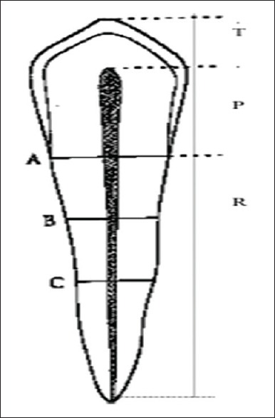
Levels of measurement
The statistical analysis was done using SPSS Version 16.0 statistical analysis software. SPSS Inc. Released 2007 Chicago. The statistical formulas used were mean, standard deviation (SD), correlation, regression, and coefficient of determination. To test the significance of two means, Student's t-test was used. P value was defined as P > 0.05 not significant, P < 0.05 as significant, and P < 0.01 as highly significant.
Results
The age of the 152 subjects, namely, 96 males and 56 females included in the study ranged 14-56 years with a mean of 29.20 years, and the subjects were divided into five groups [Table 1].
Table 1.
Distribution of number of subjects (n=152) in various age groups

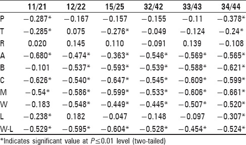
Apart from the ratios mentioned above, the following were also calculated: Mean of all six ratios (M), mean of width ratios B and C (W), mean of length ratiosP and R (L), and difference between width ratio “W” and length ratio “L”(W ~ L). Correlation was carried out between age and the ratios of measurement from each tooth using SPSS (Version 16) software [Table 2].
Table 2.
The correlation between chronological age and the ratios of measurement
Correlation was also calculated between chronological age and ratios for all six teeth, maxillary teeth, and mandibular teeth taken together [Table 3].
Table 3.
Correlation between age and M, W, L, and W~L for all study teeth, three maxillary teeth, and three mandibular teeth

After correlation, regression analysis was run to formulate regression equations for assessment of age. Regression equations for age were derived for all six study teeth, three maxillary teeth, three mandibular teeth, and all six teeth considered together with age as the dependent variable and “M” as the first predictor and “W ~ L” as the second predictor [Tables 4 and 5].
Table 4.
Regression analysis with coefficient of determination (R2) for six study teeth
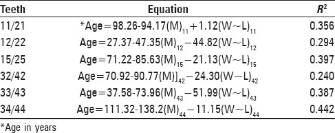
Table 5.
Regression equations for age for three maxillary teeth, three mandibular teeth, and all the six teeth considered together

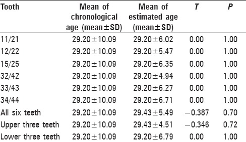
It was observed that the coefficient of determination R2 was highest (0.453) in the lower three teeth considered together. The age of the subjects was estimated by substituting the values of “M” and “W ~ L” in the derived regression equations, and this estimated age was compared with the chronological age using Student's t-test. No significant difference between the mean of the chronological age and the estimated age was observed in all teeth taken individually, three maxillar teeth taken together, three mandibular teeth taken together, and all the six teeth taken together.
Statistical analysis also calculated the standard error of the calculated ages [Table 6].
Table 6.
Comparison of estimated age with the actual age for study teeth individually
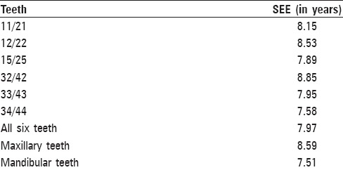
Lowest standard error of estimate (SEE) was seen with the lower three teeth taken together followed by the lower first premolar [Table 7].
Table 7.
Standard error of estimate (SEE) in years for six teeth individually, all the six teeth together, three maxillary teeth, and three mandibular teeth
The next objective of the present study was to explore if the previously presented linear regression formulae as given by Kvaal et al. could lead to statistically sound results and to evaluate the repeatability when applied on the current study sample. However, values of the mean age estimated by using regression equations as given in the study of Kvaal et al. showed negative values. The age estimated by using Kvaal's regression formulae in the present study sample significantly underestimated the chronological age.
Discussion
Dental age estimation has gained acceptance because it is less variable when compared to other skeletal and sexual maturity indicators.[3] Gustafson developed the first systematic dental method for the estimation of age in adults. Tore Solheim measured the amount of secondary dentin deposition in histological sections of teeth and the results were found to be strongly correlated to the age of subjects. Secondary dentin deposition as an age indicator was tested in full-mouth periapical radiographs by Kvaal et al. with significant results.[4] A similar method was employed by Bosman et al.[7] and Paewinsky et al. on panoramic radiographs.[9] Panoramic radiographs avoid the need for taking multiple intraoral radiographs, can be applied to living persons, and do not require extraction of teeth. It allows the age estimation of individuals older than 21 years of age, at the end of skeletal growth and development. The accuracy of this method depends on the precision of the measurements and the quality of the panoramic radiographs. Factors that may interfere would be caries, dental fillings, and intra- and inter observer errors.[10] The present study was conducted on digital panoramic radiographs. In the last decades, digital systems have improved considerably and are now considered an acceptable technology for clinical use in dentistry.[10] However, some previous authors pointed out the difficulties in identifying the reference points on digital images as viewed on the monitor screen; therefore, the defining of the relative distance between two different points, the quantification of which is in pixels, is needed.[9]
The development of each individual can be affected by genetic, racial, nutritional, climatic, hormonal, and environmental factors.[10] This study was carried out on the Indian population residing in and around Muradnagar, Uttar Pradesh. It is possible that the lower socioeconomic group was overrepresented because of the location of the institute in a rural area and the relative cost-effectiveness of the treatments rendered. The variation in the distribution of the subjects in different age groups was due to the inclusion criteria; there were a lower number of older patients having healthy teeth.
In accordance with the study of Kvaal et al., teeth from either left or right side were selected, whichever were best suited for measurement.[2] To compensate for the errors due to magnification and angulations, the various ratios as given by Kvaal et al. were calculated between the length and width measurements and were correlated to chronological age.[2] In agreement with previous studies, the width of the pulp was found to be a better indicator of age than the length.[2] Stronger correlation to age was obtained by employing mean value of all the ratios (M), which may be an expression of the overall size of the pulp.
Following correlation, regression analysis was done using M and W ~ L as the first and the second predictor, respectively, to obtain regression equations. It was observed that the coefficient of determination (R2) was highest when three mandibular teeth were considered together (R2 = 0.453). Individually, the strongest correlation was seen with the lower first premolar similar to the study of Sharma et al.[11] When the calculated age was compared with the chronological age in the present study, no significant difference was observed between the two. These results established the applicability of regression equations calculated based on Kvaal's method on digital panoramic radiographs for calculation of age.
For testing the applicability of the original Kvaal's regression equations in the current study subjects, the age of the subjects was estimated by substituting the values of “M” and “W ~ L” in the regression equations as given by Kvaal.[4] The mean difference between the chronological age and the estimated age showed consistent gross underestimation of age similar to a study by Meinl et al.[12] that clearly indicated the inapplicability of the regression equations of Kvaal et al. on the study population. A study by Patil in 2014 also concluded that the formula derived from the Norwegian population is not applicable to the Indian population, establishing a possible variation in ethnicity and restricting its use in a sample Indian population.[13] The required length measurements in Kvaal's study were obtained on conventional periapical radiographs by using vernier calipers and stereomicroscope but in the present study digital, panoramic radiographs were acquired to obtain the measurements using a standardization procedure. The other reason for the underestimation of age could be the racial difference as the original study was carried out on the Caucasian population and the present study was based on the Asian population in the Indian subcontinent. Also, it could be possible that the formation rate of secondary dentin formation does underlie differences.
Conclusion
From the current study, it can be concluded that width ratios are better correlated than length ratios and “M” (mean value of all ratios) and “W ~ L” (difference between “W” and “L”) were the best predictors for age estimation. Age could be estimated with greater accuracy by taking three mandibular teeth together, followed by mandibular first premolar and maxillary second premolar. The least accuracy was shown by the mandibular lateral incisor taken individually. Also, the derived regression equations from the present study using Kvaal's method could be used to estimate the chronological age in the study population. From the underestimation of age by using the original regression equations as given by Kvaal et al., it can be concluded that the applicability of Kvaal's equation was invalid in the study population. The results of the study give inference for the feasibility of this technique by calculation of regression equations on digital panoramic radiographs. However, it negates the applicability of same regression equations as given by Kvaal et al. on the study population. This study suggests that future studies be done on a large sample size with adequate representation of samples from different age groups, ethnicities, and sexes.
Financial support and sponsorship
Nil.
Conflicts of interest
There are no conflicts of interest.
Footnotes
Source of Support: Nil
Conflict of Interest: None declared
References
- 1.Acharya AB, Sivapathasundaram B. Forensic odontology. In: Shafer, Hine M, Levy B, editors. Textbook of Oral Pathology. 6th ed. Reed Elsevier India Private Limited; 2009. pp. 871–72. [Google Scholar]
- 2.Kvaal SI, Kolltveit KM, Thomsen IO, Solheim T. Age estimation of adults from dental radiographs. Forensic SciInt. 1995;74:175–85. doi: 10.1016/0379-0738(95)01760-g. [DOI] [PubMed] [Google Scholar]
- 3.Stavrianos CH, Mastagas D, Stavrianou I, Karaiskou O. Dental age estimation of adults: A review of methods and principles. Research J Med Sci. 2008;2:258–68. [Google Scholar]
- 4.Singh A, Gorea RK, Singla U. Age estimation from the physiological changes of teeth. JIAFM. 2004;26:94–6. [Google Scholar]
- 5.Lucy D, Pollard AM, Roberts CA. A comparison of three dental techniques for estimating age at death in humans. J Arch Sci. 1995;22:417–28. [Google Scholar]
- 6.Nanci A. 6th ed. Mosby St. Louis Missouri: 2003. Ten Cate's Oral Histology: Development, Structure and Function. [Google Scholar]
- 7.Bosmans N, Ann P, Aly M, Willems G. The application of Kvaal's dental age calculation technique on panoramic dental radiographs. Forensic Sci Int. 2005;153:208–12. doi: 10.1016/j.forsciint.2004.08.017. [DOI] [PubMed] [Google Scholar]
- 8.Landa MI, Garamendi PM, Botella MC, Alemán I. Application of method of Kvaal et al. to digital orthopantamograms. Int J Legal Med. 2009;123:123–8. doi: 10.1007/s00414-008-0268-9. [DOI] [PubMed] [Google Scholar]
- 9.Solheim T. Amount of secondary dentin as an indicator of age. Scand J Dent Res. 1992;100:193–9. doi: 10.1111/j.1600-0722.1992.tb01740.x. [DOI] [PubMed] [Google Scholar]
- 10.Paewinsky E, Pfeiffer H, Brinkmann B. Quantification of secondary dentine formation from orthopantamograms - A contribution to forensic age estimation methods in adults. Int J Legal Med. 2005;119:27–30. doi: 10.1007/s00414-004-0492-x. [DOI] [PubMed] [Google Scholar]
- 11.Sharma R, Srivastava A. Radiographic evaluation of dental age of adults using Kvaal's method. J Forensic Dent Sci. 2010;2:22–6. doi: 10.4103/0974-2948.71053. [DOI] [PMC free article] [PubMed] [Google Scholar]
- 12.Meinl A, Tangl S, Pernicka E, Fenes C, Watzek G. On the applicability of secondary dentin formation to radiological age estimation in young adults. J Forensic Sci. 2007;52:438–41. doi: 10.1111/j.1556-4029.2006.00377.x. [DOI] [PubMed] [Google Scholar]
- 13.Patil SK, Mohankumar KP, Donoghue M. Estimation of age by Kvaal's technique in sample Indian population to establish the need for local Indian-based formulae. J Forensic Dent Sci. 2014;6:166–70. doi: 10.4103/0975-1475.137050. [DOI] [PMC free article] [PubMed] [Google Scholar]


