ABSTRACT
With the rapid development of high voltage direct current transmission, the possibility of health effects associated with static electric field (SEF) has caused wide public concern. To examine the effects of long-lasting, full-body exposure to SEF on cognition, Institute of Cancer Research mice were exposed to SEF for 35 d. The intensities of SEF in experimental group I (EG-I), experimental group II (EG-II) and control group (CG) were 2.30∼15.40 kV/m, 9.20∼21.85 kV/m and 0 kV/m, respectively. The performance in learning and memory of mice were tested by Morris water maze (MWM) on days 2∼6, 16∼20 and 30∼34 during the exposure period. The concentrations of hippocampal amino acid neurotransmitters were evaluated on days 7, 21 and 35. Results showed that the latency in the MWM test had no significant difference among the EG-I, EG-II and CG (P > 0.05) during the exposure period. The percentage of time spent in the target quadrant was significantly decreased in the EG-II on day 34 during the exposure period (P < 0.05), whereas the percentage of time spent in the opposite quadrant increased markedly (P < 0.01). The glutamate and gamma-aminobutyric acid concentrations showed no significant differences among the EG-I, EG-II and CG (P > 0.05) during the exposure period. These results indicated that long-lasting, full-body exposure to SEF with certain intensity would not cause significant influence on learning ability, but it might associate with memory impairment of receptors. Meanwhile, this effect of memory impairment was dose-dependent and not causally linked to the glutamate and gamma-aminobutyric acid levels in the hippocampus.
KEYWORDS: cognition, glutamate, gamma-aminobutyric acid, Morris water maze, static electric field
Introduction
High voltage direct current (HVDC) transmission is a more efficient way to deliver electrical power in long distance and large capacity than alternating current transmission and develops rapidly in China. Synchronously, the possibility of health effects related to the electrical field from HVDC transmission lines has drawn more and more attentions.
Most studies regarding the health effects of electromagnetic from transmission lines focus on time-varying electromagnetic field. The extremely low-frequency electromagnetic field (ELF-EMF) has been controversial suggested to affect cognitive function. Jadidi et al.1 found that exposure to an ELF-EMF with parameters of 50 Hz, 8 mT for 20 min can impair spatial memory. Zhao et al.2 reported the association between ELF-EMF exposure and damage of recognition memory. However, little research has investigated the impact of static electric field on learning and memory.
The Morris water maze (MWM) test, firstly described by Morris more than 30 y ago,3 is one of the widest used test for learning and memory.4-6 Although amino acid neurotransmitters play significant roles in learning and memory,7,8 whether the effects of static electric field on cognition are mediated by these neurotransmitters is still unclear. In this study, to investigate the effects of long-lasting, full-body exposure to static electric field on learning and memory in mice, neural behaviors were tested by MWM. To understand the relation between the effects of static electric field on cognitive and amino acid neurotransmitters, the levels of glutamate (Glu) and gamma-aminobutyric acid (GABA) were measured using o-phthalaldehyde (OPA)-high performance liquid chromatography (HPLC).
Results
Performance of morris water maze test
The results of MWM test in the 3 groups in particular days of exposure cycle are presented in Table 1 and Table 2. During the hidden platform acquisition training, 2-way repeated measures ANOVA showed that static electric field exposure did not influence the latency of reaching the platform for mice (P > 0.05). The main effect of days of training was significant (P < 0.01), indicating that the mice improved with training. During the stage of probe test, as shown in Table 2, the numbers of crossing in the EG-II were always smaller than those in the CG on days 6, 20 and 34 during the period of exposure, but not significant (P > 0.05). One-way ANOVA showed that on days 34 during the exposure period, the percentage of time spent in the target quadrant of EG-II was significantly lower than that of the CG and EG-I (P < 0.05), while the percentage of time spent in the opposite quadrant of the EG-II was significantly higher than that of the CG and EG-I (P < 0.01).
Table 1.
Results from hidden platform acquisition training in the control and different experimental groups at particular days of exposure period (values are presented as mean ± SEM, n = 10 for each group).
| Latency (s) |
||||
|---|---|---|---|---|
| Exposure duration | Days of training | CG | EG-I | EG-II |
| 2∼5d | 1d | 80.6 ± 3.1 | 83.8 ± 2.8 | 82.3 ± 3.1 |
| 2d | 77.6 ± 5.7 | 76.9 ± 6.2 | 76.6 ± 4.3 | |
| 3d | 72.6 ± 6.2 | 60.8 ± 6.0 | 73.7 ± 5.6 | |
| 4d | 68.0 ± 8.7 | 59.2 ± 5.7 | 72.2 ± 4.9 | |
| 16∼19d | 1d | 80.8 ± 4.5 | 85.7 ± 2.8 | 74.8 ± 6.1 |
| 2d | 79.8 ± 3.8 | 75.4 ± 5.5 | 67.2 ± 6.7 | |
| 3d | 77.4 ± 5.3 | 70.6 ± 7.5 | 66.8 ± 7.2 | |
| 4d | 55.8 ± 5.2 | 66.2 ± 9.1 | 70.2 ± 8.0 | |
| 30∼33d | 1d | 82.8 ± 2.8 | 75.8 ± 4.5 | 74.6 ± 5.3 |
| 2d | 69.8 ± 6.8 | 68.0 ± 7.6 | 63.3 ± 7.2 | |
| 3d | 58.5 ± 8.1 | 67.0 ± 6.9 | 65.2 ± 6.8 | |
| 4d | 54.4 ± 10.0 | 46.1 ± 8.8 | 57.0 ± 8.8 | |
Table 2.
Results from probe test in the control and different experimental groups at particular days of exposure period (values are presented as mean ± SEM, n = 10 for each group).
| Number of crossing |
% Time in the target quadrant |
% Time in the opposite quadrant |
|||||||
|---|---|---|---|---|---|---|---|---|---|
| Exposure duration | CG | EG-I | EG-II | CG | EG-I | EG-II | CG | EG-I | EG-II |
| 6d | 0.9 ± 0.4 | 1.5 ± 0.5 | 0.4 ± 0.2 | 24.3 ± 3.9 | 25.9 ± 3.5 | 22.8 ± 4.9 | 33.9 ± 4.0 | 26.8 ± 4.2 | 31.1 ± 4.0 |
| 20d | 1.3 ± 0.4 | 0.9 ± 0.3 | 0.6 ± 0.4 | 27.8 ± 3.6 | 35.5 ± 5.3 | 27.1 ± 4.2 | 28.4 ± 4.5 | 27.6 ± 6.6 | 28.5 ± 4.4 |
| 34d | 2.0 ± 0.6 | 1.9 ± 0.5 | 1.3 ± 0.5 | 36.8 ± 4.8 | 41.7 ± 4.5 | 24.0 ± 3.7* | 15.8 ± 1.7 | 15.1 ± 2.8 | 30.7 ± 4.1** |
Indicates significant difference compared with the CG and EG-I, P < 0.05;
Indicates significance compared with the CG and EG-I, P < 0.01.
Concentrations of hippocampal amino acid neurotransmitters
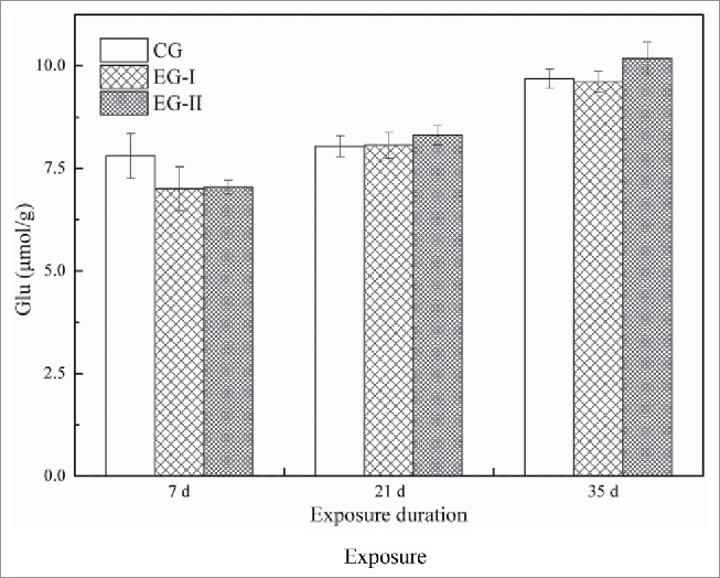
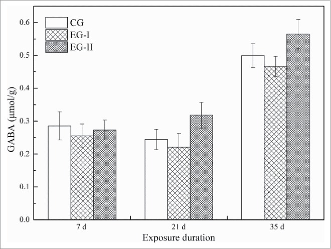
The average levels of Glu and GABA in the hippocampus of mice from control and different experimental groups are shown in Fig. 1 and Fig. 2. The average concentrations of both Glu and GABA in both exposed groups showed no significant difference compared with the CG s(P > 0.05) during the entire period of exposure.
Figure 1.

Mean levels of Glutamate (Glu) in the control and different experimental groups at particular days of exposure period (values are presented as mean ± SEM, n = 10 for each group).
Figure 2.

Mean levels of gamma-aminobutyric acid (GABA) in the control and different experimental groups at particular days of exposure period (values are presented as mean ± SEM, n = 10 for each group).
Discussion
Signal transmission among neurons is the basis of learning and memory formation. Given that an electric signal is a neuronal signal transduction pathway, an electric field can drive a current in the conducting body and may impact cognitive function.9 The present study investigated the effects of exposure to environment static electric field with different intensities of 2.30∼15.40 kV/m (EG-I) and 9.20∼21.85 kV/m (EG-II) on the performance of learning and memory, as well as the amino acid neurotransmitter levels in the hippocampus. It was found that long-lasting exposure to static electric field with a strong intensity may impair memory ability in mice. However, the impaired performance was not causally linked to the Glu and GABA levels in the hippocampus.
In the MWM test, spatial learning and memory was specifically assessed by hidden platform acquisition training and probe test, respectively.5 It is generally accepted that the learning ability of animals is negatively correlated with the latency of reaching the platform in the hidden platform acquisition training.3,4 Cielar et al.10 exposed male Wistar albino rats to electric field with parameter of 25 kV/m for 56 d (8 h daily) and evaluated the performance of rats at specific days of exposure period (7th, 14th, 21st, 28th, 42nd and 56th day). They found that there was no significant difference in latency between rats from the experimental group and the control group. Results in this study were in line with the previous studies showing insignificant difference in latency at different exposure period, which indicated that static electric field of certain intensity would not influence learning ability. In general, in the probe test, the memory ability of animals is positive correlated with the number of crossing and the percentage of time animals spent in the target quadrant, whereas it is negatively correlated with the percentage of time animals spent in the opposite quadrant.3 In our study, the percentage of time spent in the target quadrant in the EG-II was significantly decreased compared with the EG-I and CG at the exposure time of 34 d, whereas the percentage of time spent in the opposite quadrant increased markedly. These results suggested that exposure to static electric field might impair memory ability of receptors and this effect was dose-dependent.
In the central nervous system, Glu and GABA are the primary excitatory and inhibitory neurotransmitters, respectively.5,7 They can act on their receptors and then regulate appropriate levels of excitatory signals and inhibitory signals essential for plasticity of synapses, which is the basis for learning and memory. At present study, the Glu and GABA levels did not significantly change during the exposure period, which was somewhat inconsistent with the performance of MWM. Li et al.6 found that chronic exposure (14 and 28 d) to a 50 Hz, 0.5 mT ELF-EMF differently influenced the AMPAR and NMDAR (2 kinds of glutamate receptors) subunit expressions in distinct brain regions. It can be hypothesized that exposure to static electric field would not cause changes in Glu and GABA levels, but might influence expressions of relevant receptors and might further result in memory impairment. Further studies need to be conducted to clarify this hypothesis.
Conclusions
The presented study investigated the impact of exposure to environmental static electric field with intensities of 2.3∼15.4 kV/m and 9.2∼21.85 kV/m for 35 d on the cognition of mice. It was found that long-lasting, full-body exposure to static electric field with certain intensity did not induce significant influences on learning ability, but might induce impairments in memory ability of receptors. In addition, this effect of memory impairment was dose-dependent and was not causally linked to the Glu and GABA levels in the hippocampus.
Materials and methods
Animals
90 four-week-old male Institute of Cancer Research (ICR) mice weighing 15∼20 g were used as subjects in this study. They were purchased from Experimental Animal Center of Zhejiang Province (Hangzhou, China) and randomly assigned into 3 groups: the control group (CG, n = 30), the experimental group I (EG I, n = 30), and the experimental group II (EG II, n = 30). Ten mice were housed in each plastic cage which was specially designed and supplied with regular food and water ad libitum. All procedures performed in this study were in accordance with the Quality Management Approach to Laboratory Animals.
Static electric field exposure
The exposure of environmental static electric field was carried out in autumn whose climate was most suitable for survival of animals in the open air. The animals were exposed to the environmental static electric field which was generated by the HVDC transmission lines for 35 d. Three sites with the same environmental conditions except the intensity of static electric field were chosen to place different groups. The animals from EG-I were exposed to the static electric field whose intensity was 2.30∼15.40 kV/m, the animals from EG-II were exposed to a relatively strong electric field with intensity of 9.20∼21.85 kV/m, and the animals from CG were placed in the site whose intensity of static electric field was near 0 kV/m. The three groups were exposed for 24 h per day except raining days and low-temperature days. The actual exposure time and intensity of environmental static electric field under different weather conditions were recorded exactly.
Morris water maze test
Ten mice from each group were selected randomly and to which the MWM test were carried out on days 2∼6, 16∼20 and 30∼34 during the period of static electric field exposure. A black circular pool (120 cm in diameter, 42 cm in height) was used in the MWM test. The pool was filled with water (24 ± 1°C) to a depth of 26 cm and the water was made opaque by adding nontoxic ink. The pool was surrounded by blue curtains on which distinctive visual cues were hung. The pool was divided into 4 imaginary quadrants and a black platform (6 cm in diameter) was placed 1 cm below the surface of water in the middle of a fixed quadrant during training. The quadrant in which the platform placed was named the target quadrant. During hidden platform acquisition training, each mouse received 4 trials per day over 4 continuous days. On each trial, the mice were placed into the water facing the wall of pool at one of 4 start locations that alternated pseudo-randomly (N, E, NW, or SE). The movements of mice were recorded by a video camera. A maximum of 90 s was allowed before the animals were moved to the platform for 15 s. The latency of reaching the platform was measured in each trial. On the 5th day, a single probe test was performed to each mouse without the platform. The mice were placed into the pool at the start position of NE for 90 s. The percentage of time mice spent in the target quadrant and the opposite quadrant as well as the number of crossing the previous platform position were analyzed.
Amino acid neurotransmitters assays
The mice were sacrificed by decapitation one day after the MWM test (days 7, 21 and 35 during static electric field exposure). In order to avoid circadian rhythm induced variations, hippocampi sampling was carried out at the same time of the day. After sacrificed, the mice hippocampi were separated rapidly on an ice-cold plate. Hippocampal samples were homogenized with ice-cold saline and centrifuged for 15 min (14000 rpm, 4°C). The supernatant affiliated with same volume of 0.4 M perchloric acid was kept in ice-bath for 20 min to make the proteins fully subside and again centrifuged for 15 min. 20 µL of the supernatant was injected into an OPA-HPLC system (Agilent Eclipse AAA column; 150 mm × 4.6 mm, 5 μm) to measure the levels of Glu and GABA.
Statistical analysis
All data are presented as mean ± SEM. Data obtained from hidden platform acquisition training were analyzed by 2-way repeated-measures ANOVA, considering the factors: different exposure groups × days of training. Data of probe test and amino acid neurotransmitters assays were analyzed by one-way ANOVA. All analyses were carried out using software SPSS 20.0. P < 0.05 was considered to be statistically significant.
Disclosure of potential conflicts of interest
No potential conflicts of interest were disclosed.
Funding
This work was supported by the Science and Technology Funds from the State Grid Corporation of China (Contract No. SGTYHT/14-JS-188).
References
- [1].Jadidi M, Firoozabadi SM, Rashidy-Pour A, Sajadi AA, Sadeghi H, Taherian AA. Acute exposure to a 50 Hz magnetic field impairs consolidation of spatial memory in rats. Neurobiol Learn Mem 2007; 88:387-92; PMID:17768075; http://dx.doi.org/ 10.1016/j.nlm.2007.07.010 [DOI] [PubMed] [Google Scholar]
- [2].Zhao QR, Lu JM, Yao JJ, Zhang ZY, Ling C, Mei YA. Neuritin reverses deficits in murine novel object associative recognition memory caused by exposure to extremely low-frequency (50 Hz) electromagnetic fields. Sci Rep 2015; 5:11768; PMID:26138388; http://dx.doi.org/ 10.1038/srep11768 [DOI] [PMC free article] [PubMed] [Google Scholar]
- [3].Morris R. Developments of a water-maze procedure for studying spatial-learning in the rat. J Neurosci Meth 1984; 11:47-60; http://dx.doi.org/ 10.1016/0165-0270(84)90007-4 [DOI] [PubMed] [Google Scholar]
- [4].Andersen JD, Pouzet B. Spatial memory deficits induced by perinatal treatment of rats with PCP and reversal effect of D-serine. Neuropsychopharmacol 2004; 29:1080-90; http://dx.doi.org/ 10.1038/sj.npp.1300394 [DOI] [PubMed] [Google Scholar]
- [5].Cui B, Wu M, She X. Effects of chronic noise exposure on spatial learning and memory of rats in relation to neurotransmitters and NMDAR2B alteration in the hippocampus. J Occup Health 2009; 51:152-8; PMID:19225220; http://dx.doi.org/ 10.1539/joh.L8084 [DOI] [PubMed] [Google Scholar]
- [6].Li C, Xie ML, Luo FL, He C, Wang JL, Tan G, Hu ZA. The extremely low-frequency magnetic field exposure differently affects the AMPAR and NMDAR subunit expressions in the hippocampus, entorhinal cortex and prefrontal cortex without effects on the rat spatial learning and memory. Environ Res 2014; 134:74-80; PMID:25046815; http://dx.doi.org/ 10.1016/j.envres.2014.06.025 [DOI] [PubMed] [Google Scholar]
- [7].Zhao L, Peng RY, Wang SM, Wang LF, Gao YB, Dung J, Li X, Su ZT. Relationship between cognition function and hippocampus structure after long-term microwave exposure. Biomed Environ Sci 2012; 25:182-8; PMID:22998825 [DOI] [PubMed] [Google Scholar]
- [8].Buchanan RJ, Gjini K, Darrow D, Varga G, Robinson JL, Nadasdy Z. Glutamate and GABA concentration changes in the globus pallidus internus of Parkinson patients during performance of implicit and declarative memory tasks: A report of two subjects. Neurosci Lett 2015; 589:73-8; PMID:25596441; http://dx.doi.org/ 10.1016/j.neulet.2015.01.028 [DOI] [PubMed] [Google Scholar]
- [9].WHO-World Health Organization Environmental health criteria 238: extremely low frequency (ELF) fields. Geneva: WHO Press; c2007. Chapter 1, Summary and recommendations for further study; p. 1-13 [Google Scholar]
- [10].Cielar G, Mrowiec J, Sowa P, Kasperczyk S, Sieron A. Effect of exposure to static, high voltage electric field generated nearby HVDC transmission lines on behavior of rats. In: Progress in Electromagnetics Research Symposium (PIERS 2009 Moscow); 2009 Aug 12–21; Moscow. Cambridge (USA): Electromagnetics Acad; 2009. p. 1097-101 [Google Scholar]


