Abstract
Heat stress is associated with death and other maladaptions including muscle dysfunction and impaired growth across species. Despite this common observation, the molecular effects leading to these pathologic changes remain unclear. The purpose of this study was to determine the extent to which heat stress disrupted redox balance and initiated an inflammatory response in oxidative and glycolytic skeletal muscle. Female pigs (5–6/group) were subjected to thermoneutral (20 °C) or heat stress (35 °C) conditions for 1 or 3 days and the semitendinosus removed and dissected into red (STR) and white (STW) portions. After 1 day of heat stress, relative abundance of proteins modified by malondialdehyde, a measure of oxidative damage, was increased 2.5-fold (P < 0.05) compared with thermoneutral in the STR but not the STW, before returning to thermoneutral conditions following 3 days of heat stress. This corresponded with increased catalase and superoxide dismutase-1 gene expression (P < 0.05) and superoxide dismutase-1 protein abundance (P < 0.05) in the STR but not the STW. In the STR catalase and total superoxide dismutase activity were increased by ~30% and ~130%, respectively (P < 0.05), after 1 day of heat stress and returned to thermoneutral levels by day 3. One or 3 days of heat stress did not increase inflammatory signaling through the NF-κB pathway in the STR or STW. These data suggest that oxidative muscle is more susceptible to heat stress-mediated changes in redox balance than glycolytic muscle during chronic heat stress.
Keywords: free radicals, inflammation, mitochondria, oxidative, NF-kB, pig
Introduction
Despite advances in cooling technologies and strategies heat stress (HS), the inability of an organism to properly dissipate thermal energy produced,1,2 continues to represent a serious health concern. In 2012, heat-related illnesses resulted in the largest number of weather-related fatalities in the US due to heat stroke3 and also caused additional morbidities related to excess heat load.4 These negative effects of heat stress are often more pronounced in agricultural species as they frequently are held under environmental conditions with a limited capacity for cooling and are bred for rapid growth. Given that climate variability has, and may continue to result in increasing temperatures,5 these problems will likely become more frequent and severe.
The extent to which and mechanism by which HS causes pathologic changes in specific organ systems is less clear. In skeletal muscle hyperthermia has been used as a therapeutic intervention to spare muscle from loss due to disuse6,7 as well as augment regrowth following atrophy.8,9 In these studies therapeutic hyperthermia was also found to decrease oxidative damage. These hyperthermic interventions were generally brief in nature lasting approximately 30 min. In contrast, prolonged exposure to an excessive heat load results in HS, which negatively impacts muscle growth10,11 and has been associated with increased production of reactive oxygen species (ROS) in avian skeletal muscle.12-14 Oxidative stress can lead to protein degradation through increased proteolysis and autophagy15-19 as well as impair protein synthesis by preventing translation.20,21
It seems likely that HS would also lead to increased inflammatory signaling in skeletal muscle through activation of the NF-κB pathway. While there are a number of molecular triggers that lead to increased NF-κB signaling, within the context of heat stressed skeletal muscle increased ROS and endotoxemia seem to be likely candidates. Importantly, free radicals have been identified as initiators of NF-κB signaling22,23 and are increased following HS in avian skeletal muscle.12-14 Likewise, HS may lead to increased intestinal permeability and subsequent endotoxemia.24,25 Bacterial lipopolysaccharide (LPS) that has entered the circulation can be recognized by TLR4 receptors on muscle cell membranes26 and initiate an inflammatory response via NF-κB signaling.27 Of note, activation of the NFKB transcription factor has been associated with increased production of ubiquitin ligases, as well as muscle atrophy.28,29
It is apparent that there are fundamental differences that distinguish therapeutic hyperthermia and heat stress, however, these have not been identified. Further, the mechanisms by which heat stress leads to muscle dysfunction are also unclear. Our long-term goal is to understand the mechanisms caused by HS that lead to muscle loss. Given that goal, the purpose of this investigation was to determine the extent to which HS led to increased oxidative stress and NF-κB activation in oxidative and glycolytic mammalian skeletal muscle. We hypothesized that HS would result in a progressive increase in oxidative stress and NF-κB pathway activation in oxidative and glycolytic skeletal muscle.
Materials and Methods
Ethical approval
All animal experiments were approved by the Iowa State University Institutional Animal Care and Use Committee and complied with rules and guidelines for animal use established by the USDA.
Study design and animal treatments
Due to their anatomical and physiological similarities with humans30 and high sequence homology to the human genome,31 pigs have long been used as biomedical models.32-34 Prior data from animals used in this investigation and a detailed study design have been previously reported.24 Female pigs (35 ± 4 kg; n = 5–6/group) were kept at thermal-neutral (TN) conditions (20 ± 1 °C; 35–50% relative humidity) as recommended by FASS35 or exposed to constant heat stress (HS) (35 ± 1 °C; 20–35% relative humidity) for a period of 1 or 3 d. Pigs were housed in individual pens with ad libitum access to food and water in either a TN or HS room with concrete floors. Temperature and humidity were recorded in continuous 30 min intervals using a data logger (Lascar). Animals were euthanized by the captive bolt technique and exsanguination at the end of the respective treatment period. At this time, the semitendinosus (ST) muscle was removed and dissected into red (STR) and white (STW) muscle. The red and white portions of the pig ST muscle are visually apparent. Two gram sections were collected for each portion and frozen in liquid nitrogen for further analyses.
Protein abundance
Muscle was powdered on dry ice and divided into portions used for measurement of relative protein and gene abundance. In order to measure protein abundance, approximately 50 mg of muscle powder was homogenized in 1.5 mL of protein extraction buffer (10 mM sodium phosphate, pH 7.0, and 2% SDS) using a Dounce homogenizer. The sample was then centrifuged at 1500 × g for 15 min at 20 °C to remove cellular debris. Protein concentration was determined (Pierce® BCA microplate protein assay kit, Pierce) and samples were diluted to 4 mg/mL in loading buffer (62.5 mM Tris (pH 6.8), 1.0% SDS, 0.01% bromophenol blue, 15.0% glycerol, and 5% β-mercaptoethanol). Ten microliters (40 μg of protein) of each sample were loaded into 4–20% precast gradient gels and proteins were separated at room temperature for 30 min at 60 V followed by 50 min at 120 V. Afterward, proteins were transferred (90 min; 90 V) to a nitrocellulose membrane with a pore diameter of 0.2 μm. Membranes were blocked in 5% dehydrated milk TTBS (Tris-buffered saline containing 0.1% Tween 20) solution for 1 h and exposed to primary antibody overnight at 4 °C in 1% dehydrated milk TTBS solution as follows: catalase (Sigma, primary 1:1000, cat. no C0979, secondary 1:2000), Malondialdehyde (MDA; Abcam; primary 1:5000, cat. no ab27642, secondary 1:2000), MnSOD (Abcam; primary 1:5000, cat. no ab13533, secondary 1:2000), NF-κB p-65 (Abcam; primary 1:1000, cat. no ab7970, secondary 1:2000), IL-6 (Abcam; primary 1:1000, cat. no ab6672, secondary 1:2000), phosphorylated-NF-κB p65 (Thermo Scientific; primary 1:1000, cat. no MA5–15160, secondary 1:2000), TNF-α (Abcam; primary 1:1000, cat. no ab6671, secondary 1:2000), IκB-α (Santa Cruz Biotechnology; primary 1:1000, cat. no SC-371, secondary 1:2000). After three 10 min washes with TTBS, membranes were exposed to secondary antibody (as noted above) for 1 h at room temperature in 1% dehydrated milk TTBS solution. Membranes were washed again three times for 10 min with TTBS and detection was performed by enhanced chemiluminescence and X-ray film. X-ray film was then scanned and blot signal was quantified through the use of Kodak software. Optical density was determined and values for each group were normalized to the mean of TN samples on each membrane. Values are reported relative to TN. All membranes were stained with Ponceau S to assure equal loading. We found that Ponceau S staining was similar for all groups for all membranes.
mRNA transcript abundance
RNA was isolated using the TRIzol reagent according to the manufacturer’s instructions (Invitrogen; cat. no 15596). Briefly, powdered muscle was homogenized with TRIzol reagent, centrifuged and extracted with chloroform, and precipitated with ethanol. Total RNA was DNase treated (RNaseFree DNase set, Qiagen Inc.; cat. no 79254) to remove potential genomic DNA contamination and purified using a column (RNeasy kit, Qiagen Inc.; cat. no 74106). RNA concentration and purity was determined by measuring absorbance at 260 nm and 280 nm with a Nanodrop (Thermo Scientific). Total RNA (1μg) was then reverse transcribed (Qiagen Inc.; cat. no 205311) and gene expression measured through qRT-PCR using SYBR green (Qiagen Inc.; cat. no 204056). Transcript abundance was determined by the delta CT method using 18S rRNA as the control gene, and fold change calculated from the delta delta CTs. Transcript abundance is presented as fold changes relative to TN. Sequences of primer pairs can be found in Table 1.
Table 1. Primer sequences used to measure relative mRNA abundance.
| Gene of Interest | Forward Primer | Reverse Primer |
|---|---|---|
| TNF-α (TNF) | GCCCTTCCAC CAACGTTTTC | TCCCAGGTAGA TGGGTTCGT |
| IL-1β (IL1B) | AAGATAACAC GCCCACCCTG | TGTCAGCTTC GGGGTTCTTC |
| IL-15 (IL15) | CAGAAGCAAC CTGGCAGCAC G | ACGCGTAACT CCAGGAGAAAG CA |
| Catalase (CAT) | CAGCTTTAGT GCTCCCGAAC | AGATGACCCG CAATGTTCTC |
| MnSOD (SOD2) | CGCTGAAAAA GGGTGATGTT | AGCGGTCAAC TTCTCCTTGA |
| CuZnSOD (SOD1) | CGAGCTGAAG GGAGAGAAGA | AGTCACATTG CCCAGGTCTC |
Enzymatic activities
Catalase activity was measured according to manufacturer instructions (Catalase Assay Kit, Cayman Chemical Company, Item No. 707002). Catalase activity is measured colorimetrically from tissue homogenates (50 mM potassium phosphate, pH 7.0, 1mM EDTA) and determined by linear regression using a standard curve. The principle of the assay is based on the peroxidatic function of catalase. Catalase reacts with methanol to produce formaldehyde, which is then measured spectrophotometrically. Catalase activity is expressed as nmol/min/mL/mg of protein. Total superoxide dismutase (SOD) activity was measured using a commercially available kit (Superoxide Dismutase Assay Kit, Cayman Chemical Company, Item No. 706002) according to manufacturer instructions. Like above, measured activity was determined colorimetrically from tissue homogenates (20 mM HEPES buffer, pH 7.2, 1 mM EGTA, 210 mM mannitol, 70 mM sucrose) using linear regression from a standard curve. The principle of the assay is based on the dismutation of superoxide radicals produced by xanthine oxidase and hypoxanthine by all types of SOD. Total SOD activity is expressed as U/mL/mg-of protein.
Statistics
In preliminary experiments we determined that TN groups were similar for all measures, as we anticipated. Because of this the TN group used for analyses was comprised of representatives from both the 1 d TN and 3 d TN groups chosen randomly for each measurement (n = 3–5 from each group/measure). To determine the extent to which heat stress altered variables over time data from TN, 1 d HS, and 3 d HS animals were compared using an ANOVA followed by a Newman–Keuls post hoc test when appropriate. To determine statistical significance, α level was set at P < 0.05. Values are displayed as means ± SEM unless otherwise noted.
Results
Oxidative stress
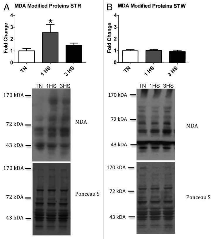
To determine the extent to which HS caused free radical damage in skeletal muscle, abundance of proteins modified by malondialdehyde (MDA), a marker of lipid peroxidation, was measured in STR and STW. In STR the relative abundance of proteins containing MDA adducts was increased 2.5-fold compared with TN (P < 0.05) after 1 d of HS (Fig. 1A), however, returned to TN levels in the 3 HS group. More detailed analyses (single band analysis, band subgroups) demonstrated that this increase was due to a subtle increase in abundance of most or all bands rather than significant modifications in 1 or a small subset of proteins. HS did not increase MDA accumulation in STW (Fig. 1B). To assure that the band at approximately 45 kDa was not the result of nonspecific binding from our secondary antibody we probed a membrane with secondary only and found that no such band was present. This supports the notion that the band detected is the result of MDA modified protein. Regardless, MDA abundance was similar between groups in the STW independent of inclusion or exclusion of the 45 kDa band.
Figure 1. Heat stress increased oxidative injury in oxidative, but not glycolytic muscle. Oxidative stress was measured by quantifying the relative abundance of MDA modified proteins, a marker of lipid peroxidation. (A) MDA modified proteins increased 2.5-fold compared with TN after 1 d of HS in the STR (TN, n = 8; 1 HS, n = 5; 3 HS, n = 5) (B) but remained unchanged in the STW (TN, n = 6; 1 HS, n = 5; 3 HS, n = 4). Representative blots are shown as well as corresponding Ponceau S staining to demonstrate equal loading. *indicates significantly different from TN; P < 0.05.
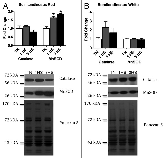
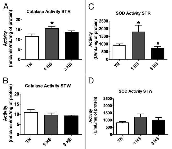
Given that free radical injury can be caused by a failure of the antioxidant system to adequately respond to an oxidative insult we measured transcript abundance of select antioxidant enzymes. In STR, mRNA transcript abundance of CAT and SOD2 was increased by 4-fold and 1.5-fold, respectively, compared with TN (P < 0.05) following 1 d of HS (Fig. 2A). This increase in transcript abundance was transient as abundance for both CAT and SOD2 returned to TN levels following 3 d of HS. Following that general pattern of a rise and subsequent fall, transcript abundance of SOD1, was statistically similar to TN after 1 d of HS, however, was decreased by 25% (P < 0.05) after 3 d of HS compared with TN (Fig. 2A). As changes in mRNA transcript abundance of CAT and SOD2 closely mirrored that of oxidative damage we measured protein abundance of catalase and MnSOD. In STR catalase protein abundance was similar between all treatment groups (Fig. 3A), however MnSOD was increased 1.5-fold (P < 0.05) after 1 and 3 d of HS compared with TN (Fig. 3A). In the STR, activities of both catalase (Fig. 4A) and SOD (Fig. 4C) were increased by 30% and 130% (P < 0.05), respectively, following 1 d of HS compared with TN but returned to TN levels after 3 d of HS.
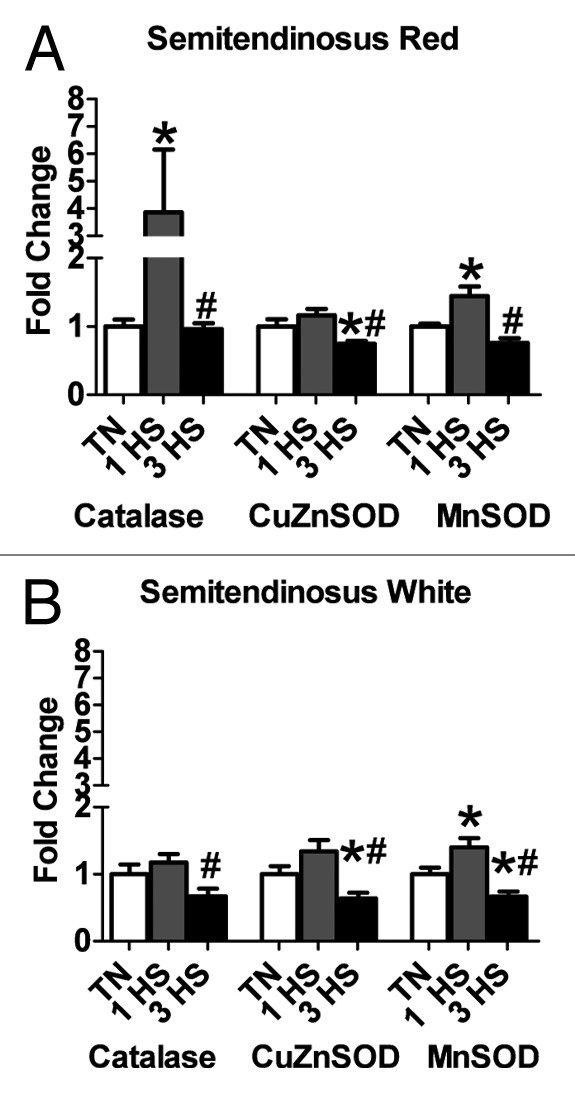
Figure 2. Gene expression of antioxidant enzymes following heat stress. (A) HS increased gene expression of both CAT (TN, n = 6; 1 HS, n = 6; 3 HS, n = 6) and SOD2 (TN, n = 6; 1 HS, n = 5; 3 HS, n = 6) following 1 d of HS, but not that of SOD1 (TN, n = 6; 1 HS, n = 5; 3 HS, n = 6) in the STR. Expression of all antioxidant enzymes was decreased after 3 d of HS compared with 1 d of HS in the STR. (B) HS increased SOD2 gene expression after 1 d of HS compared with TN, though expression of all antioxidant enzymes was decreased by day 3 in the STW (TN, n = 6; 1 HS, n = 6; 3 HS, n = 6). *indicates significantly different from TN; # indicates significantly different from 1-HS; P < 0.05.
Figure 3. Protein expression of catalase and SOD following heat stress. (A) Catalase protein content was unchanged with HS in the STR. However, MnSOD protein content was increased after 1 and 3 d of HS in the STR. (B) Catalase and MnSOD protein content remain unchanged with HS in the STW. Representative blots are shown as well as corresponding Ponceau S staining to demonstrate equal loading. * indicates significantly different from TN; P < 0.05.
Figure 4. Antioxidant enzyme activities following HS treatment in porcine skeletal muscle. (A) One day of HS increased catalase activity in STR (TN, n = 6; 1 HS, n = 5; 3 HS, n = 6) (B) but not STW (TN, n = 6; 1 HS, n = 6; 3 HS, n = 6) compared with TN. (C) Likewise, SOD activity was increased after 1 d of HS in the STR (TN, n = 10; 1 HS, n = 6; 3 HS, n = 6) (D) but remained similar in STW (TN, n = 10; 1 HS, n = 6; 3 HS, n = 6). *Indicates significantly different from TN; # indicates significantly different from 1-HS; P < 0.05.
An absence of apparent oxidative damage in STW could stem from a robust response in antioxidant enzyme expression and activity serving to mitigate ROS. Therefore, we measured antioxidant transcript and protein abundance and activity in STW. In general, the pattern of change observed in antioxidant enzyme transcript abundance in the STW followed closely that observed in STR in that there was a shift toward increased abundance following 1 d of HS followed by a reduction in antioxidant enzyme transcript abundance following 3 d of HS (Fig. 2B). Specifically, in the STW CAT transcript abundance was similar following 1 d of HS, however was decreased 40% (P < 0.05) after 3 d of HS compared with the 1 d HS group, however failed to reach significance compared with TN. SOD1 was also decreased after 3 d of HS by 40% and 50% compared with TN and 1 d of HS, respectively (P < 0.05). Finally, SOD2 transcript abundance was increased by 40% (P < 0.05) after 1 d of HS compared with TN, but decreased (P < 0.05) after 3 d of HS by 30% and 50% compared with TN and 1 d of HS, respectively (Fig. 2B). Protein abundance of both catalase and MnSOD (as determined by western blot) were similar between all treatment groups (Fig. 3B).
Enzymatic activities of catalase (Fig. 4B) and SOD (Fig. 4D) were similar between all treatment groups in STW. Transcript abundance of antioxidant enzymes in the STW (Fig. 2B) supports the numerical increase observed in SOD activity in STW (Fig. 4D), though it failed to reach statistical significance.
Inflammatory response
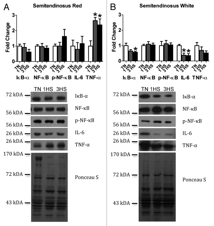
We have previously shown that HS resulted in a systemic increase in LPS,24 which has the potential to trigger an inflammatory response in tissues with a TLR4 receptor, including skeletal muscle.27 Further, independent of systemic pro-inflammatory factors, oxidative stress can also initiate an inflammatory response.22,36,37 Hence, in skeletal muscle subjected to HS two independent mechanisms may drive inflammatory signaling. To determine the extent to which skeletal muscle contributes to systemic inflammation we evaluated NF-κB pathway activation, a major pathway involved in inflammatory signaling. To assess inflammatory signaling we measured relative protein abundance of NF-κB (p65 subunit), activated phosphorylated- NF-κB, the NF-κB inhibitor, IkB-α, and the pathway product IL-6.38 We found that the relative abundance of NF-κB, phospho- NF-κB, IkB-α and IL-6 was similar between all treatment groups in STR (Fig. 5A). Further supporting pathway quiescence, mRNA transcript abundance of NF-κB pathway products TNF, IL15, and IL1B were similar between all groups in STR (Fig. 6A). Of interest, in STR, relative protein abundance of TNF-α was increased 2.5-fold (P < 0.05) after 1 and 3 d of HS compared with TN (Fig. 5B).
Figure 5. Protein expression of inflammatory signaling molecules in the NF-κB pathway. (A) In STR, IkB-α, NF-κB, phospho-NF-κB and IL-6 protein abundance were similar between all treatment groups in the STR, however, TNF-α was increased 2.5-fold following 1 and 3 d of HS (n = 5–6/group). (B) In the STW, TNF-α, NF-κB and phospho-NF-κB protein content remain unchanged, however, HS decreased IkB-α and IL-6 protein content (n = 5–6/group). Representative blots are shown as well as corresponding Ponceau S staining to demonstrate equal loading. *indicates significantly different from TN; P < 0.05.
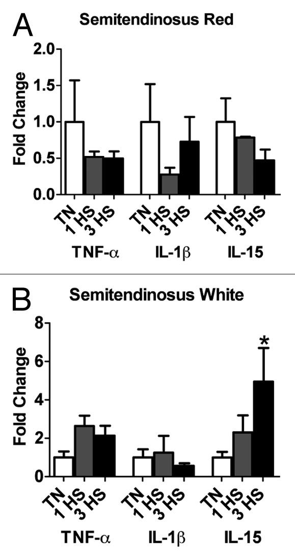
Figure 6. Gene expression of inflammatory cytokines in skeletal muscle following HS. (A) Gene expression of TNF, IL1B, and IL15 was similar between all treatment groups in STR (TNF and IL1B n = 6/group; IL15 TN n = 8, 1 HS = 3, 3 HS = 5). (B) Gene expression of TNF and IL1B, was similar between all treatment groups, but IL15 was increased after 3 d of HS in STW (TNF and IL1B n = 6/group; IL15 TN n = 8, 1 HS = 5, 3 HS = 4). *Indicates significantly different from TN; P < 0.05.
In the STW, relative protein abundance of TNF-α, NF-κB and phosphorylated- NF-κB were similar between all treatment groups (Fig. 5B). IkB-α protein abundance was decreased by 40% (P < 0.05) after 3 d of HS compared with TN (Fig. 5B). Similarly, IL-6 protein abundance was decreased by approximately 60% after 1 and 3 d of HS (P < 0.05) compared with TN (Fig. 5B). TNF and IL1B transcript abundance were similar between all treatment groups, but IL15 was significantly increased (P < 0.05) after 3 d of HS (Fig. 6B).
Discussion
Heat stress is well known to have deleterious consequences to human and animal health,1,39 including death; however, little is known about HS-mediated alterations to skeletal muscle physiology. In addition to health considerations, skeletal muscle is an interesting tissue to consider as hyperthermia has been used previously to attenuate muscle loss due to disuse6,9 and augment muscle regrowth following atrophy.7,8 Conversely, HS has been demonstrated to blunt muscle growth raising the possibility of dysfunction due to an increased heat load. It is necessary to improve our understanding of HS-mediated changes to skeletal muscle physiology so that mitigation strategies for associated pathologies can be developed. In this investigation we tested the hypotheses that HS caused oxidative stress and increased inflammatory signaling via the NF-κB pathway in skeletal muscle. Our data show that HS selectively caused free radical damage in oxidative muscle, but not glycolytic muscle. Counter to our hypothesis, we also found that HS does not appear to initiate an inflammatory signaling response via NF-κB in skeletal muscle of either fiber type.
Our finding of increased free radical injury is in agreement with published reports in avian and amphibian skeletal muscle.12,14,40,41 Previously, HS was found to increase the production of free radicals after 12, 16, and 18 h13,14 of HS in avian skeletal muscle. Further, the time of exposure resulting in that injury suggests a rapid onset of changes that led to a pro-oxidant intracellular environment, similar to our investigation. We also found that oxidative injury was transient, however, the persistence of oxidative injury in the previous investigations is unknown as only a single or early time points were included. In mammals, heat stressed dairy cows had increased markers of oxidative stress in plasma,42 though the effect of on HS on skeletal muscle was not addressed. Clarity regarding the onset and persistence of oxidative damage in skeletal muscle is hindered by the surprising lack of similar studies in mammalian skeletal muscle.
The increase in free radical injury was well countered by corresponding changes in antioxidant enzyme expression and activity. Given the close timing of increased free radical damage following 1 d of HS and the corresponding increase in antioxidant enzymes, it raises the possibility of greater damage occurring at an earlier time point where injury and antioxidant enzyme expression may be uncoupled. Of note, we were unable to detect increased oxidant injury or increased antioxidant enzyme activity following 3 d of HS. This suggests that the rate of free radical production returned to TN conditions, which is likely indicative of a significant shift in cellular physiology.
Of interest then is the source of free radical production. A common trigger of free radical production in skeletal muscle is loss of Ca2+ homeostasis. Indeed, sarcoplasmic reticulum Ca2+ ATPase (SERCA) function is decreased in HS conditions43-45 potentially contributing to a loss of Ca2+ homeostasis. Increased free Ca2+ within a cell can decrease mitochondrial membrane potential,46 and lead to electron leakage, primarily in complexes I and III, ultimately culminating in increased ROS production.47,48 Supporting this notion, dysfunction has been previously reported in mitochondria isolated from HS avian muscle12 along with increased free radical production.12,14,49,50 Further, increased MnSOD expression, as was found in this investigation, is indicative of increased free radical production from the mitochondria.51 Also pointing to mitochondria as the source of free radicals is the observed metabolic shift toward glycolysis for ATP production.52,53 This shift away from mitochondrial flux for ATP production may serve to mitigate production of these radicals. Lastly, this change was observed in oxidative muscle, which is known to have a great mitochondrial density while we failed to detect increased oxidative injury in glycolytic muscle, which has a low mitochondrial density. Considering this evidence, we postulate that the mitochondria are the primary source of free radical production during HS in mammalian skeletal muscle, however, acknowledge that cytosolic sources of free radical production should also be considered as potential contributing factors.
Regardless of source, findings in this investigation and those cited above lead us to propose the following mechanism. We hypothesize that the pro-oxidant intracellular environment leads to oxidation of myoglobin-bound iron from Fe2+ (ferrous) to Fe3+ (ferric), which impairs its oxygen binding capacity. While sufficient oxygen is likely delivered to the muscle via circulating hemoglobin, oxygen delivery to the mitochondria fails because of the oxidation state of myoglobin-bound iron. This, in turn, leads to cellular hypoxia, which leads to induction of HIF-1α and metabolic dysregulation. Indeed, increased abundance of HIF-1α and downstream activation has been previously reported in C. elegans,54 mouse testes,55 and rat hearts56 during heat stress. Evidence supporting metabolic dysregulation has been previously discussed.52,53 In this proposed mechanism a transient increase in oxidative stress would be expected to have persistent effects via activation of HIF-1α.
Despite increased LPS,24 TNF-α, and ROS in tissues taken from these animals, and counter to our hypothesis, HS resulted in an inconsistent array of changes associated with NF-κB signaling in oxidative and glycolytic skeletal muscle. For example, in oxidative muscle at the protein level the summative evidence points toward NF-κB pathway activation, however, relative abundance of genes driven by this pathway is suppressed. In glycolytic muscle protein and gene expression data comprise an inconsistent data set with some factors increasing and others decreasing in abundance. In total, these data suggest quiescence of the NF-κB pathway in oxidative and glycolytic muscle in response to HS. Such findings are in good agreement with studies in heat-stressed myotubes57 and LPS-treated, hyperthermic mice.58 These findings are also consistent with studies that that found expression of inflammatory markers was independent of NF-κB signaling during HS.59,60
The noted increase in IL-15 abundance in glycolytic muscle is interesting considering the apparent lack of NF-κB signaling and further implicates a role of alternative transcription factors. The promoter region of porcine IL-15 contains a number of binding sites for transcription factors that have also been associated with heat and other cellular stresses.61 It appears likely that these alternative transcription factors are responsible for driving increased IL-15 abundance and speculatively, more wide-spread changes associated with heat stressed skeletal muscle. However, identification of the factor distinguishing this response in oxidative and glycolytic muscle is not evident, though may be related to the pro-oxidant intracellular environment or differing hemoglobin content and potential for subsequent downstream changes as proposed above.
Also, the noted increase in TNF-α protein abundance in STR (Fig. 5) is puzzling. Speculatively, there are 2 likely sources for this additional TNF-α including delivery by the circulatory system or production by muscle cells. If indeed, TNF-α is entering skeletal muscle via the circulation, increased blood flow in oxidative muscle compared with glycolytic muscle62-64 may allow increased accumulation of TNF-α. Alternatively, TNF-α may have been differentially produced by oxidative and glycolytic muscle as the intracellular milieu may favor an innate or stress-induced cytokine response,60 which is compounded by the duration of chronic HS. Indeed, regulation of TNF-α expression is complex and involves multiple suppression and stimulatory factors acting in concert.65
In summary, the first 24 h of HS resulted in increased oxidative stress in the STR but not STW. This insult was quickly compensated by an antioxidant response, resulting in the resolution of oxidative damage by 72 h. Further, selective increases in MnSOD and oxidative stress in the STR, but not the STW, strongly suggest an involvement of the mitochondria in the HS response.51 However, contrary to our expectations, HS did not seem to play a role in the initiation of an inflammatory response via NF-κB signaling in porcine skeletal muscle.
Glossary
Abbreviations:
- HS
heat stress
- IL
interleukin
- LPS
lipopolysaccharide
- MDA
malondialdehyde
- ROS
reactive oxidative species
- STR
semitendinosus red
- STW
semitendinosus white
- TN
thermoneutral
- TTBS
Tris buffered saline containing tween-20
Disclosure of Potential Conflicts of Interest
No potential conflicts of interest were disclosed.
Acknowledgments
This work was supported in part by USDA grants 2011–6700330007 (L.H.B.) and 2013–02000 (J.T.S.) and the Martin Fund (J.T.S.).
Note
This paper was accepted through Temperature’s Accelerated Track. For more information, visit:
https://www.landesbioscience.com/journals/temperature/guidelines/#policies/peer_review
References
- 1.St-Pierre NR, Cobanov B, Schnitkey G. . Economic losses from heat stress by US livestock industries. Journal of Dairy Science 2003; 86:Suppl E52 - 77; http://dx.doi.org/ 10.3168/jds.S0022-0302(03)74040-5 [DOI] [Google Scholar]
- 2.Baumgard LH, Rhoads RP. . Effects of Heat Stress on Postabsorptive Metabolism and Energetics. Annual Review of Animal Biosciences 2013; 1:311 - 37; http://dx.doi.org/ 10.1146/annurev-animal-031412-103644 [DOI] [PubMed] [Google Scholar]
- 3.Natural Hazard Statistics [Internet]. Silver Spring (MD): National Weather Service Office of Climate, Water, and Weather Services: c2014. Available from: http://nws.noaa.gov/om/hazstats.html
- 4.Occupational Heat Exposure: Heat-related Illnesses and First Aid [Internet]. Washington (DC): United States Department of Labor OSaHA. Available from: https://www.osha.gov/SLTC/heatstress/heat_illnesses.html
- 5.Pachauri RKR. A. Climate Change 2007: Synthesis Report In: IPCC, ed. Contribution of Working Groups I, II and III to the Fourth Assessment Report of the Intergovernmental Panel on Climate Change. Geneva, Switzerland, 2007. [Google Scholar]
- 6.Naito H, Powers SK, Demirel HA, Sugiura T, Dodd SL, Aoki J. . Heat stress attenuates skeletal muscle atrophy in hindlimb-unweighted rats. J Appl Physiol (1985) 2000; 88:359 - 63; PMID: 10642402 [DOI] [PubMed] [Google Scholar]
- 7.Selsby JT, Dodd SL. . Heat treatment reduces oxidative stress and protects muscle mass during immobilization. Am J Physiol Regul Integr Comp Physiol 2005; 289:R134 - 9; http://dx.doi.org/ 10.1152/ajpregu.00497.2004; PMID: 15761186 [DOI] [PubMed] [Google Scholar]
- 8.Goto K, Honda M, Kobayashi T, Uehara K, Kojima A, Akema T, Sugiura T, Yamada S, Ohira Y, Yoshioka T. . Heat stress facilitates the recovery of atrophied soleus muscle in rat. Jpn J Physiol 2004; 54:285 - 93; http://dx.doi.org/ 10.2170/jjphysiol.54.285; PMID: 15541206 [DOI] [PubMed] [Google Scholar]
- 9.Selsby JT, Rother S, Tsuda S, Pracash O, Quindry J, Dodd SL. . Intermittent hyperthermia enhances skeletal muscle regrowth and attenuates oxidative damage following reloading. J Appl Physiol (1985) 2007; 102:1702 - 7; http://dx.doi.org/ 10.1152/japplphysiol.00722.2006; PMID: 17110516 [DOI] [PubMed] [Google Scholar]
- 10.Close WH, Mount LE. . Energy retention in the pig at several environmental temperatures and levels of feeding. Proc Nutr Soc 1971; 30:33A - 4A; PMID: 5090484 [PubMed] [Google Scholar]
- 11.Verstegen MW, Close WH, Start IB, Mount LE. . The effects of environmental temperature and plane of nutrition on heat loss, energy retention and deposition of protein and fat in groups of growing pigs. Br J Nutr 1973; 30:21 - 35; http://dx.doi.org/ 10.1079/BJN19730005; PMID: 4720731 [DOI] [PubMed] [Google Scholar]
- 12.Azad MA, Kikusato M, Sudo S, Amo T, Toyomizu M. . Time course of ROS production in skeletal muscle mitochondria from chronic heat-exposed broiler chicken. Comp Biochem Physiol A Mol Integr Physiol 2010; 157:266 - 71; http://dx.doi.org/ 10.1016/j.cbpa.2010.07.011; PMID: 20656050 [DOI] [PubMed] [Google Scholar]
- 13.Kikusato M, Toyomizu M. . Crucial role of membrane potential in heat stress-induced overproduction of reactive oxygen species in avian skeletal muscle mitochondria. PLoS One 2013; 8:e64412; http://dx.doi.org/ 10.1371/journal.pone.0064412; PMID: 23671714 [DOI] [PMC free article] [PubMed] [Google Scholar]
- 14.Mujahid A, Pumford NR, Bottje W, Nakagawa K, Miyazawa T, Akiba Y, Toyomizu M. . Mitochondrial oxidative damage in chicken skeletal muscle induced by acute heat stress. Jpn Poult Sci 2007; 44:439 - 45; http://dx.doi.org/ 10.2141/jpsa.44.439 [DOI] [Google Scholar]
- 15.McClung JM, Judge AR, Talbert EE, Powers SK. . Calpain-1 is required for hydrogen peroxide-induced myotube atrophy. Am J Physiol Cell Physiol 2009; 296:C363 - 71; http://dx.doi.org/ 10.1152/ajpcell.00497.2008; PMID: 19109522 [DOI] [PMC free article] [PubMed] [Google Scholar]
- 16.McClung JM, Judge AR, Powers SK, Yan Z. . p38 MAPK links oxidative stress to autophagy-related gene expression in cachectic muscle wasting. Am J Physiol Cell Physiol 2010; 298:C542 - 9; http://dx.doi.org/ 10.1152/ajpcell.00192.2009; PMID: 19955483 [DOI] [PMC free article] [PubMed] [Google Scholar]
- 17.Smuder AJ, Hudson MB, Nelson WB, Kavazis AN, Powers SK. . Nuclear factor-κB signaling contributes to mechanical ventilation-induced diaphragm weakness*. Crit Care Med 2012; 40:927 - 34; http://dx.doi.org/ 10.1097/CCM.0b013e3182374a84; PMID: 22080641 [DOI] [PMC free article] [PubMed] [Google Scholar]
- 18.Whidden MA, Smuder AJ, Wu M, Hudson MB, Nelson WB, Powers SK. . Oxidative stress is required for mechanical ventilation-induced protease activation in the diaphragm. J Appl Physiol (1985) 2010; 108:1376 - 82; http://dx.doi.org/ 10.1152/japplphysiol.00098.2010; PMID: 20203072 [DOI] [PMC free article] [PubMed] [Google Scholar]
- 19.Dodd SL, Gagnon BJ, Senf SM, Hain BA, Judge AR. . Ros-mediated activation of NF-kappaB and Foxo during muscle disuse. Muscle Nerve 2010; 41:110 - 3; http://dx.doi.org/ 10.1002/mus.21526; PMID: 19813194 [DOI] [PMC free article] [PubMed] [Google Scholar]
- 20.Zhang L, Kimball SR, Jefferson LS, Shenberger JS. . Hydrogen peroxide impairs insulin-stimulated assembly of mTORC1. Free Radic Biol Med 2009; 46:1500 - 9; http://dx.doi.org/ 10.1016/j.freeradbiomed.2009.03.001; PMID: 19281842 [DOI] [PMC free article] [PubMed] [Google Scholar]
- 21.Shenton D, Smirnova JB, Selley JN, Carroll K, Hubbard SJ, Pavitt GD, Ashe MP, Grant CM. . Global translational responses to oxidative stress impact upon multiple levels of protein synthesis. J Biol Chem 2006; 281:29011 - 21; http://dx.doi.org/ 10.1074/jbc.M601545200; PMID: 16849329 [DOI] [PubMed] [Google Scholar]
- 22.Ji LL. . Antioxidant signaling in skeletal muscle: a brief review. Exp Gerontol 2007; 42:582 - 93; http://dx.doi.org/ 10.1016/j.exger.2007.03.002; PMID: 17467943 [DOI] [PubMed] [Google Scholar]
- 23.Li YP, Schwartz RJ, Waddell ID, Holloway BR, Reid MB. . Skeletal muscle myocytes undergo protein loss and reactive oxygen-mediated NF-kappaB activation in response to tumor necrosis factor alpha. FASEB J 1998; 12:871 - 80; PMID: 9657527 [DOI] [PubMed] [Google Scholar]
- 24.Pearce SC, Gabler NK, Ross JW, Escobar J, Patience JF, Rhoads RP, Baumgard LH. . The effects of heat stress and plane of nutrition on metabolism in growing pigs. J Anim Sci 2013; 91:2108 - 18; http://dx.doi.org/ 10.2527/jas.2012-5738; PMID: 23463563 [DOI] [PubMed] [Google Scholar]
- 25.Pearce SC, Mani V, Boddicker RL, Johnson JS, Weber TE, Ross JW, Baumgard LH, Gabler NK. . Heat stress reduces barrier function and alters intestinal metabolism in growing pigs. J Anim Sci 2012; 90:Suppl 4 257 - 9; http://dx.doi.org/ 10.2527/jas.52339; PMID: 23365348 [DOI] [PubMed] [Google Scholar]
- 26.Drummond MJ, Timmerman KL, Markofski MM, Walker DK, Dickinson JM, Jamaluddin M, Brasier AR, Rasmussen BB, Volpi E. . Short-term bed rest increases TLR4 and IL-6 expression in skeletal muscle of older adults. Am J Physiol Regul Integr Comp Physiol 2013; 305:R216 - 23; http://dx.doi.org/ 10.1152/ajpregu.00072.2013; PMID: 23761639 [DOI] [PMC free article] [PubMed] [Google Scholar]
- 27.Janeway CA Jr., Medzhitov R. . Innate immune recognition. Annu Rev Immunol 2002; 20:197 - 216; http://dx.doi.org/ 10.1146/annurev.immunol.20.083001.084359; PMID: 11861602 [DOI] [PubMed] [Google Scholar]
- 28.Cai D, Frantz JD, Tawa NE Jr., Melendez PA, Oh BC, Lidov HG, Hasselgren PO, Frontera WR, Lee J, Glass DJ, et al. . IKKbeta/NF-kappaB activation causes severe muscle wasting in mice. Cell 2004; 119:285 - 98; http://dx.doi.org/ 10.1016/j.cell.2004.09.027; PMID: 15479644 [DOI] [PubMed] [Google Scholar]
- 29.Hunter RB, Kandarian SC. . Disruption of either the Nfkb1 or the Bcl3 gene inhibits skeletal muscle atrophy. J Clin Invest 2004; 114:1504 - 11; http://dx.doi.org/ 10.1172/JCI200421696; PMID: 15546001 [DOI] [PMC free article] [PubMed] [Google Scholar]
- 30.Tumbleson ME, Schook LB. Advances in Swine in Biomedical Research. Springer Publisher Corp., 1996. [Google Scholar]
- 31.Humphray SJ, Scott CE, Clark R, Marron B, Bender C, Camm N, Davis J, Jenks A, Noon A, Patel M, et al. . A high utility integrated map of the pig genome. Genome Biol 2007; 8:R139; http://dx.doi.org/ 10.1186/gb-2007-8-7-r139; PMID: 17625002 [DOI] [PMC free article] [PubMed] [Google Scholar]
- 32.Fiedler VB, Seuter F, Perzborn E. . Effects of the novel thromboxane antagonist Bay U 3405 on experimental coronary artery disease. Stroke 1990; 21:Suppl IV149 - 51; PMID: 2260140 [PubMed] [Google Scholar]
- 33.Most AS, Williams DO, Millard RW. . Acute coronary occlusion in the pig: effect of nitroglycerin on regional myocardial blood flow. Am J Cardiol 1978; 42:947 - 53; http://dx.doi.org/ 10.1016/0002-9149(78)90680-X; PMID: 103419 [DOI] [PubMed] [Google Scholar]
- 34.Beguiristain JL, De Salis J, Oriaifo A, Cañadell J. . Experimental scoliosis by epiphysiodesis in pigs. Int Orthop 1980; 3:317 - 21; http://dx.doi.org/ 10.1007/BF00266028; PMID: 7399773 [DOI] [PubMed] [Google Scholar]
- 35.Guide for the care and use of agricultural animals in research and teaching [Internet]. Champaign (IL): Federation of Animal Science Societies; 2010. Chapter 11, Swine; p. 37-57. Available from: http://www.fass.org/docs/agguide3rd/Ag_Guide_3rd_ed.pdf
- 36.Bulua AC, Simon A, Maddipati R, Pelletier M, Park H, Kim KY, Sack MN, Kastner DL, Siegel RM. . Mitochondrial reactive oxygen species promote production of proinflammatory cytokines and are elevated in TNFR1-associated periodic syndrome (TRAPS). J Exp Med 2011; 208:519 - 33; http://dx.doi.org/ 10.1084/jem.20102049; PMID: 21282379 [DOI] [PMC free article] [PubMed] [Google Scholar]
- 37.Zhou R, Yazdi AS, Menu P, Tschopp J. . A role for mitochondria in NLRP3 inflammasome activation. Nature 2011; 469:221 - 5; http://dx.doi.org/ 10.1038/nature09663; PMID: 21124315 [DOI] [PubMed] [Google Scholar]
- 38.Cronin JG, Turner ML, Goetze L, Bryant CE, Sheldon IM. . Toll-like receptor 4 and MYD88-dependent signaling mechanisms of the innate immune system are essential for the response to lipopolysaccharide by epithelial and stromal cells of the bovine endometrium. Biol Reprod 2012; 86:51; http://dx.doi.org/ 10.1095/biolreprod.111.092718; PMID: 22053092 [DOI] [PMC free article] [PubMed] [Google Scholar]
- 39.Centers for Disease Control and Prevention (CDC). . Heat-related deaths--United States, 1999-2003. MMWR Morb Mortal Wkly Rep 2006; 55:796 - 8; PMID: 16874294 [PubMed] [Google Scholar]
- 40.Mujahid A, Akiba Y, Toyomizu M. . Olive oil-supplemented diet alleviates acute heat stress-induced mitochondrial ROS production in chicken skeletal muscle. Am J Physiol Regul Integr Comp Physiol 2009; 297:R690 - 8; http://dx.doi.org/ 10.1152/ajpregu.90974.2008; PMID: 19553496 [DOI] [PubMed] [Google Scholar]
- 41.Young KM, Cramp RL, Franklin CE. . Each to their own: skeletal muscles of different function use different biochemical strategies during aestivation at high temperature. J Exp Biol 2013; 216:1012 - 24; http://dx.doi.org/ 10.1242/jeb.072827; PMID: 23197095 [DOI] [PubMed] [Google Scholar]
- 42.Bernabucci U, Ronchi B, Lacetera N, Nardone A. . Markers of oxidative status in plasma and erythrocytes of transition dairy cows during hot season. J Dairy Sci 2002; 85:2173 - 9; http://dx.doi.org/ 10.3168/jds.S0022-0302(02)74296-3; PMID: 12362449 [DOI] [PubMed] [Google Scholar]
- 43.Davidson GA, Berman MC. . Mechanism of thermal uncoupling of Ca2+-ATPase of sarcoplasmic reticulum as revealed by thapsigargin stabilization. Biochim Biophys Acta 1996; 1289:187 - 94; http://dx.doi.org/ 10.1016/0304-4165(95)00155-7; PMID: 8600972 [DOI] [PubMed] [Google Scholar]
- 44.Lepock JR, Rodahl AM, Zhang C, Heynen ML, Waters B, Cheng KH. . Thermal denaturation of the Ca2(+)-ATPase of sarcoplasmic reticulum reveals two thermodynamically independent domains. Biochemistry 1990; 29:681 - 9; http://dx.doi.org/ 10.1021/bi00455a013; PMID: 2140054 [DOI] [PubMed] [Google Scholar]
- 45.Schertzer JD, Green HJ, Tupling AR. . Thermal instability of rat muscle sarcoplasmic reticulum Ca(2+)-ATPase function. Am J Physiol Endocrinol Metab 2002; 283:E722 - 8; PMID: 12217889 [DOI] [PubMed] [Google Scholar]
- 46.Duchen MR, Leyssens A, Crompton M. . Transient mitochondrial depolarizations reflect focal sarcoplasmic reticular calcium release in single rat cardiomyocytes. J Cell Biol 1998; 142:975 - 88; http://dx.doi.org/ 10.1083/jcb.142.4.975; PMID: 9722610 [DOI] [PMC free article] [PubMed] [Google Scholar]
- 47.Dröse S, Brandt U. . Molecular mechanisms of superoxide production by the mitochondrial respiratory chain. Adv Exp Med Biol 2012; 748:145 - 69; http://dx.doi.org/ 10.1007/978-1-4614-3573-0_6; PMID: 22729857 [DOI] [PubMed] [Google Scholar]
- 48.Herrero A, Barja G. . ADP-regulation of mitochondrial free radical production is different with complex I- or complex II-linked substrates: implications for the exercise paradox and brain hypermetabolism. J Bioenerg Biomembr 1997; 29:241 - 9; http://dx.doi.org/ 10.1023/A:1022458010266; PMID: 9298709 [DOI] [PubMed] [Google Scholar]
- 49.Mujahid A, Yoshiki Y, Akiba Y, Toyomizu M. . Superoxide radical production in chicken skeletal muscle induced by acute heat stress. Poult Sci 2005; 84:307 - 14; http://dx.doi.org/ 10.1093/ps/84.2.307; PMID: 15742968 [DOI] [PubMed] [Google Scholar]
- 50.Mujahid A, Sato K, Akiba Y, Toyomizu M. . Acute heat stress stimulates mitochondrial superoxide production in broiler skeletal muscle, possibly via downregulation of uncoupling protein content. Poult Sci 2006; 85:1259 - 65; http://dx.doi.org/ 10.1093/ps/85.7.1259; PMID: 16830867 [DOI] [PubMed] [Google Scholar]
- 51.Kondo H, Nakagaki I, Sasaki S, Hori S, Itokawa Y. . Mechanism of oxidative stress in skeletal muscle atrophied by immobilization. Am J Physiol 1993; 265:E839 - 44; PMID: 8279538 [DOI] [PubMed] [Google Scholar]
- 52.Baumgard LH, Wheelock JB, Sanders SR, Moore CE, Green HB, Waldron MR, Rhoads RP. . Postabsorptive carbohydrate adaptations to heat stress and monensin supplementation in lactating Holstein cows. J Dairy Sci 2011; 94:5620 - 33; http://dx.doi.org/ 10.3168/jds.2011-4462; PMID: 22032385 [DOI] [PubMed] [Google Scholar]
- 53.Wheelock JB, Rhoads RP, Vanbaale MJ, Sanders SR, Baumgard LH. . Effects of heat stress on energetic metabolism in lactating Holstein cows. J Dairy Sci 2010; 93:644 - 55; http://dx.doi.org/ 10.3168/jds.2009-2295; PMID: 20105536 [DOI] [PubMed] [Google Scholar]
- 54.Treinin M, Shliar J, Jiang H, Powell-Coffman JA, Bromberg Z, Horowitz M. . HIF-1 is required for heat acclimation in the nematode Caenorhabditis elegans. Physiol Genomics 2003; 14:17 - 24; PMID: 12686697 [DOI] [PubMed] [Google Scholar]
- 55.Paul C, Teng S, Saunders PT. . A single, mild, transient scrotal heat stress causes hypoxia and oxidative stress in mouse testes, which induces germ cell death. Biol Reprod 2009; 80:913 - 9; http://dx.doi.org/ 10.1095/biolreprod.108.071779; PMID: 19144962 [DOI] [PMC free article] [PubMed] [Google Scholar]
- 56.Maloyan A, Eli-Berchoer L, Semenza GL, Gerstenblith G, Stern MD, Horowitz M. . HIF-1alpha-targeted pathways are activated by heat acclimation and contribute to acclimation-ischemic cross-tolerance in the heart. Physiol Genomics 2005; 23:79 - 88; http://dx.doi.org/ 10.1152/physiolgenomics.00279.2004; PMID: 16046617 [DOI] [PubMed] [Google Scholar]
- 57.Welc SS, Phillips NA, Oca-Cossio J, Wallet SM, Chen DL, Clanton TL. . Hyperthermia increases interleukin-6 in mouse skeletal muscle. Am J Physiol Cell Physiol 2012; 303:C455 - 66; http://dx.doi.org/ 10.1152/ajpcell.00028.2012; PMID: 22673618 [DOI] [PMC free article] [PubMed] [Google Scholar]
- 58.Pritts TA, Wang Q, Sun X, Fischer DR, Hungness ES, Fischer JE, Wong HR, Hasselgren PO. . The stress response decreases NF-kappaB activation in liver of endotoxemic mice. Shock 2002; 18:33 - 7; http://dx.doi.org/ 10.1097/00024382-200207000-00007; PMID: 12095131 [DOI] [PubMed] [Google Scholar]
- 59.Welc SS, Judge AR, Clanton TL. . Skeletal muscle interleukin-6 regulation in hyperthermia. Am J Physiol Cell Physiol 2013; 305:C406 - 13; http://dx.doi.org/ 10.1152/ajpcell.00084.2013; PMID: 23636453 [DOI] [PubMed] [Google Scholar]
- 60.Welc SS, Clanton TL, Dineen SM, Leon LR. . Heat stroke activates a stress-induced cytokine response in skeletal muscle. J Appl Physiol (1985) 2013; 115:1126 - 37; http://dx.doi.org/ 10.1152/japplphysiol.00636.2013; PMID: 23928112 [DOI] [PubMed] [Google Scholar]
- 61.Fu Y, Quan R, Zhang H, Hou J, Tang J, Feng WH. . Porcine reproductive and respiratory syndrome virus induces interleukin-15 through the NF-κB signaling pathway. J Virol 2012; 86:7625 - 36; http://dx.doi.org/ 10.1128/JVI.00177-12; PMID: 22573868 [DOI] [PMC free article] [PubMed] [Google Scholar]
- 62.Andersen P, Kroese AJ. . Capillary supply in soleus and gastrocnemius muscles of man. Pflugers Arch 1978; 375:245 - 9; http://dx.doi.org/ 10.1007/BF00582437; PMID: 567793 [DOI] [PubMed] [Google Scholar]
- 63.Hermansen L, Wachtlova M. . Capillary density of skeletal muscle in well-trained and untrained men. J Appl Physiol 1971; 30:860 - 3; PMID: 5580806 [DOI] [PubMed] [Google Scholar]
- 64.Ingjer F. . Capillary supply and mitochondrial content of different skeletal muscle fiber types in untrained and endurance-trained men. A histochemical and ultrastructural study. Eur J Appl Physiol Occup Physiol 1979; 40:197 - 209; http://dx.doi.org/ 10.1007/BF00426942; PMID: 421683 [DOI] [PubMed] [Google Scholar]
- 65.Cooper ZA, Singh IS, Hasday JD. . Febrile range temperature represses TNF-alpha gene expression in LPS-stimulated macrophages by selectively blocking recruitment of Sp1 to the TNF-alpha promoter. Cell Stress Chaperones 2010; 15:665 - 73; http://dx.doi.org/ 10.1007/s12192-010-0179-9; PMID: 20221720 [DOI] [PMC free article] [PubMed] [Google Scholar]


