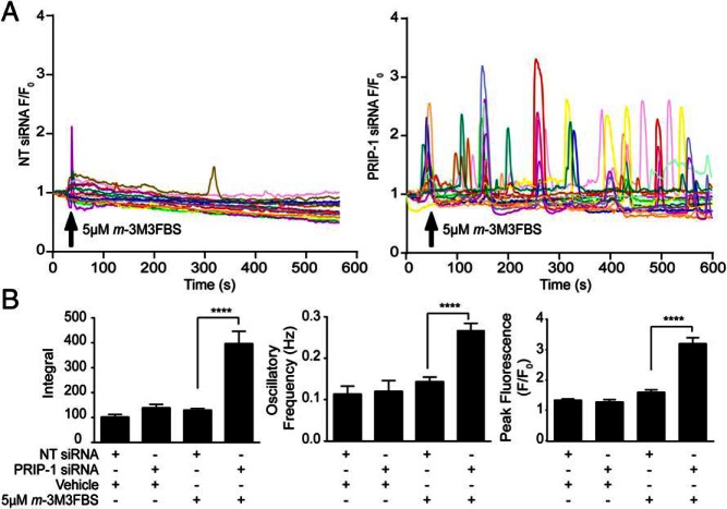
Figure 5. PRIP-1 blocks PLC-dependent Ca2+ signaling in decidual cells.
A, HESCs cultured in glass bottomed Petri dishes were transfected with NT (left panel) or PRIP-1 (right panel) siRNA and decidualized for 4 days. Cells were then loaded with 5μM Fluo-4-AM and challenged with 5μM m-3M3FBS or vehicle (data not shown) at the indicated time point. Cytosolic fluorescence recorded by confocal microscopy over 10 minutes was used as an index of [Ca2+]i. Traces showing fluorescence within individual cells, transfected either with NT (left panel) or PRIP-1 siRNA (right panel) are expressed as a fold increase over fluorescence at time 0 (F/F0). Data were obtained from 4 independent cultures. B, Traces were analyzed to assess the peak changes in fluorescence, the integral, and oscillation frequency (Hz). Data show mean ± SEM; ****, P < .0001.

