

Key Clinical Message
Sigmoid volvulus in pregnancy is a very rare condition. Despite this, clinicians should have a high index of suspicion of this condition if they encounter a pregnant woman with symptoms suggestive of bowel obstruction. Incorrect diagnosis may be catastrophic, resulting in major complications, including fetal and maternal death.
Keywords: Pregnancy, sigmoid volvulus, surgical emergency
Background
Sigmoid volvulus (SV) in pregnancy is a very rare entity which can be associated with extremely high rates of mortality and morbidity for both mother and fetus 1. The danger lies in the insidious nature of symptom development. Delay in presentation and diagnosis can result in bowel ischemia, which may require colectomy and formation of a stoma, and also put pregnancy in jeopardy 2. Maternal complications include perforation, peritonitis, and sepsis. Fetal complications include preterm delivery, intrauterine death, and neonatal sepsis. A high index of suspicion and use of modern imaging modalities are required for achieving better results for both mother and fetus 3.
Case Presentation
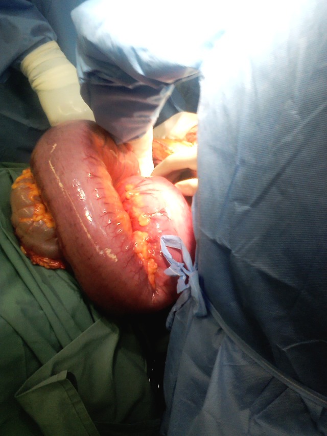
A 21‐year‐old lady, gravida 1 para 0 in the 30th week of gestation, was referred to the surgical service from the O&G team with a 2‐day history of acute onset abdominal pain, distention, and multiple episodes of diarrhea. She denied vomiting. On clinical examination she was hemodynamically stable and afebrile. Examination of her abdomen revealed moderate distension and a prominent loop of bowel palpable in the left upper quadrant. Otherwise her abdomen was soft and the upper margin of the uterus was at the level of the umbilicus. Digital rectal examination revealed an empty rectal ampulla. Her white cell count was 9000. Plain abdominal X‐ray was of low quality, thus nondiagnostic. Instead of repeating an X‐ray a decision was made to proceed to abdominal MRI. MRI revealed massive dilatation of the sigmoid colon, the diameter of which was more than 10 cm, as well as a transition point at the level of the distal sigmoid. The transverse and ascending colon were both dilated. There was no free intraperitoneal gas. An attempt was made to decompress the colon using a soft rectal tube, but was unsuccessful. The patient was transferred to the operating theater and a laparotomy was performed with the O&G team on standby. The sigmoid colon was extremely dilated and at risk of perforation. It had rotated in a clockwise direction with partial occlusion of the mesosigmoid vessels at their origin accompanied, by a big hematoma. Thankfully the sigmoid colon itself was viable. The affected part of colon was unwound and cautiously put back in place and was decompressed using a soft rectal tube. The colon was warmed and reassessed and found to be viable (Figs. 1 and 2). No resection was required and there was no need for intervention by the O&G team. The fetus appeared unaffected and the uterus was assessed to be normal for gestational age. The patient was transferred to the obstetric ward and had an unremarkable postoperative course. She was discharged from the hospital on day 4 post‐op.
Figure 1.

Sigmoid colon before decompression.
Figure 2.
Sigmoid colon after decompression.
Discussion
Sigmoid volvulus in pregnancy is an extremely uncommon condition, with only 84 cases to have been reported in the English literature. Bowel obstruction in pregnancy varies from 1 in 1500 to 1 in 66,431 deliveries, and SV is the cause of 44% of the cases. A long sigmoid colon (dolichocolon) compressed by the enlarged gravid uterus can cause sigmoid volvulus. This might explain the increased incidence of SV in the third trimester of gestation 2, 3.
The diagnosis of SV in pregnancy is often delayed because the symptoms mimic typical pregnancy‐associated complaints. The literature suggests to suspect the diagnosis of SV when a pregnant patient presents with abdominal distention, pain, and absolute constipation. The patient will vomit and not tolerate oral intake of food or water 4. Our patient did not present exactly as described, as there was pain and distention, but no vomiting, and additionally there were multiple episodes of diarrhea. While the obstruction was incomplete, the dilatation of the large intestine was quite marked, as revealed on the MRI scan and at laparotomy.
Imaging options for the diagnosis of SV in pregnancy are controversial given the rarity of this condition in pregnancy. It is widely accepted that exposure of the pregnant patient to radiation should be avoided due to the danger of chromosomal mutations during the first two trimesters and the increased risk for hematological abnormalities such as leukemia in the third trimester. The safe radiation exposure limit is between 5 and 10 rad. Although the radiation dose of an abdominal CT scan is thought to be within this limit, many authors still believe that CT should be avoided. This was the reason we decided to proceed to MRI option, given that it combines diagnostic accuracy and is also considered to be safe for the fetus 2, 5, 6.
The management of SV in the pregnant patient involves aggressive fluid resuscitation, decompression of the proximal bowel and recognition of this entity as an acute surgical emergency 7. The treatment options include endoscopic reduction of the volvulus and surgery.
There are case reports indicating that endoscopy and decompression can have good results in the treatment of SV during pregnancy in cases where bowel necrosis or vascular occlusion has been ruled out. In the absence of peritoneal signs or mucosal ischemia, it would seem reasonable to attempt detorsion and decompression via sigmoidoscopic placement of a soft rectal tube, volvulus distortion through a flexible sigmoidoscopy, or colonoscopy 2, 3, 7.
In our case, the extremely dilated sigmoid colon prevented us from attempting endoscopic reduction, as we did not want to increase the possibility of an iatrogenic rupture of the colon. The intraoperative findings confirmed our suspicions, as the intestinal wall appeared to be extremely thin and prone to perforation. In cases with dead intestine, resection and formation of a stoma are the necessary actions which must be taken. Even though many surgeons attempt primary anastomosis in cases with uncomplicated sigmoid volvulus, this requires further thought in pregnant patients as an anastomotic leak can result in major problems to the gravid uterus and fetus 2, 3, 8. In this case, the intestinal wall showed no signs of vascular compromise, so after unwinding and decompression, the sigmoid colon was put back in place. A healthy baby was delivered 9 weeks after following a normal labor.
Conflict of Interest
The authors declare that they have no competing interests.
References
- 1. Ribeiro Nascimento, E. F. , Chechter M., Fonte F. P., Puls N., Valenciano J. S., Fernandes Filho C. L., et al. 2012. Volvulus of the sigmoid colon during pregnancy: a case report. Case Rep. Obstet. Gynecol. 2012: Article ID 641093. [DOI] [PMC free article] [PubMed] [Google Scholar]
- 2. Khan, M. R. , and Ur Rehman S.. 2012. Sigmoid volvulus in pregnancy and puerperium: a surgical and obstetric catastrophe. Report of a case and review of the world literature. World J. Emerg. Surg. 7:10. [DOI] [PMC free article] [PubMed] [Google Scholar]
- 3. Aftab, A. , Toro A., Abdelaal A., Dasovky M., Gehani S., Abdel Mola A., et al. 2014. Endoscopic reduction of a volvulus of the sigmoid colon in pregnancy: case report and a comprehensive review of the literature. World J. Emerg. Surg. 9:41. [DOI] [PMC free article] [PubMed] [Google Scholar]
- 4. Kolusari, A. , Kurdoglu M., Adali E., Yildizhan R., Sahin H. G., and Kotan C.. 2009. Sigmoid volvulus in pregnancy and puerperium: a case series. Cases J. 2:9275. [DOI] [PMC free article] [PubMed] [Google Scholar]
- 5. The American College of Radiology practice guideline for imaging pregnant or potentially pregnant adolescents and women with ionizing radiation. 2008. (Res. 26).
- 6. Palmucci, S. , Lanza M. L., Gulino F., Scilletta B., and Ettorre G. C.. 2014. Diagnosis of a sigmoid volvulus in pregnancy: ultrasonography and magnetic resonance imaging findings. J. Radiol. Case Rep. 8:54–62. [DOI] [PMC free article] [PubMed] [Google Scholar]
- 7. Wani, M. D. , and Mir S. A.. 2013. Gangrenous sigmoid volvulus complicating pregnancy. World J. Med. Surg. Case Rep. 4; http://www.npplweb.com/wjmscr/fulltext/2/23. [Google Scholar]
- 8. Akcan, A. , Akyildiz H., Artis T., Yilmaz N., and Sozuer E.. 2007. Feasibility of single‐stage resection and primary anastomosis in patients with acute noncomplicated sigmoid volvulus. Am. J. Surg. 193:421–426. [DOI] [PubMed] [Google Scholar]