Abstract
Context:
Steep learning curve is found initially in pure endoscopic procedures. Video telescopic operating monitor (VITOM) is an advance in rigid-lens telescope systems provides an alternative method for learning basics of neuroendoscopy with the help of the familiar principle of microneurosurgery.
Aims:
The aim was to evaluate the clinical utility of VITOM as a learning tool for neuroendoscopy.
Materials and Methods:
Video telescopic operating monitor was used 39 cranial and spinal procedures and its utility as a tool for minimally invasive neurosurgery and neuroendoscopy for initial learning curve was studied.
Results:
Video telescopic operating monitor was used in 25 cranial and 14 spinal procedures. Image quality is comparable to endoscope and microscope. Surgeons comfort improved with VITOM. Frequent repositioning of scope holder and lack of stereopsis is initial limiting factor was compensated for with repeated procedures.
Conclusions:
Video telescopic operating monitor is found useful to reduce initial learning curve of neuroendoscopy.
Key words: Exoscope, neuroendoscopy, video telescopic operating monitor
Introduction
Neuroendoscopy now has more widespread application in modern neurosurgery. Pure endoscopic, endoscopic assisted and minimally invasive technique is proven their results in various cranial and spinal neurosurgeries.[1,2,3,4] There is an urgent need for dedicated “endoscopic” training programs like the postresidency fellowships; along with encouragement of translational research and establishment of dedicated societies that can lay the guidelines and monitor its progress.[5,6,7]
Most of the neurosurgeons and residents are in various phases of learning the curve of neuroendoscopy. There are very few neuroendoscopy program is currently giving dedicated training in this field.[8,9]
Feedback from this program shows that for a beginner it's difficult to sudden shifting from familiar microneurosurgery to endoneurosurgery because of lack of hand-eye coordination, small-diameter delicate endoscope with a very short focal distance, unfamiliar instruments often collide with the endoscope, the lens obscuration and two-dimensional field of view.[10]
Video telescopic operating monitor (VITOM) is a recently developed video telescope operating monitor system (VITOM, Karl Storz GmBH and Co., Tuttlingen, Germany) to perform microsurgery in place of the microscope.[11,12,13,14]
Video telescopic operating monitor is a worked on the principle of telescopic surgery with familiar microsurgical instrument thus it act as a bridge between microneurosurgery and endoneurosurgery as well as it improves surgeon comfort, minimizes fatigue, and reduces surgical morbidity and patient discomfort. The best advantage of VITOM is that it improves the hand-eye coordination in learning neuroendoscopy. A description of this system and use in animal models was reported.[11] This system was also used successfully in various cranial and spinal microneurosurgery in place of the microscope as well as in another specialty.[11,12,13,14,15,16,17,18]
Its usefulness in training of neuroendoscopy is not reported so far. Since our center providing a fellowship in neuroendoscopy in which we have used VITOM to learn the basics of neuroendoscopy.[19] We now report the initial clinical experience with the VITOM and its usefulness for training in neuroendoscopy.
Materials and Methods
Description of system
The VITOM consists of a rigid-lens telescope, camera head, light source, and video display monitor. The telescope is an autoclavable rigid-lens telescope (Model 28095 VA, Karl Storz Endoscopy, Tuttlingen, Germany) with a 10 mm outer diameter and shaft length of 14 cm. The design provides high resolution, minimal distortions, and viewing angle similar to 0° endoscope with a focal distance 25-60 cm. The light source is a commercially available 300-W xenon fiberoptic light source (Xenon Nova300, Karl Storz GmBH and Co., Tuttlingen, Germany). A fiberoptic light cable lens was attached to the rod of the telescope. The camera head with optical zoom and focus features attached to a video monitor. The telescope is held in position by a mechanical endoscope holder (Karl Storz GmBH and Co., Tuttlingen, Germany) with a wide range of motion [Figure 1].
Figure 1.
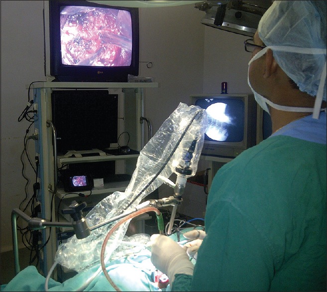
Video telescopic operating monitor positioning and set up
Clinical material
Thirty-nine patients were enrolled in a Neurosurgery Department of a tertiary care Government Medical college and hospital. The department had run 1-week neuroendoscopy fellowship program twice in the year. In this program, fellows are exposed to basics of neuroendoscopy and demonstration of live cranial and spinal neuroendoscopic procedures by senior neurosurgeon. Fellows are trained for hands on in model and cadaver. At the end of program feedback taken from each candidate and applied it for further refinement of the fellowship.
Selected patients were scheduled to undergo a standard neurosurgical procedure. Each procedure was carried out in the standard fashion. At time points when the operating microscope or endoscope is used, the VITOM was used in its place. Surgeries were carried out with standard microsurgical instruments. After each procedure, a subjective analysis was done assessing the optical quality, ease of manipulation, ease of performing surgery, hand-eye coordination and overall sense of utility of the VITOM for learning of neuroendoscopy. Because VITOM is a different system than operating microscope and neuroendoscope, it was not possible to compare between these systems. Analysis was inherently subjective. Nonetheless, it did achieve a primary goal of the study, namely to determine whether surgeons perceived any advantage or disadvantage of VITOM for learning neuroendoscopy.
Results
The VITOM was used in 39 procedures over 18 month time period, including 25 cranial and 14 spinal procedures [Tables 1 and 2]. No surgical complications could be directly attributed to the use of the VITOM. Result was analyze following point, assessing the image quality, ease of manipulation and performing surgery, hand-eye coordination, and overall utility of the VITOM for learning of neuroendoscopy. Overall subjective assessment graded as excellent, good, fair, and average depending upon surgeons comfort.
Table 1.
Cranial procedures

Table 2.
Spinal procedure
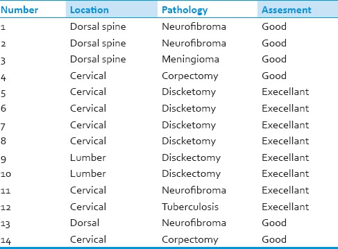
Image quality
The visual quality of the magnified images on the monitor produced by the VITOM was uniformly perceived to be of nearly equal quality to the microscope and endoscope. Quality will also determine by use of camera specification (single chip vs. three chip high definition) as well as video monitor quality (simple vs. high definition medical monitor). Most of the surgeries were performed with the basic single chip camera, and simple video monitor and image quality is comparable. Like endoscope stereoscopic vision was not available with the VITOM. With the experience, VITOM is help to operate the surgeon in two-dimensional vision with standard neurosurgical instrument, which also help later on pure endoscopic surgery. In general, the lack of stereopsis was not perceived as a significant factor.
Positioning of the video monitor
Position of the video monitors proved to be an important factor for surgeon comfort and ease of use in neuroendoscopic procedures. With the VITOM the monitor were used, in the same way, as it is using in cranial and spinal neuroendoscopic surgery that is 2-3 feet from surgeon's eye at head height and to face the surgeon to provide a very wide field of view of the monitor, subtending almost the entire visual field of the surgeon.
Position of telescope and scope holder
The VITOM hold in its position by a mechanical telescope holder with base attached to the operating table permitting a wide range of maneuverability that allowed the surgeon freedom of movement and a large working field. This is the same telescope holder what we are using routinely in neuroendoscopic procedures. By using holder surgeons get familiar with advantage and limitation of holder in endoscopic neurosurgery as holder is an integral part of certain pure endoscopic surgeries like endoscopic third ventriculostomy, pituitary and lumber disc surgery.
Hand-eye coordination
Hand-eye coordination is one of most important skill to learn in neuroendoscopy. It presented a particular challenge for beginners in a few initial cases. Nonetheless, this difficulty was easily overcome by increasing experience. This is the most important advantage of VITOM useful in learning endoscopy though in endoscopy surgeons also required coordination of movement of the scope with the instrument while fixing the gaze at the endoscopic images on the monitor. Given that the entire procedure is performed freehand, with no tactile feedback these skills are particularly important to learn neuroendoscopy.
Illustrative cases
Colloid cyst
A 46-year-old man experienced progressively worsening headaches without neurological disturbances. Examination shows bilateral papilledema. Cranial imaging revealed a well-defined anterior third ventricular lesion suggestive of colloid cyst. Patient operated in supine with 3 cm right mini frontal craniotomy and transcortical approach with the help of tubular retractor lateral ventricle entered, and VITOM was used after that and with the help of single limb instrument cyst removal done, while watching in the video monitor. Postoperative external ventricular drain was placed for 3 days. Postoperative computed tomography (CT) scan showed complete removal of the lesion [Figure 2].
Figure 2.
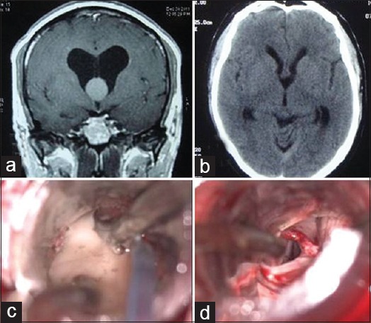
Preoperative coronal T1-weighted contrast magnetic resonance imaging showing colloid cyst (a) and postoperative computed tomography scan reveal complete excision (b), Intraoperative video telescopic operating monitor view of cyst decompression (c) and excision (d)
Cerebello pontine angle tumor
A 20-year-old female presented with gradual progressive hearing loss in left ear with headache and subjective blurred vision. Neurological examination discloses the bilateral papilledema left sided sensorineural hearing loss and left cerebellar signs. Cranial magnetic resonance imaging (MRI) demonstrating a large cerebellopontine angle lesion extending into internal acoustic meatus. Patients operated in supine with head turn position with standard retrosigmoid approach. After opening of dura, VITOM was used in place of the microscope. With standard microsurgical technique and instrument tumor was removed while watching in the video monitor. Anatomical preservation of the facial nerve done. Postoperative neurological examination did not reveal any new neurological sign. Histopathogical examination reveals benign schwanoma. CT scan 3 month after surgery showed complete tumor removal [Figure 3].
Figure 3.
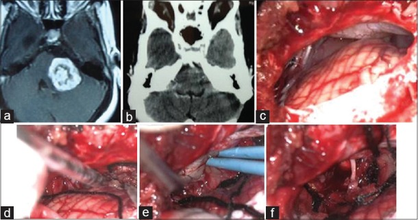
Preoperative axial T1-weighted contrast magnetic resonance imaging showing left cerebellopontine (CP) angle tumor (a), postoperative contrast computed tomography scan reveal complete removal of tumor (b), Intraoperative video telescopic operating monitor view of CP angle (c), tumor debulking (d), separation from brain stem (e) and after tumor removal and anatomical preservation of facial nerve (f)
Lumber disc
A 32-year-male patient presented with left lower limb radicular pain with extensor hallucis longus weakness. MRI lumbar spine suggestive of L5-S1 disc prolapsed. Patients operated for microdisckectomy with the VITOM. Postoperative course was uneventful with relieved of symptoms and weakness patient discharged on 3rd postoperative day [Figure 4].
Figure 4.
Preoperative T2-weighted sagittal (a) and axial (b) magnetic resonance imaging showing L5-S1 disc, Intraoperative video telescopic operating monitor view of removing lamina (c), and disc (d)
C2-3 neurofibroma
A 42-year-female presented with gradual onset progressive spastic quadriparesis more on the right side with neck pain. MRI cervical spine showed a well-defined C2-C3 intra-extra dural mass compressing the cord. Patients operated in the prone position C2-C3 laminectomy done and total excision of the tumor done with VITOM. Postoperative patient is improved to normal power in 3 month. MRI after 3 month showed complete removal of tumor [Figure 5].
Figure 5.
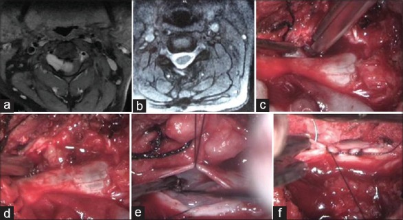
Preoperative axial T1-weighted contrast magnetic resonance imaging (MRI) showing C2-3 intra-extradural tumor (a), postoperative T2-weighted axial MRI reveal complete removal of tumor (b), Intraoperative video telescopic operating monitor view of removing C2 lamina (c), extradural component removal (d), intradural assessment (e) and dural closure (f)
Discussion
Neuroendoscopy has developed rapidly for the past 20 years. Today, it is standard in many institutions. It is logical that rod-lens technology is tested not only for “inside” inspection but also for “outside” inspection.[18,19]
Learning neuroendoscopy in the initial phase has certain difficulties.
First, neuroendoscope has a long, narrow-diameter (1-4 mm) rod-lens. These endoscopes typically have a focal distance of 3-20 mm from the operative site and thus sit adjacent to the operative field. The smaller the diameter of the rod is, the closer the endoscope must be to the operative target. However, for most neurosurgery, there is a need for a larger field of view with frequent alternation. Another limitation by frequent obscuring of the lens by blood and other tissue during surgery, with almost complete loss of visualization during significant arterial or venous bleeding.[20]
To reduce the learning curve in neuroendoscopy in early stage VITOM found to be useful in many aspects. The usefulness of VITOM in neuroendoscopy has never studied before. The first description of VITOM in neurosurgery is given by Mamelak et al.[11] Later on their subsequent report they have advocated the use of VITOM in place of operating microscope to perform open microsurgery.[11,12,13] They concluded that this device was ideally suited for most spinal surgery, but more limited for cranial applications due to the difficulty in delicate aspect of dissection.[13] Use of VITOM in other specialty is also been reported.[8,16,17,18]
Since VITOM is a telescope based system, it works on the principle of endoscopic surgery. VITOM has certain features, which helps neurosurgeons to overcome the early learning difficulties with endoscope. It has a large field of view and a long working distance and can be manipulated and positioned without the constraints of bony anatomy. The long working distance permits operating with standard instruments those neurosurgeons are familiar with and a much smaller “learning curve” than that experienced with traditional endoscopic neurosurgery.[21] Because the VITOM system is entirely extracorporeal, there are no issues with lens fogging or cleaning, problems frequently encountered with the early phase of training of traditional endoscopy.[18,22]
One of the advantages of the VITOM is that it provides an image in the monitor is seen by the operator, assistant, resident, anesthesiologists and nurses which increase the active participation in procedures, education and thus increases the patient's safety. Mamelak et al. integrated VITOM with surgical navigation systems and other video monitor-based intraoperative applications. We do not have the facility of neuronavigation, so we not have experience of that. Portability of this device due to its lightweight is another advantage of the VITOM is its size. Weighing only 1.5 lb, it is easily stored and transported from site to site.
The present study reports on a limited number of cases. In this study all type of cases has included in which certain cases are not suitable for neuroendoscopy like meneingioma, glioma or medulloblastoma. In these cases, VITOM is used as a microscope rather than endoscope. Complicated cases such cerebello pontine angle neuroma, aneurysm clipping in which delicate dissection is required around nerve and vessels should be considered only after getting sufficient experience in simple cases. In our series also we performed simple cases in the beginning along with other neuroendoscopic procedures which are routinely performed in author's institute in large numbers. Although we did not convert any of our procedure into microscopic technique, it may be required in selected patient. Also, VITOM is not useful in pure endcoscopic procedure such as endoscopic third ventriculostomy. Furthermore, it is uncertain if the scope would affect surgery. Limited manueuverality of the telescope holder is a limiting factor may require subsequent modification.
Another limitation is the lack of stereopsis. Most of us are accustomed with the microscopic three-dimensional vision so changing from this to two-dimensional telescopic visions had certain initial difficulties so during the VITOM surgery the delicate dissections using micro-scissors should not be encouraged especially for the beginners. As sufficient experience gained, one can do the difficult cases with the VITOM. This can be adjusted for with surgical experience.[17,23]
Conclusion
Video telescopic operating monitor is an addition to the neurosurgical armamentarium providing a new platform for the learning neuroendoscopy from the microscopic principle. VITOM is not a replacement of microscope or endoscope. Based on the principal of telescopic surgery it will be a useful tool to learn the neuroendoscopy and help to reduce the steep learning curve.
Footnotes
Source of Support: Nil
Conflict of Interest: None declared.
References
- 1.Yadav Y, Sachdev S, Parihar V, Namdev H, Bhatele P. Endoscopic endonasal trans-sphenoid surgery of pituitary adenoma. J Neurosci Rural Pract. 2012;3:328–37. doi: 10.4103/0976-3147.102615. [DOI] [PMC free article] [PubMed] [Google Scholar]
- 2.Yadav YR, Parihar V, Pande S, Namdev H, Agarwal M. Endoscopic third ventriculostomy. J Neurosci Rural Pract. 2012;3:163–73. doi: 10.4103/0976-3147.98222. [DOI] [PMC free article] [PubMed] [Google Scholar]
- 3.Yadav YR, Parihar V, Namdev H, Agarwal M, Bhatele PR. Endoscopic interlaminar management of lumbar disc disease. J Neurol Surg A Cent Eur Neurosurg. 2013;74:77–81. doi: 10.1055/s-0032-1333127. [DOI] [PubMed] [Google Scholar]
- 4.Yadav YR, Yadav S, Sherekar S, Parihar V. A new minimally invasive tubular brain retractor system for surgery of deep intracerebral hematoma. Neurol India. 2011;59:74–7. doi: 10.4103/0028-3886.76870. [DOI] [PubMed] [Google Scholar]
- 5.Agrawal A, Kato Y, Sano H, Kanno T. The incorporation of neuroendoscopy in neurosurgical training programs. World Neurosurg. 2013;79:S15.e11–3. doi: 10.1016/j.wneu.2012.02.028. [DOI] [PubMed] [Google Scholar]
- 6.Dias LA, Gebhard H, Mtui E, Anand VK, Schwartz TH. The use of an ultraportable universal serial bus endoscope for education and training in neuroendoscopy. World Neurosurg. 2013;79:337–40. doi: 10.1016/j.wneu.2012.06.018. [DOI] [PubMed] [Google Scholar]
- 7.Fernandez-Miranda JC, Barges-Coll J, Prevedello DM, Engh J, Snyderman C, Carrau R, et al. Animal model for endoscopic neurosurgical training: Technical note. Minim Invasive Neurosurg. 2010;53:286–9. doi: 10.1055/s-0030-1269927. [DOI] [PubMed] [Google Scholar]
- 8.Neubauer A, Wolfsberger S. Virtual endoscopy in neurosurgery: A review. Neurosurgery. 2013;72(Suppl 1):97–106. doi: 10.1227/NEU.0b013e31827393c9. [DOI] [PubMed] [Google Scholar]
- 9.Haji FA, Dubrowski A, Drake J, de Ribaupierre S. Needs assessment for simulation training in neuroendoscopy: A Canadian national survey. J Neurosurg. 2013;118:250–7. doi: 10.3171/2012.10.JNS12767. [DOI] [PubMed] [Google Scholar]
- 10.Stagno V, Cappabianca P. See one. Do one. Teach one. A perfect paradigm for education and training in neuroendoscopy. World Neurosurg. 2013;79:258–9. doi: 10.1016/j.wneu.2012.07.017. [DOI] [PubMed] [Google Scholar]
- 11.Mamelak AN, Danielpour M, Black KL, Hagike M, Berci G. A high-definition exoscope system for neurosurgery and other microsurgical disciplines: Preliminary report. Surg Innov. 2008;15:38–46. doi: 10.1177/1553350608315954. [DOI] [PubMed] [Google Scholar]
- 12.Mamelak AN, Nobuto T, Berci G. Initial clinical experience with a high-definition exoscope system for microneurosurgery. Neurosurgery. 2010;67:476–83. doi: 10.1227/01.NEU.0000372204.85227.BF. [DOI] [PubMed] [Google Scholar]
- 13.Mamelak AN, Owen TJ, Bruyette D. Transsphenoidal surgery for pituitary adenomas in canines and felines using a high definition video telescope: Methods and initial surgical results. Vet Surg. 2011;40:1067–76. doi: 10.1111/j.1532-950X.2014.12146.x. [DOI] [PubMed] [Google Scholar]
- 14.Mamelak AN, Drazin D, Shirzadi A, Black KL, Berci G. Infratentorial supracerebellar resection of a pineal tumor using a high definition video exoscope (VITOM ®) J Clin Neurosci. 2012;19:306–9. doi: 10.1016/j.jocn.2011.07.014. [DOI] [PubMed] [Google Scholar]
- 15.Shirzadi A, Mukherjee D, Drazin DG, Paff M, Perri B, Mamelak AN, et al. Use of the video telescope operating monitor (VITOM) as an alternative to the operating microscope in spine surgery. Spine (Phila Pa 1976) 2012;37:E1517–23. doi: 10.1097/BRS.0b013e3182709cef. [DOI] [PubMed] [Google Scholar]
- 16.Vercellino GF, Erdemoglu E, Kyeyamwa S, Drechsler I, Vasiljeva J, Cichon G, et al. Evaluation of the VITOM in digital high-definition video exocolposcopy. J Low Genit Tract Dis. 2011;15:292–5. doi: 10.1097/LGT.0b013e3182102891. [DOI] [PubMed] [Google Scholar]
- 17.Wiwanitkit V. VITOM in digital high-definition video exocolposcopy. J Low Genit Tract Dis. 2012;16:480. doi: 10.1097/LGT.0b013e31824b9a99. [DOI] [PubMed] [Google Scholar]
- 18.Carlucci C, Fasanella L, Ricci Maccarini A. Exolaryngoscopy: A new technique for laryngeal surgery. Acta Otorhinolaryngol Ital. 2012;32:326–8. [PMC free article] [PubMed] [Google Scholar]
- 19.Neuroendoscopy.org. Fellowship in Neuroendoscopy. NSCB Govt. Medical College Jabalpur. [Last accessed on 2013 Apr 17]. Available from: http://www.neuroendoscopyjbp.org .
- 20.Schroeder HW. Initial clinical experience with a high-definition exoscope system for microneurosurgery. Neurosurgery. 2010;67:483. doi: 10.1227/01.NEU.0000372204.85227.BF. [DOI] [PubMed] [Google Scholar]
- 21.Jagannathan J, Laws ER, Jane JA., Jr . Advantages of the endoscope and transitioning from the microscope to the endoscope for endonasal approaches. In: Kassam AB, editor. Endoscopic Approaches to the Skull Base. Progess in Neurological Surgery. 1st ed. Basel: Karger; 2012. pp. 7–20. [Google Scholar]
- 22.Snyderman CH, Pant H, Kassam AB, Carrau RL, Prevedello MD, Gardner PA. The learning curve for endonasal surgery of the cranial base: A systematic approach to training. In: Kassam AB, editor. Endoscopic Approaches to the Skull Base. Progess in Neurological Surgery. 1st ed. Basel: Karger; 2012. pp. 222–31. [Google Scholar]
- 23.Kassam AB, Gardner PA, Prevedello MD, Snyderman CH, Carrau RL. Principles of Endoneurosurgery. In: Kassam AB, editor. Endoscopic Approaches to the Skull Base. Progess in Neurological Surgery. 1st ed. Basel: Karger; 2012. pp. 21–6. [Google Scholar]


