Sir,
Melasma is a common hypermelanoses affecting the female gender. The etiology of melasma has not been clearly identified. Factors associated with melasma include exposure to ultraviolet light, pregnancy, oral contraceptives, hormone replacement therapy, thyroid autoimmunity, cosmetic ingredients, and phototoxic drugs.[1] The commonest site affected by melasma is the face and has been distinguished into three clinical patterns, namely, centrofacial, malar, and mandibular. Extrafacial melasma is a rare entity and has been very rarely reported in the literature. Extrafacial melasma should be distinguished from acquired brachial cutaneous dyschromatosis (ABCD).[2]
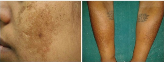
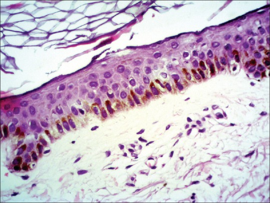
A 55-year-old woman presented with brownish pigmentation of her both forearms for past six years. She reported that initially few brown hyperpigmented asymptomatic spots appeared on her right forearm and similar pigmented spots started appearing on the other forearm too. She denied any history of inflammatory dermatoses prior to appearance of her skin problem. The patient was postmenopausal since three years. She reported that the pigmentation became more prominent on sun exposure. She denied the intake of oral contraceptive pills or hormone replacement therapy. There was no history suggestive of thyroid dysfunction. She did not give history of receiving any photosensitizing drugs. Cutaneous examination showed a large, brown irregular patch on her right forearm and a similar developing lesion on her other forearm. The patches comprised of multiple coalescing hyperpigmented macules with irregular borders with intervening areas showing normal skin. She also had malar type of facial melasma [Figure 1]. We made a provisional diagnosis of extrafacial melasma and ABCD. Potassium hydroxide mount from the patches failed to show any fungal elements, which ruled out pityriasis versicolor. Wood's lamp examination showed accentuation of pigmentation suggestive of epidermal pigmentation. Skin biopsy from the lesion showed epidermal thinning and increased melanization of basal layer [Figure 2]. Histology showed minimal epidermal atrophy and mild solar elastosis in the form of elastic fiber degeneration. No evidence of amyloid material was seen on special stains, which ruled out frictional amyloid deposition. On a review of the literature, we were able to rule out acquired brachial cutaneous dyschromatosis as there were no hypopigmented atrophic macules. On correlation, we made a diagnosis of extrafacial melasma involving dorsa of both forearms.
Figure 1.

Patient having malar type of melasma and brownish pigmentation on dorsa of both forearms
Figure 2.

H and E stained section of biopsy showing epidermal thinning and increased melanization of basal layers (×20)
Extrafacial melasma is an uncommon variant of melasma and has been very rarely reported in the literature. Extrafacial melasma shares similar clinical features as that of facial melasma with brownish hyperpigmented spots with irregular borders. Although facial and extrafacial melasma share some clinical features, they differ in incidence. Melasma occurs predominantly in the age group of 20–40 years with a mean age of 33 years and is rarely observed in menopausal females.[3] Extrafacial melasma is known to affect menopausal women in the fifth decade of life.[4]
A close differential for extrafacial melasma affecting forearm is ABCD. ABCD is characterized by dyschromatosis (hyperpigmented and hypopigmented macules) and face is never involved, whereas extrafacial melasma lacks hypopigmented atrophic macules. ABCD has a strong causal association with antihypertensive drugs particularly angiotensin converting enzyme inhibitors.[2] A striking observation in cases of ABCD is poikiloderma of the neck (Civatte), which may be incidental in cases of extrafacial melasma.
Histologically, extrafacial melasma resembles facial melasma with increased pigmentation of basal layer and epidermal atrophy. Both the entities show signs of chronic actinic damage on histology Skin biopsy in our patient showed epidermal thinning, which can be a sign of chronic actinic damage. Treatment of extrafacial melasma can be challenging with frequent relapses. Depigmenting drugs, retinoids, and sunscreen are the mainstay of therapy for extrafacial melasma.[5,6] It is premature to comment whether extrafacial melasma responds to depigmenting therapy akin to typical facial melasma.
Financial support and sponsorship
Nil.
Conflicts of interest
There are no conflicts of interest.
REFERENCES
- 1.Achar A, Rathi SK. Melasma: A clinico-epidemiological study of 312 cases. Indian J Dermatol. 2011;56:380–2. doi: 10.4103/0019-5154.84722. [DOI] [PMC free article] [PubMed] [Google Scholar]
- 2.Rongioletti F, Rebora A. Acquired brachial cutaneous dyschromatosis: A common pigmentary disorder of the arm in middle-aged women. J Am Acad Dermatol. 2000;42:680–4. [PubMed] [Google Scholar]
- 3.Sardesai VR, Kolte JN, Srinivas BN. A clinical study of melasma and a comparison of the therapeutic effect of certain currently available topical modalities for its treatment. Indian J Dermatol. 2013;58:239. doi: 10.4103/0019-5154.110842. [DOI] [PMC free article] [PubMed] [Google Scholar]
- 4.Ritter CG, Fiss DV, Borges da Costa JA, de Carvalho RR, Bauermann G, Cestari TF. Extra-facial melasma: Clinical, histopathological, and immunohistochemical case-control study. J Eur Acad Dermatol Venereol. 2013;27:1088–94. doi: 10.1111/j.1468-3083.2012.04655.x. [DOI] [PubMed] [Google Scholar]
- 5.Ikino JK, Nunes DH, Silva VP, Fröde TS, Sens MM. Melasma and assessment of the quality of life in Brazilian women. An Bras Dermatol. 2015;90:196–200. doi: 10.1590/abd1806-4841.20152771. [DOI] [PMC free article] [PubMed] [Google Scholar]
- 6.Sarkar R, Arora P, Garg VK, Sonthalia S, Gokhale N. Melasma update. Indian Dermatol Online J. 2014;5:426–35. doi: 10.4103/2229-5178.142484. [DOI] [PMC free article] [PubMed] [Google Scholar]


