
Figure 3b:
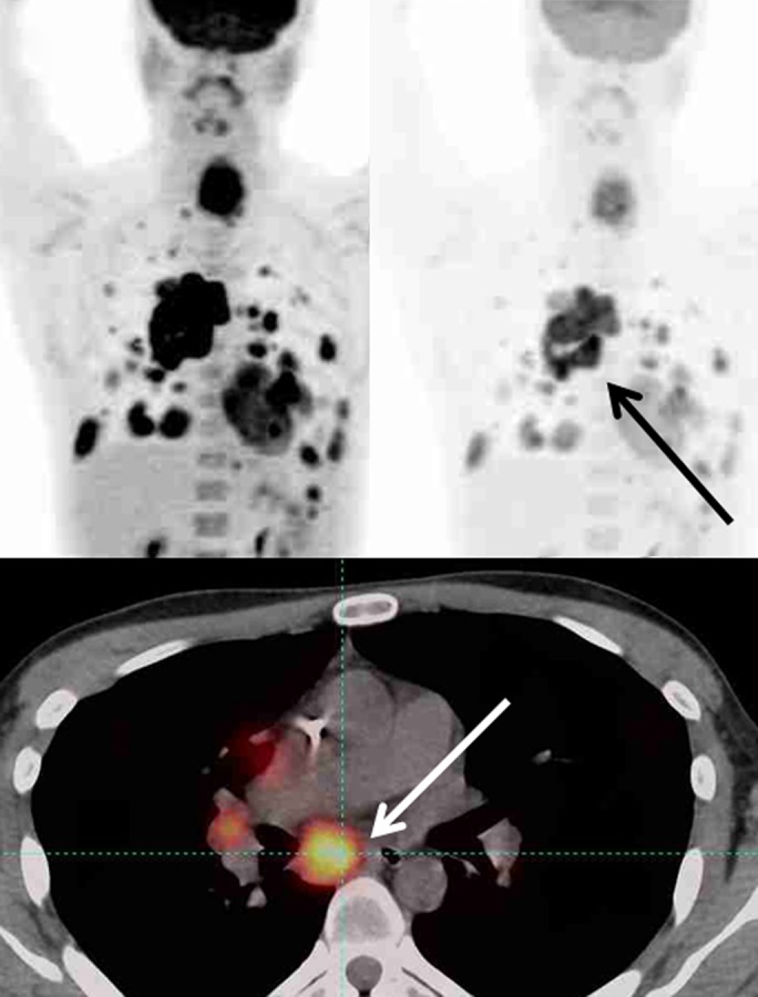
Maximum intensity projection images (top row) in two different SUL windows and axial fusion images (bottom row) in a man with sarcoma and multiple distant metastatic lesions. SULpeak was measured from the hottest tumor at each time point and can be different at baseline and follow-up. (a) Hottest tumor SULpeak focus was located in right lower paratracheal lymph node at baseline (arrows). (b) After therapy, subcarinal lymph node was hottest tumor (arrows).