Abstract
Tibial plateau fractures are common in the elderly population following a low-energy mechanism. Initial evaluation includes an assessment of the soft tissues and surrounding ligaments. Most fractures involve articular depression leading to joint incongruity. Treatment of these fractures may be complicated by osteoporosis, osteoarthritis, and medical comorbidities. Optimal reconstruction should restore the mechanical axis, provide a stable construct for mobilization, and reestablish articular congruity. This is accomplished through a variety of internal or external fixation techniques or with acute arthroplasty. Regardless of the treatment modality, particular focus on preservation and maintenance of the soft tissue envelope is paramount.
Keywords: tibial plateau fracture, elderly, osteoporosis
Introduction
Tibial plateau fractures are a complex group of periarticular fractures that require careful evaluation and preoperative planning. Most research has focused on these injuries in the younger patient population with higher energy injuries. In the elderly patients, however, lower energy mechanisms may result in similar injury patterns due to age-related changes in bone architecture.
Further, these injuries are complicated by patient comorbidities, preexisting osteoarthritis, and lower levels of preinjury functional and ambulatory status.1 The purpose of this article is to critically evaluate the differences in the elderly patient with a tibial plateau fracture and to develop an understanding of the differing treatment options to maximize postoperative return to functioning.
Anatomy
The tibia is a triangularly shaped bone in cross section that flares from the relatively narrow diaphysis to the proximal tibia. The anterior proximal third of the tibia serves as the attachment for the patellar tendon on the tibial tubercle. More proximally, the lateral aspect of the tibia is the origin of the anterior compartment musculature and the insertion of the iliotibial band onto Gerdy tubercle. Posterior to the lateral plateau, the fibular collateral ligament, the popliteal fibular ligament, and the biceps femoris attach to the fibular head, which contribute to the proximal tibiofibular joint. In a normal knee, 60% of the load is predominantly borne on the tibial side. As such, the bone is more sclerotic and more resistant to failure than the lateral tibial plateau.2,3 The proximal medial tibia is covered by a thin layer of skin and subcutaneous tissue overlying the insertion of the pes anserinus tendons. These structures, in addition to the medial collateral ligament (MCL) and lateral collateral ligament (LCL), provide soft tissue balance and stability to the knee joint. Three percent of tibial plateau fractures involve injury to the MCL or LCL.4 In elderly patients, the energy imparted during fracture of the tibial plateau often spares the ligaments, as most of the energy is absorbed by the weaker bones.5
The tibial plateau articular surface is shaped with mild convexity on the lateral side and is larger and concave on the medial side. This concavity provides a smoother articulation between the medial femoral condyle and the plateau. In addition, the lateral plateau lies 2 to 3 mm proximal (superior) to the medial plateau, accounting for the subtle approximately 3 degrees of varus of the proximal tibia. This disparity enables the clinician to better delineate articular congruity on the lateral radiograph during fracture characterization. There is considerable variation in tibial slope, but one study found the average to be −3° to +10° for medial tibial slope, from 0° to +14° for lateral tibial slope, and from −1° to +6° for coronal tibial slope. Overall, females had a larger slope than males.6
On the femoral side, the femoral condyles are larger anteriorly than posteriorly. During axially directed forces such as those resulting in tibial plateau fractures, the femoral condyles impact the plateau creating depressed and/or split fractures in the anterior tibia. During knee flexion, femoral rollback allows for the smaller posterior condyles to impact the posterior plateau. Although high degrees of knee flexion are not associated with common injury mechanisms, care must be taken to scrutinize the posterior plateau on radiographs for fragments that may result in further joint instability.
Epidemiology
Tibial plateau fractures comprise 8% of all fractures in the elderly patients. Dating back to 1973, Rasmussen reported a series of 260 patients who sustained this injury. He found that 72% of patients were older than 55 years. Women had more medial or bicondylar fractures (31%) and posterior compression fractures (61%).7 In 2 large clinical series, 55% to 70% of the population with tibial plateau fractures was older than 50 years.8 As in younger patients, the lateral condyle is most often affected.1,5 This may be due in part to patients being impacted on the lateral aspect of the knee at the time of injury and that there is a natural 5 to 7° valgus alignment of the knee. However, given the higher energy fracture patterns seen in the younger population, one must be mindful that the prognostic information provided by the use of current classification systems may not be as helpful in the elderly patient with the same injury.
Mechanism of Injury
Tibial plateau fractures usually occur due to high-energy trauma, characteristically produced by varus or valgus forces coupled with axial loading. Greater axial loading leads to an increased likelihood of bicondylar involvement.3 They occur mainly after motor vehicle accidents, pedestrians struck by automobiles, or a fall from a height. Cars attempting to brake may strike patients crossing the street with the lateral side of their knee facing traffic. In doing so, the inertia forces the car’s front bumper toward the ground to the same level of the tibial plateau. This has been termed a “bumper fracture.”
Pure split fractures are more common in younger patients. With increase in age comes a corresponding decrease in the strength of cancellous bone. As a result, split depressions are more common after the fifth decade of life. The magnitude of the force producing the fracture determines not only the degree of comminution but also the degree of displacement, which is also governed by the degree of knee flexion at the time of injury.
Initial Evaluation
When an elderly patient is being evaluated with knee pain, it is paramount to obtain a thorough history and physical examination. The clinician should ascertain the context of the injury, the mechanism, and the circumstances surrounding the fall. Any prodromal symptoms such as dizziness, lightheadedness, or palpitations should warrant a further syncopal workup, as this may indicate underlying cardiovascular disease. The surgeon should also obtain information regarding the patient’s comorbidities (ie, diabetes, vascular disease, obesity, smoking status), social support, and preinjury functional status. This will aid in the decision-making process for treatment as well as identify potential barriers to soft tissue healing.
Physical examination should always begin with a complete assessment of airway, breathing, and circulation. It is the responsibility of the consulting orthopedic surgeon as well as the emergency department staff to conduct this examination looking for any associated injuries. With the rising use of oral anticoagulant medications in elderly patients for various thrombotic conditions such as atrial fibrillation or valvular heart disease, the patient’s list of medications must be scrutinized as this may have an effect on surgical timing and in-hospital anticoagulation decisions in the perioperative period.9 Once this is complete and the patient is deemed hemodynamically stable, a secondary examination of injuries is performed.
Clinical examination of the knee should begin with inspection and comparison to the uninjured side. Injury involving the knee contained within the joint capsule may result in an effusion or lipohemarthrosis, contributing to a loss of range of motion. Ecchymosis may also be present. Given the age-related transition to thinner, more tenuous soft tissues with less collagen, the condition of the skin should be scrutinized for blisters, open wounds, and devitalization.10 Examination of the knee for varus or valgus stability helps to judge fracture severity and the potential need for operative reduction and may be an indicator of average plateau depression.11 This test is sensitive but not specific because of the potential for patient guarding due to pain. Rasmussen defined 10 degrees of valgus laxity as indicative of instability.7 Likewise, a ligamentous examination of the knee may be interpreted as positive but is confounded by displacement of fracture fragments during testing. In this case, intraoperative assessment is more appropriate and other injuries may be addressed at that time. Meniscal injury may occur in 50% of tibial plateau fractures and may even be as high as 91% for lateral meniscus pathology.12,13
Lastly, motor and sensory examination should be conducted and deficits documented, with a focus on the integrity of the peroneal nerve, which has been reported to be injured in 3% of patients with tibial plateau fractures.14 If vascular injury is suspected based on the presence of unequal distal pulses, ankle–brachial indices (ABIs) should be performed, and if the value is <0.9, a computed tomography (CT) angiogram can be performed. In medial condylar fractures, which represent a functional knee dislocation, the ABI is especially important. One series found that of 38 patients with a knee dislocation, 11 had an ABI <0.9 and 100% of them had a vascular injury requiring surgery.15
Following the initial and secondary surveys, the injured limb should be placed in a posterior splint, a knee immobilizer, or a combination of the 2. The knee should be extended and the ankle in neutral. A knee immobilizer may be added for additional support and rotational stability. If axial traction is required to obtain a length-stable fracture reduction, the ABI should be repeated following this maneuver. If there is concern for evolving soft tissue swelling, serial compartment checks should be performed, especially in Schatzker IV to VI patterns. Compartment syndrome has been reported in 14% to 67% of patients with medial condylar injuries and 18% in bicondylar injuries with diaphyseal extension.16,17 Elevation and cryotherapy are important adjunctive modalities to address soft tissue insult early in treatment and mitigate rising compartment pressures.
Imaging
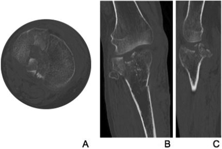
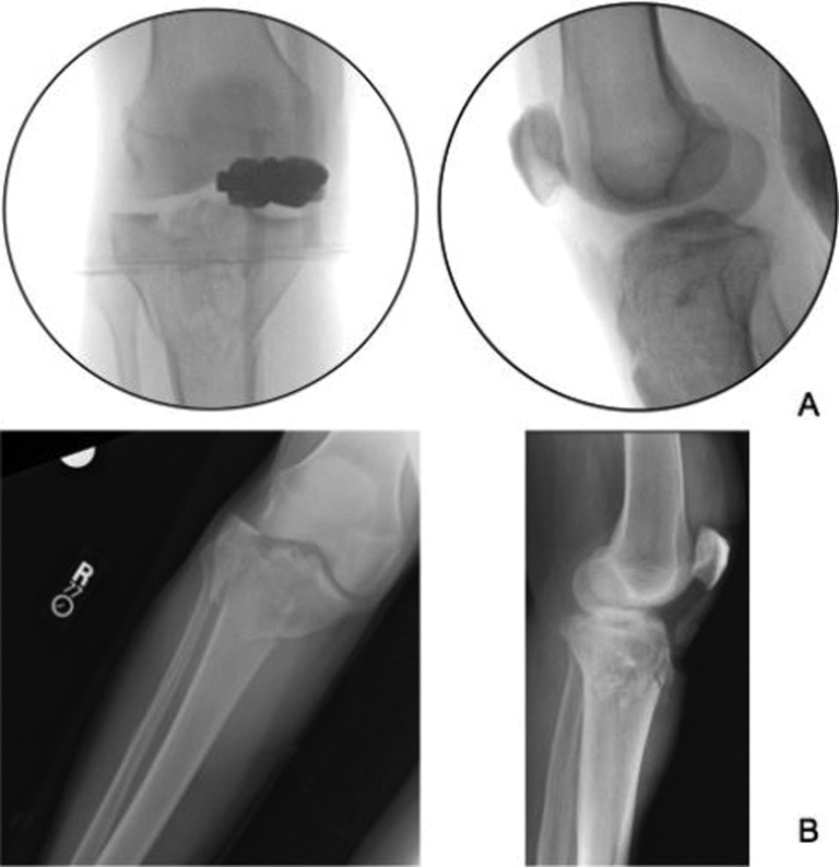
Radiographic assessment should include anteroposterior (AP), lateral, and oblique views of the knee, assessing for fracture lines, condylar widening, and articular step-off. For severely comminuted fractures, slight axial traction may help to restore the gross geometry of the proximal tibia and decrease fragment overlap. A CT scan is essential for these articular fractures (Figure 1). If the patient needs to be taken for urgent external fixation due to soft tissue disruption or fracture instability (Figure 2), a CT scan should be performed only after the fixator is applied. In this way, the scan can be very useful for preoperative planning in 3 dimensions once the fracture is length stable and all of fragments are visualized. According to a study by Chan et al, after reviewing CT scans in patients with tibial plateau fractures, surgeons changed their treatment plan >25% of the time as compared with viewing radiographs alone. Reasons for changing the operative plan included better identification of comminution, depression, or a fracture pattern not appreciated on plain films.18 One should maintain a high clinical suspicion in an elderly patient with severe knee pain but negative radiographs as this may indicate an insufficiency fracture, and a CT scan should be obtained for further evaluation.19 Thus, fractures of the tibial plateau, though not as common as distal radius fractures or compression fractures, should raise concern for osteoporosis and warrant bone mineral density scanning and nutritional supplementation to assess and improve bone health.
Figure 1.
Select axial (A), coronal (B), and sagittal (C) cut of a computed tomography scan, showing a comminuted, intraarticular tibial plateau fracture.
Figure 2.
A, Intraoperative anteroposterior and lateral fluoroscopic images following external fixation of a depressed, length unstable tibial plateau fracture, showing improved alignment compared with preoperative radiographs (B).
Classification
There are 2 widely used classification systems for tibial plateau fractures. The Arbeitsgemeinschaft für Osteosynthesefragen/Orthopaedic Trauma Association classification subdivides proximal tibia fractures into 3 main categories. Following the prefix designation 41, A-type fractures are extra-articular, comprising avulsion and metaphyseal fractures. B-type injuries are partial articular fractures described as a split, depression, or both. Type C fractures are completely articular and are further subdivided into simple or multifragmentary.
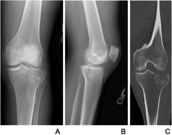
The Schatzker classification is more commonly used as a way to communicate both fracture description and severity, as well as aid in directing appropriate surgical treatment. The original series used AP radiographs in 94 patients with tibial plateau fractures.20 A type I fracture is a pure split of the lateral plateau. These are more commonly seen in younger patients with stronger bone and are frequently associated with meniscal tears as the axial load of the femoral condyle on the tibia forces the lateral tibial plateau down and out, incarcerating the lateral meniscus. Type II fractures are split depressions of the lateral plateau, where the medial articular edge is forced into the metaphysis (Figure 3). Type III injuries, the most common of the patterns in Schatzker series, involves a pure depression of the lateral articular surface.21 This pattern is more common in the elderly patients with weaker cancellous and subchondral bone and may result from a low-energy mechanism. Types IV to VI result from higher energy mechanisms and thus have a larger zone of injury with a higher incidence of associated vascular or ligamentous damage.14 Type IV is a fracture of the medial plateau, type V is bicondylar involving both plateaus, and type VI results in metaphyseal–diaphyseal dissociation.
Figure 3.
Anteroposterior (A) and lateral (B) radiographs and coronal CT (C) images of a 60-year-old male who sustained a Schatzker II tibial plateau fracture after a fall from standing. Note the degree of articular depression better appreciated on CT scan. CT indicates computed tomography.
Regardless of the classification method used, the goal for injury discussion should convey the severity and urgency of the injury. For example, describing the fracture as unicondylar or bicondylar with further characterization of condylar width, limb length discrepancy, and instability provides a substantial amount of information to develop a clinical treatment algorithm and may even be more informative than describing a fracture as, for example, a Schatzker V alone.
Treatment
Although treatment algorithms for tibial plateau fractures still remain controversial and subject to surgeon’s preference, the goals of intervention remain the same: reestablish limb mechanical alignment, provide stability, and restore articular congruity.22 Despite the articular nature of these fractures, there is a role for nonoperative management in a specific subset of patients. Patients who have medical comorbidities or acute illnesses that preclude any surgical intervention and patients who have poor or no preinjury mobility of their legs (ie, paraplegics, nonambulators) may not be surgical candidates. In addition, patients with minimally displaced fractures may not require surgery. If nonsurgical management is undertaken, treatment protocols include protection of weight bearing in a hinged knee brace with range of motion as tolerated. Continuous passive motion machines may be used to facilitate motion. Physical therapy should initially focus on isometric quadriceps exercises and subsequently progress from passive to active assist to active range of motion. For noncompliant patients, a long leg cast with slight knee flexion may replace the brace, but frequent skin checks are required. Full weight bearing can be instituted at approximately 8 to 12 weeks after the injury provided repeat radiographs show signs of healing. Fracture displacement is an indication to proceed with surgery.12
Absolute indications for fixation of tibial plateau fractures include open injuries, compartment syndrome, and injuries associated with vascular disruption. Relative indications for surgery include fracture displacement, articular step-off >3 to 5 mm, condylar widening >5 mm, and malalignment >5° to 10°. In 1973, Rasmussen defined instability of the knee as a 10° increase in lateral deviation in the extended knee as compared to the normal side.7 Each millimeter of articular depression produces approximately 2° of limb malalignment. Recommendations regarding the amount of acceptable articular depression vary from 2 mm to 1 cm. Articular cartilage of the tibial plateau has been shown to be between 3 and 4 mm in thickness on the lateral side and uniformly 2.5 mm thickness on the medial side, thus providing a rationale for a certain degree of depression.11,23 In a 20-year follow-up study of 102 patients, Landsinger et al found that fractures resulting in less than 10° of instability resulted in favorable outcomes when treated nonoperatively.24 Timing of operative treatment depends mainly on the integrity of the soft tissue envelope within the zone of injury. In most cases, waiting 7 to 10 days is sufficient to allow time for the soft tissues to recover and regain some of their normal turgor.25 Operating through a swollen soft tissue compartment may result in a higher incidence of wound complications.26
Surgical Options
Treatment of tibial plateau fractures must take into account patient age, comorbidities, bone stock, and fracture pattern. Although nonoperative treatment may be suitable for minimally displaced fractures, displaced fractures typically require surgical fixation. The first decision point in operative treatment is whether the patient requires external fixation as a temporizing measure until the soft tissues are amenable to internal fixation. If external fixation is performed to maintain a length-stable limb, this affords the surgical team the opportunity to examine the wound daily and assess for fracture blisters, skin breakdown, and wrinkling. Strict elevation is very helpful in the perioperative period to decrease swelling. Axial traction imparted by the reduction in the external fixator may lead to increased compartment pressures in the leg. However, this effect may only be transient. Egol et al evaluated 25 patients with a mean age of 52 years who underwent external fixation for tibial plateau fractures and found that 40% had a transient rise in pressures, but none met criteria for fasciotomy (▵P <30 mm Hg) 5 minutes after the reduction.27
Hybrid ring fixation is another less invasive strategy that can be used definitively for elderly patients. This method uses external fixation to provide ligamentotaxis reduction force and maintains the reduction as a neutralization device for interfragmentary screws.28 Extensive soft tissue exposure during plate fixation with infection rates ranging from 20% to 80% has prompted some authors to adopt this technique.29 The advantages of circular frame fixation (with or without percutaneous lag screw fixation) include minimal soft tissue disruption, the ability to correct deformity in multiple planes, early knee motion, and the option of spanning the knee in patients with concomitant ligamentous injury.30 The ring fixator produces beam loading creating even support for the joint and fracture site in a controlled mechanical environment, minimizing unwanted motion that should decrease the chance of infection and loss of fracture reduction.31
The fine wires under tension provide adequate purchase in softer, osteoporotic bone and can allow for early weight bearing. Disadvantages of the ring fixator include increased risk of pin tract infection and valgus malunion requiring reoperation.32
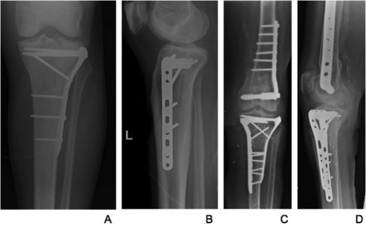
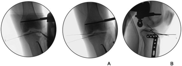
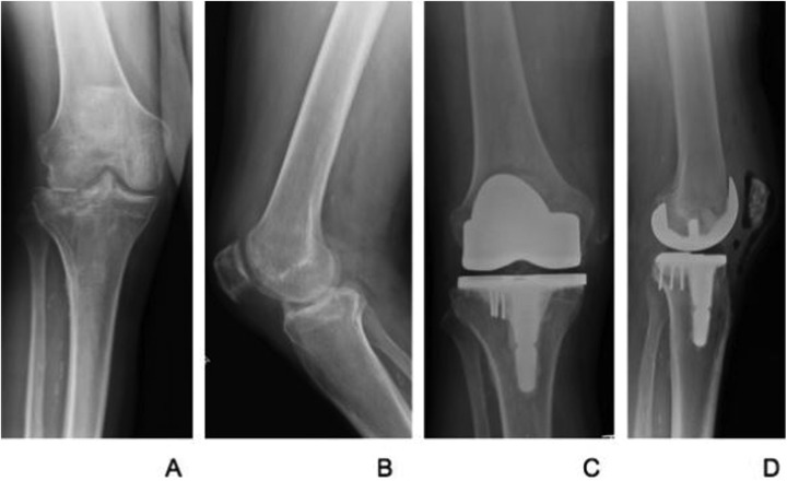
Open reduction with internal fixation has been shown to be more advantageous than external fixation in elderly patients with comminuted intraarticular fractures because their osteoporotic bones are vulnerable to severe comminution.5 Fixation strategies are governed by the fracture pattern and degree of comminution. Careful soft tissue handling is paramount due to the paucity of soft tissue around the knee, all in an effort to reconstruct the articular surface, provide stable fixation, and allow for early mobilization. For fractures confined to the lateral plateau, fixation may be accomplished by two or three 6.5- or 7.0-mm cannulated lag screws over guide wires through small stab incisions. For more extensile exposure, the anterolateral approach or a dual medial and lateral approach to the tibia is commonly used (Figure 4). After reducing the articular surface, minimally invasive percutaneous plate osteosynthesis serves as a way to spare periosteal stripping and disruption of the bony vascular supply.33 Other ways to reduce soft tissue violation include the use of a femoral distractor, percutaneous reduction clamps, and Kirschner wires (Figure 5). Buttress plates can support lag screw fixation if the fracture is more comminuted. In younger patients with good bone stock, the use of locking plate technology has little role. However, there are certain indications to consider locked plating. Laterally based locking plates provide increased stability in the presence of metaphyseal or metadiaphyseal comminution and severe osteoporosis and may offer an alternative to an additional medial plate or external fixator for added support of the medial column if a conventional plate is used for a Schatzker V fracture pattern.33,34 This allows fixation through a single lateral incision, potentially avoiding wound complications. Although not without its own complications, dual plating is considered to be the most mechanically stable fixation construct.31 In cases where internal fixation may have a higher incidence of failure, such as with substantial bone loss and extensive comminution, or in patients with preexisting arthritis, acute total knee arthroplasty (TKA) has become an option for elderly patients with tibial plateau fractures (Figure 6). Although infection and hardware failure risks still exist, arthroplasty offers patients the ability to immediately bear weight and eliminates fracture-healing issues. In addition, the use of augments, stemmed implants, bone graft substitute, and cementation is able to address bone defects and add to stability.35,36 More recently, Kini et al used computer navigation to assess limb alignment and component positioning during the procedure and reported good results, with correction to within 1.7° of the calculated mechanical axis.37 Wasserstein et al defined the rate of conversion to TKA after prior fixation of a tibial plateau fracture in a matched cohort. Ten years after tibial plateau fracture surgery, 7.3% of the patients had had a TKA, a 5.3 times increase in likelihood compared with a matched group from the general population. For older patients, TKA was also more likely.38 Initial open reduction internal fixation and restoration of bone stock may allow for an easier conversion arthroplasty, however. By allowing for bony healing and potential bone stock reconstitution, one may find that performing a TKA at a later time may obviate the need for augments. These patients are also subject to further complications following TKA.39 After prior surgery, their soft tissue envelope is not as robust as a native knee. Earlier onset of posttraumatic arthritis following a plateau fracture leads to an earlier indication for TKA and thus higher likelihood of revision due to increased wear. Lastly, alteration in the knee joint biomechanics can make soft tissue balancing in TKA more difficult.40
Figure 4.
Postoperative anteroposterior (A) and lateral (B) radiographs of a patient who underwent an anterolateral approach to the tibia for a Schatzker III tibial plateau fracture. Postoperative anteroposterior (C) and lateral (D) radiographs of an osteoporotic 74-year-old female with a prior distal femur fracture who sustained a Schatzker VI tibial plateau fracture and underwent a dual incision approach to support the medial and lateral columns.
Figure 5.
A, Intraoperative fluoroscopic images using Kirschner wires and femoral distraction to obtain provisional fixation before applying lateral tibial plate (B).
Figure 6.
Preoperative anteroposterior (A) and lateral (B) radiographs of a 62-year-old male with preexisting right knee osteoarthritis who sustained a lateral tibial plateau split depression fracture after a fall. He underwent an acute total knee arthroplasty with supplemental screw fixation. Postoperative anteroposterior (C) and lateral (D) images.
Outcomes
In elderly patients, fixation of tibial plateau fractures poses unique challenges due to poor bone stock, comminution. Available literature on the outcomes of elderly patients with tibial plateau fractures is somewhat limited.1,35 Some are often eliminated from study protocols due to medical comorbidities or are treated nonoperatively, whereas for others, strict indications for surgery sometimes do not apply. Frattini et al retrospectively looked at 49 patients aged 66 to 88 years who had sustained tibial plateau fractures and found that 75% experienced satisfactory results both functionally and radiographically at 2 to 11 years of follow-up if treated operatively.41 Similarly, Hsu et al reported on 20 patients older than 60 years followed for an average of approximately 50 months who underwent buttress plating with or without supplemental interfragmentary screw fixation. All of the patients regained more than 120 degrees of knee flexion and residual malalignment less than 5 degrees.5 Rasmussen estimated the progression of osteoarthritis on follow-up radiographs of tibial plateau fractures to be 17% to 21%.7 More recently following tibial plateau fractures, progression to osteoarthritis by one grade or more occurred in 59.5% of patients using the Resnick and Niwayama grading scale for osteoarthrosis (n = 38).8 Another study estimated the risk to be 23%.42 After 7.6 years of follow-up, Honkonen found radiologic progression (joint space narrowing) of secondary osteoarthritis in 37% of patients who sustained a tibial plateau fracture, as long as the meniscus was in tact. The incidence increased slightly with age.42 Failure to achieve and maintain reduction in the depressed fragments is one of the most important predisposing factors of accelerated degeneration, and progression to TKA may occur as early as 1 year following fixation.8,40,43 However, it is important to let the patient’s functional status guide your treatment algorithm, as the severity of radiographic osteoarthritis may not correlate with pain and functional limitations.44,45
Saleh et al looked at 15 patients with a previous plateau fracture at an average of 6.2 years after TKA. They found that although TKA decreased pain and improved functional range of motion, there was a much higher rate of complications (3 deep infections, 2 patellar tendon disruptions, 3 patients requiring closed manipulation) in patients with a history of fracture.46 Although Weiss et al reported 78% good outcomes for TKA following prior plateau fracture, they emphasized unique complications and risk of the procedure. Regardless of treatment, the underlying goals of tibial plateau fracture surgery are meticulous soft tissue handling, fracture reduction, and maintenance of range of motion following fracture treatment. In patients who subsequently undergo TKA, these were the most important preoperative factors in patient outcomes.40
Conclusions
Tibial plateau fractures in the elderly patients require special attention. Preexisting osteoporosis and osteoarthritis may require alternative treatment options so preoperative planning is very important. A thorough evaluation of the patient’s preoperative functional status and comorbidities must be performed prior to consideration of surgical intervention. There are a subset of patients in this group with nondisplaced fractures that have generally good outcomes following nonoperative treatment and immobilization. Operative treatment focuses on restoring the articular surface, maintaining mechanical alignment of the leg, and preventing further complications associated with infection, loss of fixation, or failure to restore sufficient range of motion. Equally important in this patient population is the preservation of the delicate soft tissue envelope in order to improve healing potential.
Footnotes
Declaration of Conflicting Interests: The author(s) declared no potential conflicts of interest with respect to the research, authorship, and/or publication of this article.
Funding: The author(s) received no financial support for the research, authorship, and/or publication of this article.
References
- 1. Biyani A, Reddy NS, Chaudhury J, Simison AJ, Klenerman L. The results of surgical management of displaced tibial plateau fractures in the elderly. Injury. 1995;26(5):291–297. [DOI] [PubMed] [Google Scholar]
- 2. Marsh JL. Tibial plateau fractures In: Bucholz RW, Heckman JD, Court-Brown CM, Tornetta P, eds. Rockwood and Green’s Fractures in Adults. 7th ed Philadelphia, PA: Lippincott Williams & Wilkins; 2010:1780–1831. [Google Scholar]
- 3. Langford JR, Jacofsky DJ, Haidukewych GJ. Tibial plateau fractures In: Scott WN, ed. Insall & Scott Surgery of the Knee. 5th ed Philadelphia, PA: Elsevier Churchill Livingstone; 2010:773–785. [Google Scholar]
- 4. Abdel-Hamid MZ, Chang CH, Chan YS, et al. Arthroscopic evaluation of soft tissue injuries in tibial plateau fractures: retrospective analysis of 98 cases. Arthroscopy. 2006;22(6):669–675. [DOI] [PubMed] [Google Scholar]
- 5. Hsu CJ, Chang WN, Wong CY. Surgical treatment of tibial plateau fracture in elderly patients. Arch Orthop Trauma Surg. 2001;121(1-2):67–70. [DOI] [PubMed] [Google Scholar]
- 6. Hashemi J, Chandrashekar N, Gill B, et al. The geometry of the tibial plateau and its influence on the biomechanics of the tibiofemoral joint. J Bone Joint Surg. 2008;90(12):2724–2734. [DOI] [PMC free article] [PubMed] [Google Scholar]
- 7. Rasmussen PS. Tibial condylar fractures: impairment of knee joint stability as an indication for surgical treatment. J Bone Joint Surg. 1973;55(7):1331–1350. [PubMed] [Google Scholar]
- 8. Su EP, Westrich GH, Rana AJ, Kapoor K, Helfet DL. Operative treatment of tibial plateau fractures in patients older than 55 years. Clin Orthop Relat Res. 2004;(421):240–248. [DOI] [PubMed] [Google Scholar]
- 9. Robert-Ebadi H, Le Gal G, Righini M. Use of anticoagulants in elderly patients: practical recommendations. Clin Interv Aging. 2009;4:165–177. [DOI] [PMC free article] [PubMed] [Google Scholar]
- 10. Castelo-Branco C, Davila J. Menopause and aging skin in the elderly In: Firage MA, Miller KW, Woods NF, Maibach HI, eds. Skin, Mucosa, and Menopause: Management of Clinical Issues. 1st ed. Berlin Heidelberg: Springer-Verlag; 2015:345–357. [Google Scholar]
- 11. Moore TM, Patzakis MJ, Harvey JP. Tibial plateau fractures: definition, demographics, treatment rationale, and long-term results of closed traction management or operative reduction. J Orthop Trauma. 1987;1(2):97–119. [PubMed] [Google Scholar]
- 12. Koval KJ, Helfet DL. Tibial plateau fractures: evaluation and treatment. J Am Acad Orthop Surg. 1995;3(2):86–94. [DOI] [PubMed] [Google Scholar]
- 13. Gardner MJ, Yacoubian S, Geller D, et al. The incidence of soft tissue injury in operative tibial plateau fractures: a magnetic resonance imaging analysis of 103 patients. J Orthop Trauma. 2005;19(2):79–84. [DOI] [PubMed] [Google Scholar]
- 14. Bennett WF, Browner B. Tibial plateau fractures: a study of associated soft tissue injuries. J Orthop Trauma. 1994;8(3):183–188. [PubMed] [Google Scholar]
- 15. Mills WJ, Barei D, McNair P. The value of the ankle-brachial index for diagnosing arterial injury after knee dislocations: a prospective study. J Trauma. 2004;56(6):1261–1265. [DOI] [PubMed] [Google Scholar]
- 16. Stark E, Stucken C, Trainer G, Tornetta P., III Compartment syndrome in Schatzker type VI plateau fractures and medial condylar fracture-dislocations treated with temporary external fixation. J Orthop Trauma. 2009;23(7):502–506. [DOI] [PubMed] [Google Scholar]
- 17. Wahlquist M, Iaguilli N, Ebraheim N, Levine J. Medial tibial plateau fractures: a new classification system. J Trauma. 2007;63:1418–1421. [DOI] [PubMed] [Google Scholar]
- 18. Chan PS, Klimkiewicz JJ, Luchetti WT, et al. Impact of CT scan on treatment plan and fracture classification of tibial plateau fractures. J Orthop Trauma. 1997;11(7):484–489. [DOI] [PubMed] [Google Scholar]
- 19. Prasad N, Murray JM, Kumar D, Davies SG. Insufficiency fracture of the tibial plateau: an often missed diagnosis. Acta Orthop Belg. 2006;72(5):587–591. [PubMed] [Google Scholar]
- 20. Zeltser DW, Leopold SS. Classifications in brief: Schatzker classification of tibial plateau fractures. Clin Orthop Relat Res. 2013;471(2):371–374. [DOI] [PMC free article] [PubMed] [Google Scholar]
- 21. Schatzker J, McBroom R, Bruce D. The tibial plateau fracture. The Toronto experience 1968–1975. Clin Orthop Relat Res. 1979;(138):94–104. [PubMed] [Google Scholar]
- 22. Henry P, Wasserstein D, Paterson M, Kreder H, Jenkinson R. Risk factors for reoperation and mortality after the operative treatment of tibial plateau fractures in Ontario, 1996–2009. J Orthop Trauma. 2015;29(4):182–188. [DOI] [PubMed] [Google Scholar]
- 23. Kaab MJ, Gwynn IAP, Notzli HP. Collagen fiber arrangement in the tibial plateau articular cartilage of man and other mammalian species. J Anat. 1998;193(pt 1):23–34. [DOI] [PMC free article] [PubMed] [Google Scholar]
- 24. Landsinger O, Bergman B, Korner L, Andersson GB. Tibial condylar fractures. A twenty-year follow-up. J Bone Joint Surg. 1986;68(1):13–19. [PubMed] [Google Scholar]
- 25. Borrelli J. Management of soft tissue injuries associated with tibial plateau fractures. J Knee Surg. 2014;27(1):5–10. [DOI] [PubMed] [Google Scholar]
- 26. Young MJ, Barrack RL. Complications of internal fixation of tibial plateau fractures. Orthop Rev. 1994;23(2):149–154. [PubMed] [Google Scholar]
- 27. Egol KA, Bazzi J, McLaurin TM, Tejwani NC. The effect of knee-spanning external fixation on compartment pressures in the leg. J Orthop Trauma. 2008;22(10):680–685. [DOI] [PubMed] [Google Scholar]
- 28. Watson JT, Ripple S, Hoshaw SJ, Fhyrie D. Hybrid external fixation for tibial plateau fractures. Orthop Clin N Am. 2002;33(1):199–209. [DOI] [PubMed] [Google Scholar]
- 29. Ali AM, Burton M, Hashmi M, Saleh M. Outcome of complex fractures of the tibial plateau treated with a beam-loading ring fixation system. J Bone Joint Surg Br. 2003;85(5):691–699. [PubMed] [Google Scholar]
- 30. Canadian Orthopaedic Trauma Society. Open reduction and internal fixation compared with circular fixator application for bicondylar tibial plateau fractures. Results of a multicenter, prospective, randomized clinical trial. J Bone Joint Surg Am. 2006;88(12):2613–2623. [DOI] [PubMed] [Google Scholar]
- 31. Ali AM, Yang L, Hashmi M, Saleh M. Bicondylar tibial plateau fractures managed with the Sheffield Hybrid Fixator. Biomechanical study and operative technique. Injury. 2001;32(suppl 4):86–91. [DOI] [PubMed] [Google Scholar]
- 32. Ali AM, Burton M, Hashmi M, Saleh M. Treatment of displaced bicondylar tibial plateau fractures (OTA-41C2&3) in patients older than 60 years of age. J Orthop Trauma. 2003;17(5):346–352. [DOI] [PubMed] [Google Scholar]
- 33. Musahl V, Tarkin I, Kobbe P. New trends and techniques in open reduction and internal fixation of fractures of the tibial plateau. J Bone Joint Surg Br. 2009;91(4):426–433. [DOI] [PubMed] [Google Scholar]
- 34. Smith WR, Ziran BH, Anglen JO, Stahel PF. Locking plates: tips and tricks. J Bone Joint Surg. 2007;89(10):2298–2307. [DOI] [PubMed] [Google Scholar]
- 35. Bansal MR, Bhagat SB, Shukla DD. Bovine cancellous xenograft in the treatment of tibial plateau fractures in elderly patients. Int Orthop. 2009;33(3):779–784. [DOI] [PMC free article] [PubMed] [Google Scholar]
- 36. Nau T, Pflergl E, Erhart J, Vecsei V. Primary total knee arthroplasty for periarticular fractures. J Arthroplasty. 2003;18(8):968–971. [DOI] [PubMed] [Google Scholar]
- 37. Kini SG, Sathappan SS. Role of navigated total knee arthroplasty for acute tibial fractures in the elderly. Arch Orthop Trauma Surg. 2013;133(8):1149–1154. [DOI] [PubMed] [Google Scholar]
- 38. Wasserstein D, Henry P, Paterson JM, Kreder HJ, Jenkinson R. Risk of total knee arthroplasty after operatively treated tibial plateau fracture: a matched-population based cohort study. J Bone Joint Surg Am. 2014;96(2):144–150. [DOI] [PubMed] [Google Scholar]
- 39. Scott CE, Davidson E, MacDonald DJ, White TO, Keating JF. Total knee arthroplasty following tibial plateau fracture: a matched cohort study. Bone Joint J. 2015;97-B(4):532–538. [DOI] [PubMed] [Google Scholar]
- 40. Weiss NG, Parvizi J, Trousdale RT, Bryce RD, Lewallen DG. Total knee arthroplasty in patients with a prior fracture of the tibial plateau. J Bone Joint Surg Am. 2003;85(2):218–221. [DOI] [PubMed] [Google Scholar]
- 41. Frattini M, Vaienti E, Soncini G, Pogliacomi F. Tibial plateau fractures in elderly patients. Chir Organi Mov. 2009;93(3):109–114. [DOI] [PubMed] [Google Scholar]
- 42. Honoken SE. Degenerative arthritis after tibial plateau fractures. J Orthop Trauma. 1995;9(4):273–277. [DOI] [PubMed] [Google Scholar]
- 43. Lachiewicz PF, Funcik T. Factors influencing the results of open reduction and internal fixation of tibial plateau fractures. Clin Orthop Relat Res. 1990;(259):210–215. [PubMed] [Google Scholar]
- 44. Larsson AC, Petersson I, Ekdahl C. Functional capacity and early radiographic osteoarthritis in middle-aged people with chronic knee pain. Physiother Res Int. 1998;3(3):153–163. [DOI] [PubMed] [Google Scholar]
- 45. Cubukcu D, Sarsan A, Alkan H. Relationships between pain, function and radiographic findings in osteoarthritis of the knee: a cross-sectional study. Arthritis. 2012;2012:984060. [DOI] [PMC free article] [PubMed] [Google Scholar]
- 46. Saleh KJ, Sherman P, Katkin P, et al. Total knee arthroplasty after open reduction and internal fixation of fractures of the tibial plateau: a minimum five-year follow-up study. J Bone Joint Surg Am. 2001;83A(8):1144–1148. [DOI] [PubMed] [Google Scholar]


