

Summary:
We report on the use of free fat grafting as a means of redistributing normal and shear stress after healing of plantar diabetic foot wounds. Although fat augmentation (lipofilling) has been described previously as an approach to supplement defects and prevent atrophy, including use as an adjunct to wound healing and to mitigate pain in the foot, we are unaware of any reports in the medical literature that have described its use in the high-risk diabetic foot in remission. An active 37-year-old man with type 2 diabetes and neuropathy presented with gangrene of his fifth ray, which was amputated. He subsequently developed a chronic styloid process ulceration that progressed despite treatment. We performed a tibialis anterior tendon transfer and total contact casting. He went on to heal but with residual fat pad atrophy and recalcitrant preulcerative lesions. We then used autologous fat grafting for the plantar atrophy. The patient was able to successfully transition to normal shoe gear after 4 weeks with successful engraftment without complication or recurrence of the wound at 6 weeks. This therapy may provide a promising adjunct to increase ulcer-free days to the patient in diabetic foot remission.
Lower extremity complications of diabetes constitute a common, complex, and costly condition with morbidity rivaling many forms of cancer.1 The term “remission” is commonly used after healing to better describe the high likelihood of recurrence in this population.2,3 As a strategy to reduce the severity of recurrence and extend ulcer-free days of activity, investigators have explored a variety of approaches to augment the plantar fat pad.4 Regional fat augmentation or “lipofilling” using autologous fat has been previously described as a means of supplementing defects and preventing regional atrophy throughout the body, including its uses as an adjunct to wound healing and to mitigate pain in the foot.5,6–8 However, we are unaware of any reports in the literature that have described its use in the high-risk diabetic foot in remission. Herein, we report on the use of free fat grafting as a means of redistributing normal and shear stress after healing of plantar diabetic foot wounds and a review of the literature of this approach. We successfully performed autologous fat grafting from the abdomen to augment the plantar fat pad without apparent adverse effects or recurrence.
CASE REPORT
An active 37-year-old man with type 2 diabetes presented for care with a chronic wound under the styloid process of his right fifth metatarsal. He had undergone a partial fifth ray resection for wet gangrene several months earlier. The wound had not healed despite total contact casting and serial surgical debridement. The patient subsequently underwent a tibialis anterior tendon transfer followed by 1 month of total contact casting (Fig. 1). This successfully healed the plantar wound (Fig. 2), but the site had residual fat pad atrophy coupled with preulcerative hyperkeratosis and frank ulceration. This atrophy was confirmed with magnetic resonance imaging. Similarly, the first metatarsophalangeal joint area, now bearing more weight, developed hyperkeratosis.
Fig. 1.
Tibialis anterior tendon transfer to reduce supinatory force after fifth ray amputation
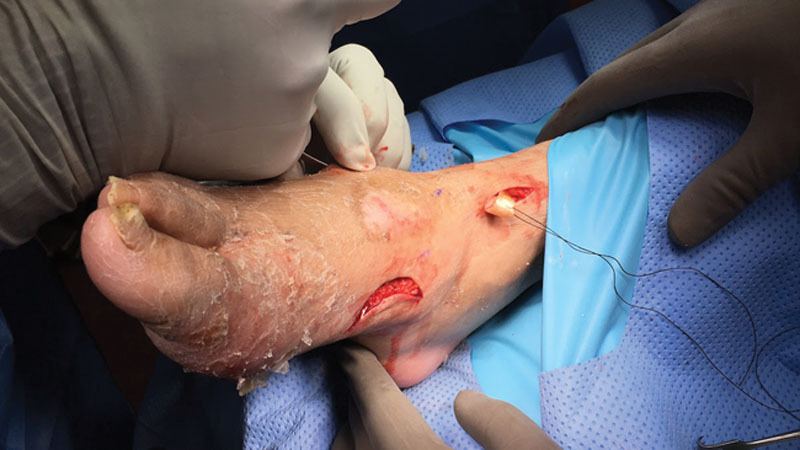
Fig. 2.
Wound after partial fifth ray amputation and tibialis anterior tendon transfer before healing.
The patient underwent an autologous fat pad transfer with lipofilling under the fifth metatarsal base and the first metatarsal head. Twenty-five milliliters of fat graft was harvested from the anterior abdomen and injected with multiple stab incisions into the plantar forefoot and plantar lateral mid-foot underlying areas of bulk loss of soft tissue (Fig. 3). The patient was placed into a posterior splint and instructed to remain nonweight bearing.
Fig. 3.
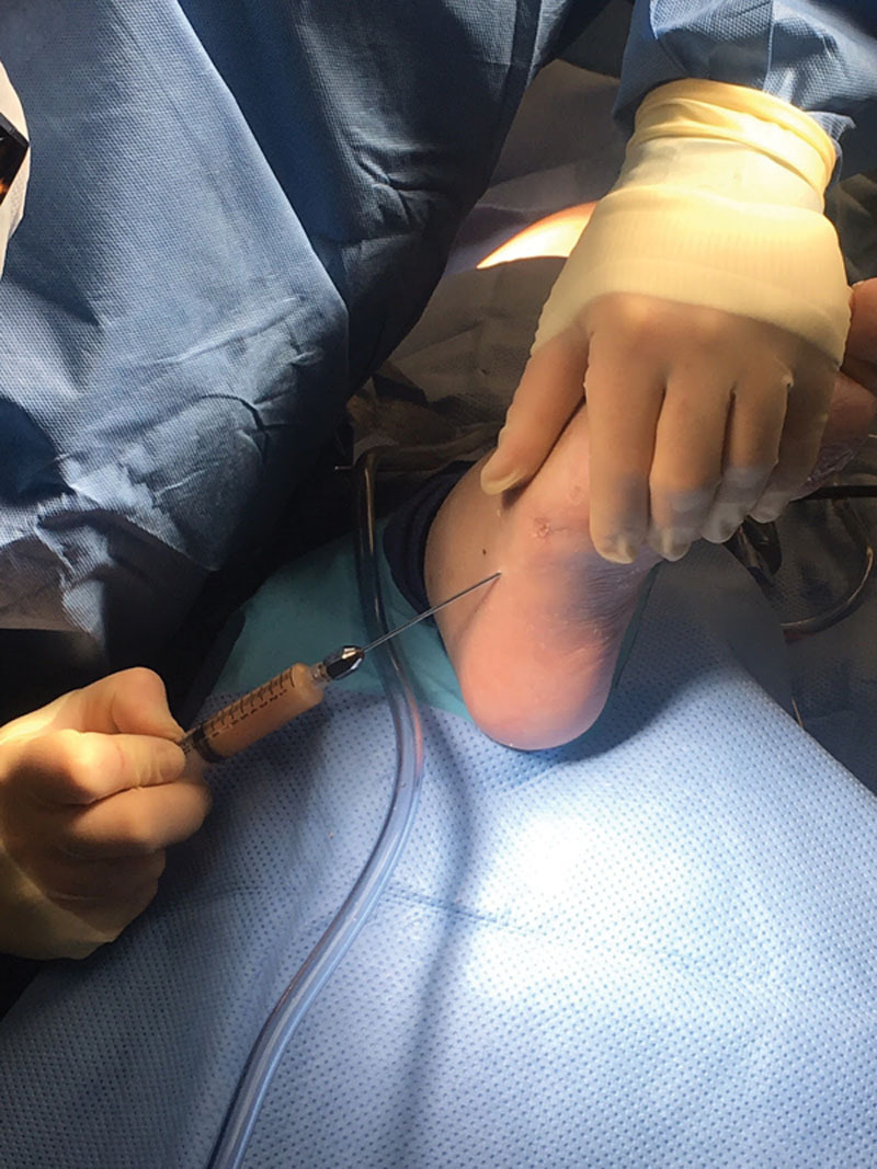
Twenty-five milliliters of fat graft was harvested from the anterior abdomen and delivered into the lateral midfoot and plantar forefoot in areas of soft-tissue loss.
He returned to the clinic 3 weeks after the procedure, and the wound remained healed, with visible take of the fat graft and supple soft tissue underlying the areas of injection without any complications. He was instructed to remain nonweight bearing in the splint for an additional week and to transition to weight bearing as tolerated in normal shoe gear.
At 6 weeks postoperatively, plantar fat pad thickness and the fat pad plantar to the fifth metatarsal base were found to be preserved on magnetic resonance imaging. Clinically, the plantar aspect of the foot continued to be supple without any new wounds or new hyperkeratosis (Fig. 4).
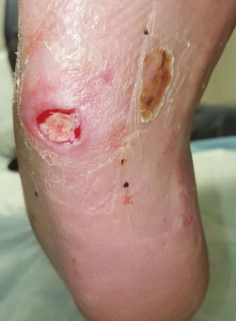
Fig. 4.
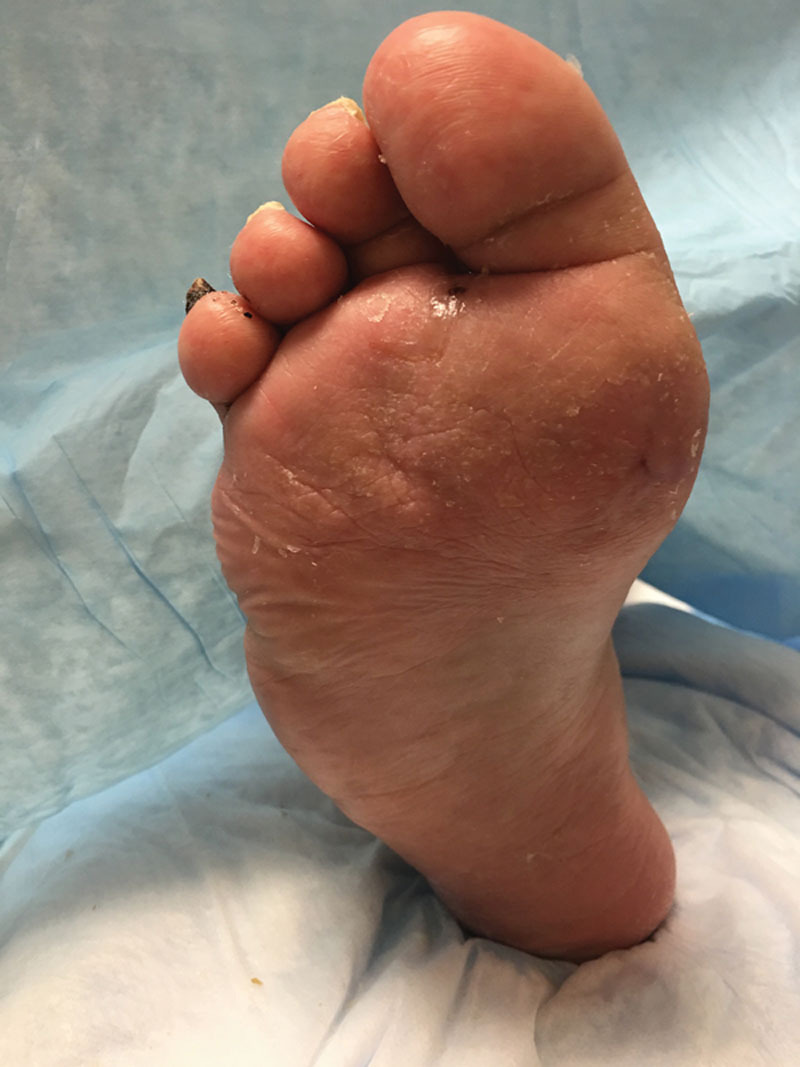
Six weeks after fat engraftment with good tissue quality.
DISCUSSION
Autologous fat grafting has been described as a methodology of tissue augmentation for over a century with varying degrees of success.9 Complications related to autologous fat grafting include absorption of fat; postoperative erythema, edema, and bleeding; fat necrosis; and infection although newer techniques and approaches have provided more predictable and reproducible results. Chairman6 transferred adipose from the calf, ankle, or abdomen in 50 patients to the plantar fat pad, and all patients reported less or no callus formation. Forty-eight of the patients felt more padding, and only 1 required a second procedure.10 However, fat grafting was performed concurrently with other procedures such as bunionectomies and metatarsal osteotomies.
Recently, a randomized controlled trial in the United States has been launched to evaluate the potential efficacy of lipofilling/fat grafting for plantar heel pain.10 Other than this study, we are aware of only 1 other report in the literature that has addressed the use of this technique in the high-risk foot. Stasch et al8 treated 26 patients with chronic open wounds (mean age, 16.7 months) and used surgical debridement along with lipofilling (mean amount, 7.1 mL) as a wound healing treatment. Although this novel treatment is similar in concept to that of ours, it has important differences. First, the surgeons’ goal was for the fat graft to serve as a trigger for wound healing, not protection from stress and extension of diabetic foot remission (ulcer-free days). Second, the amount used was significantly less than that in our technique (7 vs 25 mL). Third, a postoperative shoe was used by Stasch et al8 rather than a posterior splint for 5 days rather than 4 weeks. We think wound healing and “diabetic foot remission” care to be coequal partners in this area.2,7 For this reason, we find these indications synergistic. Future work to build on the Stasch study might include using more aggressive off-loading such as total contact casting or other irremovable devices. In that way, we think that their technique may serve 2 purposes—as a promoter of healing and possibly also as a bridge to remission. Although we think that additional fat grafting may be required after healing to better manage plantar surface normal and shear stresses, we also think that further study is required to allow for systematic refinement of this technique.
CONCLUSIONS
In summary, we present the initial evaluation of lipofilling and autologous fat grafting for the treatment of the diabetic foot in remission or as a bridge to remission. This appears to be a safe and potentially effective procedure. Complications associated with fat grafting, including fat absorption and necrosis, were not yet encountered. In the future, we hope to measure peak forefoot pressures and plantar fat pad thickness (through ultrasound), both before and after fat grafting. We look forward to future efforts that can further elucidate the efficacy and safety of this approach for broad clinical applicability in the management of the high-risk diabetic foot.
Footnotes
Disclosure: The authors have no financial interest to declare in relation to the content of this article. The Article Processing Charge was paid for by the authors.
REFERENCES
- 1.Barshes NR, Sigireddi M, Wrobel JS, et al. The system of care for the diabetic foot: objectives, outcomes, and opportunities. Diabet Foot Ankle. 2013;4 doi: 10.3402/dfa.v4i0.21847. [DOI] [PMC free article] [PubMed] [Google Scholar]
- 2.Armstrong DG, Mills JL. Toward a change in syntax in diabetic foot care: prevention equals remission. J Am Podiatr Med Assoc. 2013;103:161–162. doi: 10.7547/1030161. [DOI] [PubMed] [Google Scholar]
- 3.Miller JD, Salloum M, Button A, et al. How can i maintain my patient with diabetes and history of foot ulcer in remission? Int J Low Extrem Wounds. 2014;13:371–377. doi: 10.1177/1534734614545874. [DOI] [PubMed] [Google Scholar]
- 4.Bowling FL, Metcalfe SA, Wu S, et al. Liquid silicone to mitigate plantar pedal pressure: a literature review. J Diabetes Sci Technol. 2010;4:846–852. doi: 10.1177/193229681000400412. [DOI] [PMC free article] [PubMed] [Google Scholar]
- 5.Lam SM, Glasgold RA, Glasgold MJ. Fat harvesting techniques for facial fat transfer. Facial Plast Surg. 2010;26:356–361. doi: 10.1055/s-0030-1265016. [DOI] [PubMed] [Google Scholar]
- 6.Chairman EL. Restoration of the plantar fat pad with autolipotransplantation. J Foot Ankle Surg. 1994;33:373–379. [PubMed] [Google Scholar]
- 7.Nicoletti G, Brenta F, Jaber O, et al. Lipofilling for functional reconstruction of the sole of the foot. Foot (Edinb) 2014;24:21–27. doi: 10.1016/j.foot.2014.02.003. [DOI] [PubMed] [Google Scholar]
- 8.Stasch T, Hoehne J, Huynh T, et al. Debridement and Autologous Lipotransfer for Chronic Ulceration of the Diabetic Foot and Lower Limb improves Wound Healing (the DEALT Method). Plast Reconstr Surg. 2015;136:1357–1366. doi: 10.1097/PRS.0000000000001819. [DOI] [PubMed] [Google Scholar]
- 9.Godse K, Abhyankar S, Patil S, et al. Fat ful′fill′ment: A review of autologous fat grafting. J Cutan Aesthet Surg. 2013;6:132. doi: 10.4103/0974-2077.118402. [DOI] [PMC free article] [PubMed] [Google Scholar]
- 10.Gusenoff JA. Fat Grafting and Retention for Heel Fat Pad Atrophy. Available at: https://clinicaltrials.gov/ct2/show/NCT02465333View-ClinicalTrials.gov. Accessed February 26, 2016. [Google Scholar]