Abstract
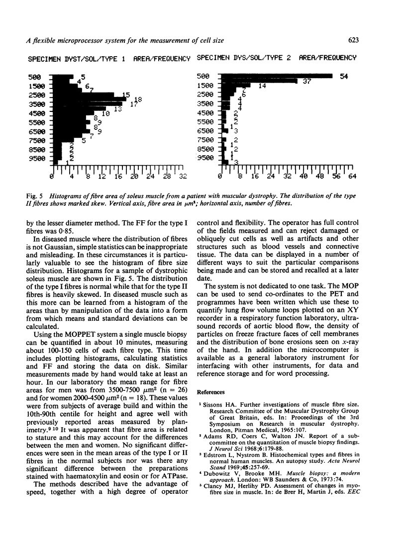
A flexible system for the measurement of length and area is described. The system consists of the Reichert Jung MOP-1 area measuring device interfaced with a Commodore PET computer. Its use is illustrated by the planimetric measurement of cross sectional areas in histochemical preparations of normal and diseased muscle. While measurements are being made data can be displayed on the computer screen either in numerical form or as a frequency histogram together with simple statistical analyses. Hard copy can be obtained from an attached printer. Mean values for fibre area in normal human skeletal muscle are reported. An alternative, widely used method of calculating fibre area from the lesser diameter was found to give a consistent underestimate of approximately 30% when compared with our planimetric method. In diseased muscle with abnormally shaped fibres the discrepancy is even larger; such fibres can be identified using a "form factor" which relates the area of a cell to its perimeter. This rapid, accurate and flexible system is also suitable for the measurement of many different types of graphical record.
Full text
PDF
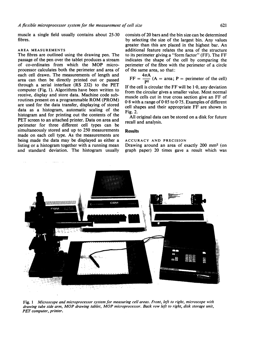
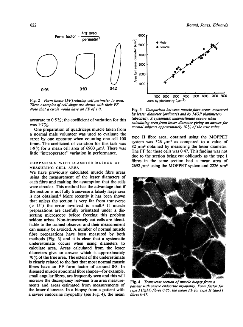
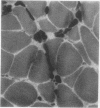
Images in this article
Selected References
These references are in PubMed. This may not be the complete list of references from this article.
- Edström L., Nyström B. Histochemical types and sizes of fibres in normal human muscles. A biopsy study. Acta Neurol Scand. 1969;45(3):257–269. doi: 10.1111/j.1600-0404.1969.tb01238.x. [DOI] [PubMed] [Google Scholar]
- Edwards R., Young A., Wiles M. Needle biopsy of skeletal muscle in the diagnosis of myopathy and the clinical study of muscle function and repair. N Engl J Med. 1980 Jan 31;302(5):261–271. doi: 10.1056/NEJM198001313020504. [DOI] [PubMed] [Google Scholar]
- Gollnick P. D., Armstrong R. B., Saltin B., Saubert C. W., 4th, Sembrowich W. L., Shepherd R. E. Effect of training on enzyme activity and fiber composition of human skeletal muscle. J Appl Physiol. 1973 Jan;34(1):107–111. doi: 10.1152/jappl.1973.34.1.107. [DOI] [PubMed] [Google Scholar]
- Report of a sub-committee on quantitation of muscle biopsy findings. Appendix B to the Minutes of the Meeting of the Research Group on Neuromuscular Diseases, held in Montreal, Canada, on 21 September, 1967. J Neurol Sci. 1968 Jan-Feb;6(1):179–188. [PubMed] [Google Scholar]
- Round J. M., Matthews Y., Jones D. A. A quick, simple and reliable histochemical method for ATPase in human muscle preparations. Histochem J. 1980 Nov;12(6):707–710. doi: 10.1007/BF01012026. [DOI] [PubMed] [Google Scholar]


