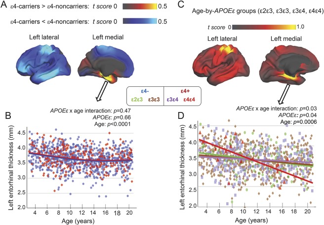
Figure 4. Cortical thickness differences between ε4 carriers and non–ε4 carriers.
These analyses were performed using the same grouping as prior studies.5 (A) On the vertex-based analyses (covaried for age, sex, scanner device, socioeconomic status, and genetic ancestry factor), compared to ε4 noncarriers (668 ε3ε3+ and 124 ε2ε3), ε4 carriers (235 ε3ε4+ and 21 ε4ε4) showed nonsignificantly thicker right precuneus (data not shown) and left entorhinal cortices (red-yellow regions), and nonsignificantly thinner left postcentral (parietal) and left lateral temporal cortices (blue areas). (B) Region-of-interest analysis of the left entorhinal region verified the nonsignificantly thicker cortex in ε4 carriers (red dots) than non–ε4 carriers (blue dots). (C) The 4 genotype groups showed a trend for group differences (p = 0.13) on age-dependent changes in cortical thickness in the left entorhinal and parahippocampal regions as well as the left postcentral cortex (yellow regions). (D) Region-of-interest analyses verified the age-dependent group differences; however, the ε4ε4 children (red dots) showed steepest age-dependent thinning in this brain region, with thicker cortices in the younger children but thinner cortices in the older children (>11 years).

