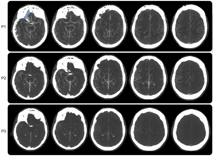
Figure 1. Case study 1.
Axial multiphase CT angiography showing 3 phases as P1 (late arterial phase), P2 (midvenous phase), and P3 (late-venous phase). P1 images are degraded by motion artifacts. There is evidence of a right MCA mid-M1 loss of contrast opacification, indicating an occlusion (blue arrow). P2 and P3 show asymmetry in pial arteries in the right MCA distribution with delayed washout of the contrast, confirming the vascular occlusion despite motion artifact. MCA = middle cerebral artery.

