Abstract
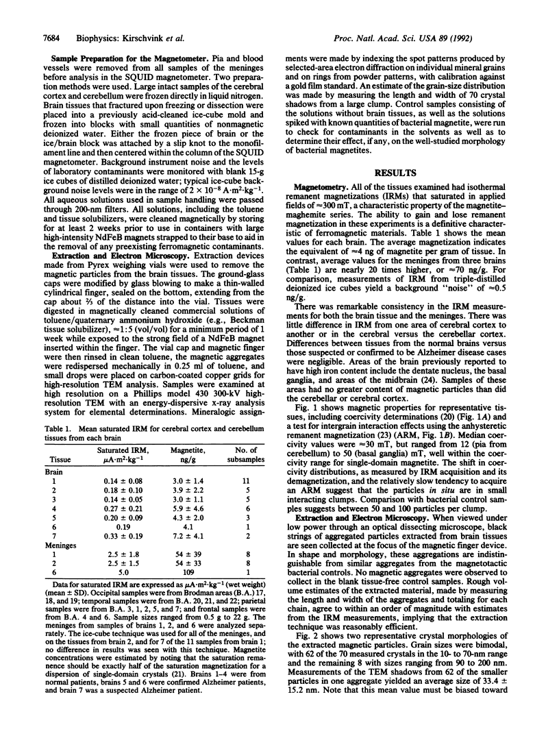
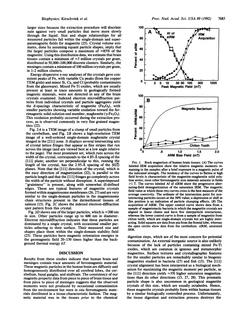
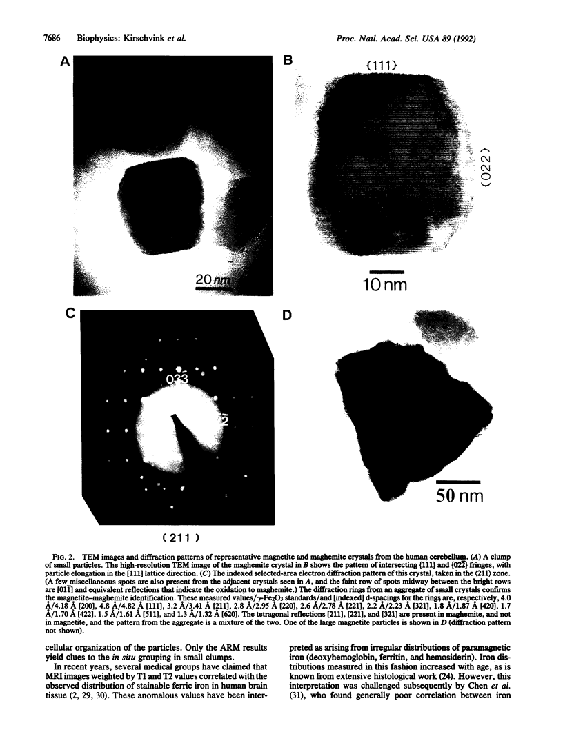
Although the mineral magnetite (Fe3O4) is precipitated biochemically by bacteria, protists, and a variety of animals, it has not been documented previously in human tissue. Using an ultrasensitive superconducting magnetometer in a clean-lab environment, we have detected the presence of ferromagnetic material in a variety of tissues from the human brain. Magnetic particle extracts from solubilized brain tissues examined with high-resolution transmission electron microscopy, electron diffraction, and elemental analyses identify minerals in the magnetite-maghemite family, with many of the crystal morphologies and structures resembling strongly those precipitated by magnetotactic bacteria and fish. These magnetic and high-resolution transmission electron microscopy measurements imply the presence of a minimum of 5 million single-domain crystals per gram for most tissues in the brain and greater than 100 million crystals per gram for pia and dura. Magnetic property data indicate the crystals are in clumps of between 50 and 100 particles. Biogenic magnetite in the human brain may account for high-field saturation effects observed in the T1 and T2 values of magnetic resonance imaging and, perhaps, for a variety of biological effects of low-frequency magnetic fields.
Full text
PDF
Images in this article
Selected References
These references are in PubMed. This may not be the complete list of references from this article.
- Adair RK. Constraints on biological effects of weak extremely-low-frequency electromagnetic fields. Phys Rev A. 1991 Jan 15;43(2):1039–1048. doi: 10.1103/physreva.43.1039. [DOI] [PubMed] [Google Scholar]
- Baker R. R., Mather J. G., Kennaugh J. H. Magnetic bones in human sinuses. Nature. 1983 Jan 6;301(5895):79–80. doi: 10.1038/301096b0. [DOI] [PubMed] [Google Scholar]
- Bizzi A., Brooks R. A., Brunetti A., Hill J. M., Alger J. R., Miletich R. S., Francavilla T. L., Di Chiro G. Role of iron and ferritin in MR imaging of the brain: a study in primates at different field strengths. Radiology. 1990 Oct;177(1):59–65. doi: 10.1148/radiology.177.1.2399339. [DOI] [PubMed] [Google Scholar]
- Brittenham G. M., Farrell D. E., Harris J. W., Feldman E. S., Danish E. H., Muir W. A., Tripp J. H., Bellon E. M. Magnetic-susceptibility measurement of human iron stores. N Engl J Med. 1982 Dec 30;307(27):1671–1675. doi: 10.1056/NEJM198212303072703. [DOI] [PubMed] [Google Scholar]
- Chen J. C., Hardy P. A., Clauberg M., Joshi J. G., Parravano J., Deck J. H., Henkelman R. M., Becker L. E., Kucharczyk W. T2 values in the human brain: comparison with quantitative assays of iron and ferritin. Radiology. 1989 Nov;173(2):521–526. doi: 10.1148/radiology.173.2.2798884. [DOI] [PubMed] [Google Scholar]
- Drayer B. P., Olanow W., Burger P., Johnson G. A., Herfkens R., Riederer S. Parkinson plus syndrome: diagnosis using high field MR imaging of brain iron. Radiology. 1986 May;159(2):493–498. doi: 10.1148/radiology.159.2.3961182. [DOI] [PubMed] [Google Scholar]
- Frankel R. B., Blakemore R. P., Wolfe R. S. Magnetite in freshwater magnetotactic bacteria. Science. 1979 Mar 30;203(4387):1355–1356. doi: 10.1126/science.203.4387.1355. [DOI] [PubMed] [Google Scholar]
- Gomori J. M., Grossman R. I., Goldberg H. I., Zimmerman R. A., Bilaniuk L. T. Intracranial hematomas: imaging by high-field MR. Radiology. 1985 Oct;157(1):87–93. doi: 10.1148/radiology.157.1.4034983. [DOI] [PubMed] [Google Scholar]
- Gorby Y. A., Beveridge T. J., Blakemore R. P. Characterization of the bacterial magnetosome membrane. J Bacteriol. 1988 Feb;170(2):834–841. doi: 10.1128/jb.170.2.834-841.1988. [DOI] [PMC free article] [PubMed] [Google Scholar]
- Gould J. L., Kirschvink J. L., Deffeyes K. S. Bees have magnetic remanence. Science. 1978 Sep 15;201(4360):1026–1028. doi: 10.1126/science.201.4360.1026. [DOI] [PubMed] [Google Scholar]
- HALLGREN B., SOURANDER P. The effect of age on the non-haemin iron in the human brain. J Neurochem. 1958 Oct;3(1):41–51. doi: 10.1111/j.1471-4159.1958.tb12607.x. [DOI] [PubMed] [Google Scholar]
- Kirschvink J. L. Ferromagnetic crystals (magnetite?) in human tissue. J Exp Biol. 1981 Jun;92:333–335. doi: 10.1242/jeb.92.1.333. [DOI] [PubMed] [Google Scholar]
- London S. J., Thomas D. C., Bowman J. D., Sobel E., Cheng T. C., Peters J. M. Exposure to residential electric and magnetic fields and risk of childhood leukemia. Am J Epidemiol. 1991 Nov 1;134(9):923–937. doi: 10.1093/oxfordjournals.aje.a116176. [DOI] [PubMed] [Google Scholar]
- Mann S., Sparks N. H., Walker M. M., Kirschvink J. L. Ultrastructure, morphology and organization of biogenic magnetite from sockeye salmon, Oncorhynchus nerka: implications for magnetoreception. J Exp Biol. 1988 Nov;140:35–49. doi: 10.1242/jeb.140.1.35. [DOI] [PubMed] [Google Scholar]
- Pool R. Electromagnetic fields: the biological evidence. Science. 1990 Sep 21;249(4975):1378–1381. doi: 10.1126/science.2402634. [DOI] [PubMed] [Google Scholar]
- Savitz D. A., Wachtel H., Barnes F. A., John E. M., Tvrdik J. G. Case-control study of childhood cancer and exposure to 60-Hz magnetic fields. Am J Epidemiol. 1988 Jul;128(1):21–38. doi: 10.1093/oxfordjournals.aje.a114943. [DOI] [PubMed] [Google Scholar]
- Walcott C., Green R. P. Orientation of homing pigeons altered by a change in the direction of an applied magnetic field. Science. 1974 Apr 12;184(4133):180–182. doi: 10.1126/science.184.4133.180. [DOI] [PubMed] [Google Scholar]
- de Araujo F. F., Pires M. A., Frankel R. B., Bicudo C. E. Magnetite and magnetotaxis in algae. Biophys J. 1986 Aug;50(2):375–378. doi: 10.1016/S0006-3495(86)83471-3. [DOI] [PMC free article] [PubMed] [Google Scholar]


