Abstract
We report the case of an 84-year-old woman who came to our attention with right palpebral edema associated with pain in the omolateral fronto-orbital region. The patient underwent an MRI scan that revealed a rounded, extracerebral intradiploic cystic lesion with dyshomogeneous signal intensity. Computed tomography (CT) imaging was also performed with reformatted 3D reconstruction. Post-surgical histologic analysis confirmed the diagnosis of intradiploic dermoid cyst. We here report the case and discuss epidemiology, imaging features and work-up of this pathological entity.
Keywords: Extracerebral dermoid cyst, intradiploic cyst, dermoid cyst, skull
Introduction
Craniofacial dermoid cysts represent about 7% of all dermoids; common locations include the periorbital, nasal, scalp and postauricular region.1 They are typically present during infancy as non-tender, subcutaneous masses along embryonal skin fusion lines; on the contrary, epidermoid cysts are usually diagnosed in adulthood. The differential diagnosis of circumscribed lytic defects of the skull includes benign cysts as well as neoplasms.2 The definitive diagnosis is made only by histopathology.
We here present a case of an elderly patient with an extracerebral intradiploic lytic lesion in the right fronto-orbital region studied with computed tomography (CT) and magnetic resonance (MR) imaging that turned out to be a pure dermoid cyst.
Case report
An 84-year-old woman came to our department with a six-month history of progressively worsening right palpebral edema associated with pain in the omolateral orbital and fronto-temporal region, exacerbated by palpation.
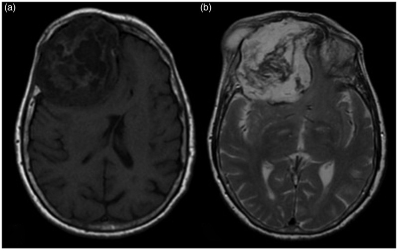
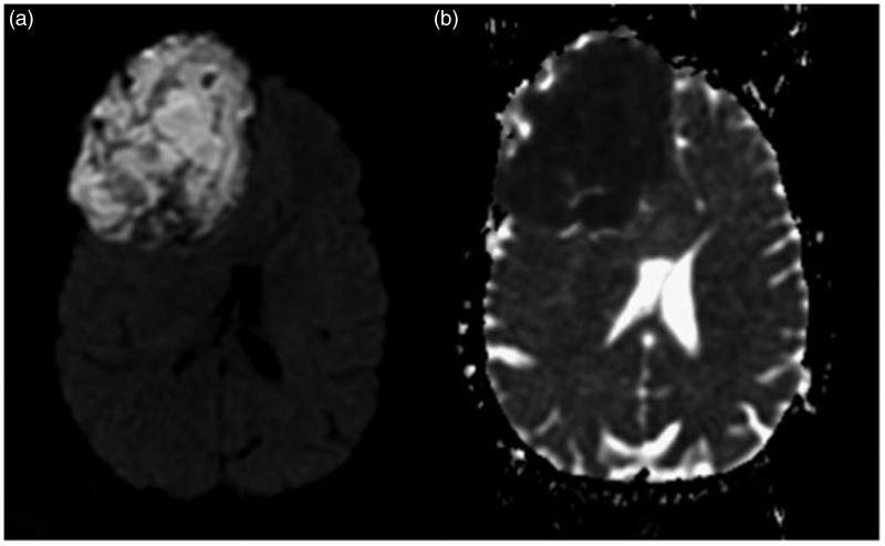
Given the symptomatology, the patient underwent MRI examination that showed the presence of an extracerebral, intradiploic lesion, measuring about 7 cm in diameter, with rounded shape and irregular margins. The lesion determined deformation and swelling of the frontal bone, with impression and posterior dislocation of the frontal lobe (Figure 1). There was also reshaping of the orbital medial wall and the orbital roof, which appeared to be interrupted in its antero-lateral portion, with leakage of the neoplastic material in the subcutaneous eyebrow region (Figure 2). The mass also invaded the frontal sinus, extending to the controlateral side and determining deviation of middle line structures of about 10 mm. The lesion showed dyshomogeneous signal intensity, from hypo- to hyperintensity on T1- and T2-weighted sequences. It demonstrated high signal intensity on diffusion-weighted imaging (DWI) and related restriction on an apparent diffusion coefficient (ADC) map (Figure 3).
Figure 1.
Axial (a) T1- and (b) T2-weighted images showing dyshomogeneous signal intensity of the lesion. Note the deformation of the frontal bone, with impression and posterior dislocation of the frontal lobe (a). The mass invaded also the frontal sinus, extending to the controlateral side (b).
Figure 2.
(a) Axial diffusion-weighted imaging (DWI) and (b) apparent diffusion coefficient (ADC) images. The lesion shows high signal intensity on DWI and related restriction on an ADC map. DWI is a valuable imaging sequence in diagnosing epidermoid cysts, since they show restricted diffusion with higher signal intensity than that of cerebrospinal fluid on DWI.
Figure 3.
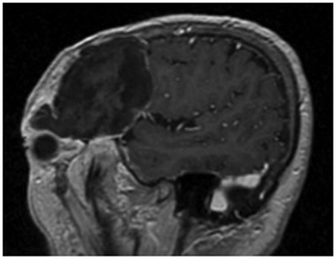
Sagittal image after intravenous gadolinium administration. The lesion showed slight peripheral enhancement, without involvement of the dura or the brain parenchyma.
After intravenous contrast medium administration (gadolinium, 0.1 mml/kg), the lesion showed slight peripheral enhancement, without involvement of the dura or the brain parenchyma (Figure 4).
Figure 4.
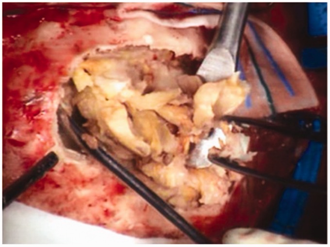
Intraoperative findings (courtesy of RJ Galzio, MD, Department of Neurosurgery, University of L’Aquila, Italy).
The MRI features suggested either dermoid cyst or mucocele.
At CT scan the lesion appeared as a well-defined, lobulated, heterogeneous, hypodense mass with intralesional calcifications.
Three days later the patient underwent neurosurgical removal of the mass to avoid further complications. Intraoperatively, the mass was well circumscribed and contained sebaceous and keratin-like material.
Histopathology examination revealed numerous keratinized squamous cells, confirming the diagnosis of dermoid cyst.
Discussion
Dermoid cysts are rare, benign, congenital ectodermal inclusion cysts comprising skin supplements surrounded by squamous epithelium in the craniofacial region; dermoid cysts are extremely rare (about 0.04–0.7% of cranial tumors) and are most commonly seen in frontonasal-sphenoidal suture lines and the periorbital region in early life.3 Nasal dermoids are a special variety of dermoids associated with the dermal sinus tract into the intracranial cavity. Orbital dermoids frequently have an intracranial extension. Extension into the frontal sinus has also been reported in the literature.4
Because of the very slow growth of the mass, the onset of signs and symptoms is often late, over a period of months to years. Typical symptoms of presentation include headaches due to erosion of the calvarium, and seizures due to local pressure. These lesions usually present as painless bony swellings under the scalp.5 The cysts may perforate the dura, rupture into the subarachnoid space resulting in chemical meningitis, or involve the brain parenchyma.
Imaging play an important role in the diagnosis; on CT scans, performed with high-performance equipment to detect bone involvement,6,7 they appear as hypodense lesions, while MR images reveal inhomogeneous T1 signal hypointensity and inhomogeneous T2-fluid-attenuated inversion recovery (FLAIR) signal hyperintensity. Dermoid cysts do not enhance after intravenous contrast medium administration; when present, contrast enhancement is minimal and peripheral.8 DWI is a valuable imaging sequence in diagnosing epidermoid cysts, since they show restricted diffusion with higher signal intensity than that of cerebrospinal fluid.
Differential diagnoses may include sebaceous cyst, dermoid cyst, hematoma with fibrosis, lipoma, and fibroma.9
Microscopically, the wall of the cystic tumor is lined by squamous epithelium and the cyst is filled with lamellated keratin content.
Complete surgical resection with careful follow-up is the treatment of choice.10
Conclusion
Few cases of pure intradiploic dermoids have been reported in the literature, mostly occurring in young individuals.11,12 Our case is of interest because the patient was an elderly female with an isolated intradiploic lesion, with no intracranial extension, fistulous tract, or orbital nasal extension despite the large size of the mass. Complete resection, which has demonstrated keratinized squamous cells in the absence of other tissue, represents another particular finding of our case. Bone reconstruction is the only treatment needed for these lesions. Our case illustrates that an intradiploic dermoid must be kept in differential diagnosis of a slow-growing, calvarial mass in an elderly person and that MR study, in particular DWI/ADC sequences, can be helpful for the correct diagnosis.
Acknowledgments
All authors contributed equally to this manuscript. Prof Splendiani is principal investigator. The other authors contributed equally to the drafting/revising of the manuscript for content, including medical writing for content, study concept or design and analysis or interpretation of the data.
Funding
This research received no specific grant from any funding agency in the public, commercial, or not-for-profit sectors.
Conflict of interest
The author(s) declared no potential conflicts of interest with respect to the research, authorship, and/or publication of this article.
References
- 1.Demir MK, Yapicier O, Onat E, et al. Rare and challenging extra-axial brain lesions: CT and MRI findings with clinico-radiological differential diagnosis and pathological correlation. Diagn Interv Radiol 2014; 20: 448–452. [DOI] [PMC free article] [PubMed] [Google Scholar]
- 2.Durmaz A, Yildizoğlu Ü, Polat B, et al. A middle cranial fossa dermoid cyst treated by an endonasal endoscopic approach. J Craniofac Surg 2015; 26: e333–e335. [DOI] [PubMed] [Google Scholar]
- 3.Nakamoto H, Kawamoto T, Suzuki S, et al. Intradiploic ciliated epithelial inclusion cyst of the skull. Neurol Med Chir (Tokyo) 2013; 53: 270–272. [DOI] [PubMed] [Google Scholar]
- 4.Pryor SG, Lewis JE, Weaver AL, et al. Pediatric dermoid cysts of the head and neck. Otolaryngol Head Neck Surg 2005; 132: 938–942. [DOI] [PubMed] [Google Scholar]
- 5.Reissis D, Pfaff MJ, Patel A, et al. Craniofacial dermoid cysts: Histological analysis and inter-site comparison. Yale J Biol Med 2014; 87: 349–357. [PMC free article] [PubMed] [Google Scholar]
- 6.D’Orazio F, Splendiani A, Gallucci M. 320-Row detector dynamic 4D-CTA for the assessment of brain and spinal cord vascular shunting malformations. A technical note. Neuroradiol J 2014; 27: 710–717. [DOI] [PMC free article] [PubMed] [Google Scholar]
- 7.Di Cesare E, Gennarelli A, Di Sibio A, et al. Assessment of dose exposure and image quality in coronary angiography performed by 640-slice CT: A comparison between adaptive iterative and filtered back-projection algorithm by propensity analysis. Radiol Med 2014; 119: 642–649. [DOI] [PubMed] [Google Scholar]
- 8.Splendiani A, Puglielli E, De Amicis R, et al. Contrast-enhanced FLAIR in the early diagnosis of infectious meningitis. Neuroradiology 2005; 47: 591–598. [DOI] [PubMed] [Google Scholar]
- 9.Patnaik A, Mishra SS, Das S, et al. Giant intradiploic dermoid cyst of the frontal bone with involvement of frontal sinus in an elderly patient. Neurol India 2012; 60: 542–543. [DOI] [PubMed] [Google Scholar]
- 10.Bahloul K, Dhouib M, Chaari I, et al. Nasal dermoid cyst with intracranial extension: Which approach? [article in French]. Neurochirurgie 2011; 57: 125–128. [DOI] [PubMed] [Google Scholar]
- 11.Gulsen S, Yilmaz C, Serhat C, et al. Ruptured intradiploic dermoid cyst overlying the torcular herophili. Neurol Neurochir Pol 2010; 44: 308–313. [DOI] [PubMed] [Google Scholar]
- 12.De Moraes Júnior LC, Wanderley EC, Montini A, et al. Intradiploic dermoid cyst of the skull: Report of a case. [article in Portuguese]. Arq Neuropsiquiatr 1984; 42: 68–71. [PubMed] [Google Scholar]


