Abstract
Non-thermal plasma is increasingly being recognized for a wide range of medical and biological applications. However, the effect of non-thermal plasma on physiological functions is not well characterized in in vivo model systems. Here we use a genetically amenable, widely used model system, Drosophila melanogaster, to develop an in vivo system, and investigate the role of non-thermal plasma in blood cell differentiation. Although the blood system in Drosophila is primitive, it is an efficient system with three types of hemocytes, functioning during different developmental stages and environmental stimuli. Blood cell differentiation in Drosophila plays an essential role in tissue modeling during embryogenesis, morphogenesis and also in innate immunity. In this study, we optimized distance and frequency for a direct non-thermal plasma application, and standardized doses to treat larvae and adult flies so that there is no effect on the viability, fertility or locomotion of the organism. We discovered that at optimal distance, time and frequency, application of plasma induced blood cell differentiation in the Drosophila larval lymph gland. We articulate that the augmented differentiation could be due to an increase in the levels of reactive oxygen species (ROS) upon non-thermal plasma application. Our studies open avenues to use Drosophila as a model system in plasma medicine to study various genetic disorders and biological processes where non-thermal plasma has a possible therapeutic application.
Introduction
Non-thermal plasma systems have been emerging as useful tools for various clinical applications [1]. Plasma is known to catalyze biochemical activities when applied on tissue and is able to regulate cellular processes such as proliferation, differentiation, and apoptosis [1]. This, in part, is due to the reactive oxygen and nitrogen species (ROS and RNS) generated by application of non-thermal plasma [2]. Most of the plasma research has been in vitro or ex vivo, which has led to investigation of potential applications such as disinfection of surfaces [3], promotion of hemostasis [4], enhancement of tissue regeneration [5], acceleration of wound healing [6], and for anti-cancer therapy [7] [8]. However, there has not been an extensive characterization of non-thermal plasma in in vivo model organisms. In order to validate the biological effect of non-thermal plasma on in vivo systems, we used Drosophila melanogaster as a model for our study.
Drosophila provides a genetically amenable and well-established system that has been used over the past 100 years [9]. It has a short life span and is easy to maintain, making the studies rapid, efficient, and reproducible. It carries homologs for several disease-related genes, and conserved signaling mechanisms, and allows extensive genetic manipulations [10]. Time- and tissue-specific gene knockdowns or gene overexpression, clonal systems provide a better system for studying the effect of plasma on disease models especially oncogenic pathways [11,12]. Moreover, with the ease of fly husbandry and available genetic technology, it is possible to carry out large scale studies involving non-thermal plasma. There is a need to validate various effects of non-thermal plasma that have been observed in in vitro and ex vivo systems, and to establish the safety of non-thermal plasma as a feasible therapeutic option in an in vivo system. Hence, Drosophila will make an excellent candidate for investigating effects of non-thermal plasma application in an in vivo model.
In this study, we standardized application distance, treatment time, and an appropriate dose of floating electrode microsecond-pulsed dielectric barrier discharge (mspDBD) non-thermal plasma for treatment of Drosophila (both larvae and adults). Furthermore, we studied its effect on various parameters such as life span, survivability, fecundity, movement etc. We discovered that treatment at 50 Hz for 10 seconds was most efficacious in stimulating an effect without changing the life cycle or affecting the factors mentioned above. We also explored a physiological system in Drosophila modulated by ROS, i.e. hematopoietic system, and show that application of plasma can induce blood cell differentiation by increasing ROS levels in the glands.
Materials and Methods
All flies used were either Canton-S or Hemolectin Gal4 GFP (obtained from Bloomington stock center).
A. Non-thermal plasma treatment
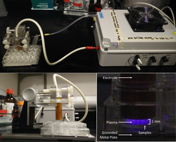
A mspDBD plasma was utilized for this study [13]. Plasma was generated by applying an alternating polarity pulse to a high-voltage, quartz-insulated electrode, 2 mm above the samples undergoing treatment “Fig 1”. Samples were placed into 24-well plates on top of a grounded metal base. The plasma pulse characteristics generated from the power supply are as follows: 30 kV amplitude pulse with rise times of 5 V/ns, pulse widths of 1.65 μs, and variable frequencies [13]. The energy per pulse of a single mspDBD discharge was established following the methods described in the previous published work [14]. Briefly, energy per pulse was calculated by measuring voltage and current, without displacement current, from a single discharge. This was found to be 0.5 mJ/pulse/cm2. The plasma treatment dose and total energy delivered to the cells during treatment, was calculated by multiplying the energy per pulse by the plasma treatment duration and frequency. All samples were treated for 10 seconds and the frequencies of pulses were 50, 850, and 1000 Hz, which corresponds to 0.012, 0.02, and 0.24 J per fly, respectively.
Fig 1. Experimental setup for plasma treatment of Drosophila.
(Top) High voltage electrode is connected to the mspDBD power supply and metal base of the z-positioner was grounded. (Bottom left) Z-positioner was used to position electrode 2 mm above the treated samples in the 24-well plate. (Bottom right) mspDBD discharge in a 24-well plate.
B. Survivability
D. melanogaster were treated as adults (day 1) and larvae (early second instar) for multiple lengths of time as well as at various frequencies (50, 830 and 1000 Hz) to determine the optimal frequency and time period for treatment in the experiment. A total of 30 adults were treated for each time period of 5, 10, 15, 20, 25, 30, 35, 40, 45, 50 seconds and then the number of survivors were recorded after 3 days. Media was changed after every three days to record their life span.
Freshly hatched flies (males and females) were treated with plasma for 10 seconds and allowed to recover for 3 days in separate vials. One female and one male group (n = 5 groups) were mated in a vial and fecundity was tested at 3 different time points.
A negative geotaxis assay was performed, 2 days after treatment, by counting the number of flies that cross 5 cm mark in 18 seconds after tapping them to the bottom of the vial (5 flies each tested for five times).
Larvae were treated as second day instars. To ensure they were all at the same age, four plates were set up with plastic cages that allowed the flies to lay eggs for four hours. After 48 hours larvae were treated with plasma for the time periods of 5, 10, 15, 20, 25, 30, 45 and 50 seconds.
C. Histochemistry
Two days after mspDBD plasma treatment, larvae were dissected and stained with DAPI (Sigma). Lymph glands were dissected in phosphate buffered saline (PBS) and then fixed for 20 minutes in 4% (w/v) paraformaldehyde (PFA). Samples were moved to PBS for 10 minutes and then washed in 0.1% (v/v) Triton-X 100 in PBS (PBS-T). Samples were incubated for 10 minutes in DAPI at 1:2000 dilutions in PBS. Tissues were washed in PBS-T for 10 minutes, and then mounted on slides with Mowiol (Sigma).
D. ROS staining
Lymph glands were dissected quickly and placed in 6 μM dihydroethidium (DHE, Molecular probes) in PBS for 3 minutes and then washed in PBS three times for 3 minutes each. The samples were the fixed in 4% (w/v) PFA for 3 minutes and then washed again in PBS twice for 2 minutes each time. The lymph gland were then mounted in PBS and immediately photographed under a Zeiss confocal microscope. ROS levels were quantified by measuring integrated intensities [15] from treated and untreated lymph glands.
E. ROS measurements
Larvae were homogenized using a pestle, and ROS was detected by amplex red using a fluorescence spectrophotometer (Hitachi F-2710) described previously [16,17]. Briefly, 5 μg horseradish peroxidase (Sigma-Aldrich) was added to the ROS buffer [mmoles/L, 20 Tris-HCl, 250 sucrose, 1 EGTA-Na2, 1 EDTA-Na2, pH 7.4 at 27°C] and the baseline fluorescence was measured (excitation at 560 nm and emission at 590 nm) for 30 minutes. Fluorescence was monitored continuously for 45 min at 5 s resolution and the rate of ROS production was calculated.
Results
A. Standardization of mspDBD plasma treatment in Drosophila
In order to standardize a dose to treat Drosophila with non-thermal plasma, we selected three frequencies (50 Hz, 830 Hz and 1 kHz) with varying time durations, using mspDBD “Fig 1”. We found that 1 kHz treatment immediately killed the flies. A 10 second treatment at 830 Hz made the flies partially immobile even after two days of recovery time. We observed that 50 Hz treatment did not affect the natural movement of flies, and hence decided to apply 50 Hz for increasing time durations to optimize time of exposure.
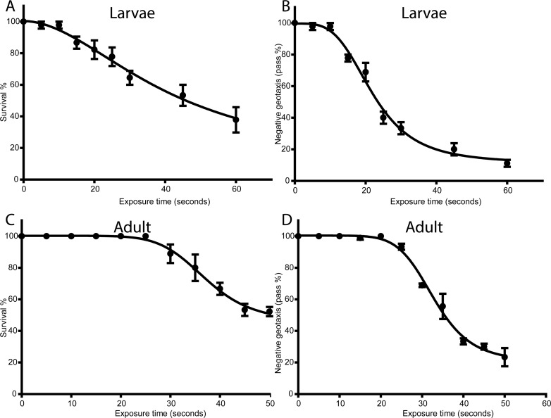
We treated adult flies and larvae (30 each in five independent experiments) with mspDBD plasma at 50 Hz, for 0, 5, 10, 15, 20, 25, 30, 35, 40, 45 and 50 seconds, respectively. After exposure, adult flies and larvae were immediately transferred and cultured in fly-media. Larvae were let to eclose into adults to carry out tests for determining the effect of plasma. Treated adult flies were also maintained in the media for two days before carrying out further tests. We found that a treatment for 10 seconds was appropriate at which both treated larvae eclosed into adults and treated flies survived similar to their wild type counterparts “Fig 2A and 2C”.
Fig 2. Standardization of non-thermal plasma doses in Drosophila.
A and C: Graphs represent percentage of treated larvae or adults (A and C, respectively) survived at different time of 50 Hz treatment. B and D: Graphs represent negative geotaxis assay performed after 50Hz treatment of larvae after turning into adults and adults after 2 days after treatment.
B. Physiology of mspDBD plasma treated Drosophila
After flies were recovered and cultured on fly media, they were tested for their physiological functions. Negative geotaxis was recorded for 15 flies (5 independent experiments) at room temperature to study their locomotory movements. “Fig 2B and 2D” shows negative geotaxis for flies (flies treated directly as well as treated larvae that eclosed into adults) after application of 50 Hz non-thermal plasma for different durations. The difference between treated and untreated groups of flies was calculated with student’s t-test. We found that application of non-thermal plasma for 10 seconds had no effect on negative geotaxis of flies.
Egg laying is a classical marker of physiological health of flies. In our fecundity test with flies treated for 10 seconds with non-thermal plasma, we found no significant difference in the number of eggs laid by controls and treated ones “S1 Fig”. Thus, we used 10 seconds as our standard treatment for all our subsequent experiments. Further, eggs from treated and untreated female flies hatched into adult flies.
Our results show that 50 Hz treatment for 10 seconds has no physiological impact on Drosophila and can be used for in vivo studies.
C. mspDBD plasma induces blood cell differentiation
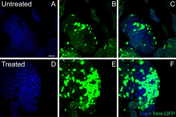
Drosophila represents an extremely powerful system which can be easily modulated for identification of genes as well as signaling pathways crucial to the hematopoietic process that are also well-conserved in the vertebrate system. Hematopoiesis in Drosophila occurs in a specialized organ known as lymph gland [18]. The lymph gland is made up of three zones- a niche, a progenitor zone that exhibits increased levels of ROS, and a differentiated zone that has relatively lower levels of ROS [19]. Blood cell differentiation in flies is governed by several genetic pathways and is highly sensitive to changes in physiological factors. Lymph glands are extremely sensitive to changes in ROS levels [19] but it is not known if non-thermal plasma treatment increases ROS in lymph glands. Hence we tested whether application of mspDBD plasma will result in hematopoiesis in the fly lymph gland, given non-thermal plasma is known to generate reactive oxygen species (ROS) [20,21]. As predicted, application of mspDBD plasma resulted in increase in the number of differentiated cells (as measured by hemolectin GFP, a marker for differentiated hemocytes, after 48 hours of treatment) [22] “Fig 3” as compared to untreated flies.
Fig 3. Plasma treatment increases blood cell differentiation.
Treatment with plasma increased blood cell differentiation. Control (upper panel) and plasma treated larval lymph glands marked with DAPI (blue) for DNA and Hemolectin-GFP (green) to mark differentiated hemocytes. Scale bar is 25 μm.
D. mspDBD plasma treatment increased ROS levels in lymph glands as well as whole larvae
To directly address if mspDBD plasma treatment results in increase in ROS levels in lymph glands, we measured ROS in these tissues by staining with DHE, a superoxide indicator [19]. We observed that the lymph glands of Drosophila had significantly higher levels of ROS upon treatment with non-thermal plasma as compared to glands of untreated flies “Fig 4A and 4B” (p≤0.05). Diffused staining with DHE was observed in the entire lymph gland, indicating that ROS was generated throughout the gland. These results show that the increased blood cell differentiation is associated with increased levels in ROS, as it has been reported in catalase or superoxide dismutase mutants, which fail to scavenge the ROS in lymph glands [19]. Our results suggest that not only genetic manipulation of ROS via eliminating scavenging enzymes increases differentiation and ROS levels in the lymph gland [19], but also by external manipulation using mspDBD plasma.
Fig 4. Plasma increased ROS levels in lymph glands and larvae.
A: DHE staining of larval lymph glands (red) shows increase in ROS in control vs. plasma treated larvae. B: Quantification of ROS in larval lymph gland from A. C: ROS measurement of whole larval extracts using amplex red for treated, untreated, boiled and amplex red only samples. Values are normalized against protein concentration for each extract. Scale bar is 25 μm.
We confirmed the increase in ROS levels by spectroflurometric measurements. To measure ROS, we prepared whole fly extracts and determined the amount of ROS in mspDBD plasma treated flies by using amplex red. As shown in the “Fig 4B”, control larvae and the boiled samples do not show detectable generation of ROS. However, larvae treated with plasma show increased ROS compared to the untreated wild type flies “Fig 4C”. These results indicate that there is a basal increase in ROS production upon treatment with mspDBD plasma.
Taken together, in this study, we establish a protocol and optimize conditions for the application of floating electrode mspDBD non-thermal plasma on Drosophila, at a level where their survival and reproduction is not affected. We tested the effect of plasma on hematopoietic organs and showed that there is an increased level of differentiation and this is due to augmented levels of ROS as a result of plasma treatment.
Discussion
Dielectric barrier discharges (DBDs) that generate non-thermal plasma have been used for years for the generation of ozone and modification of surfaces among other applications [23]. DBDs applied directly to living cells and tissues have been demonstrated to kill bacteria, induce blood coagulation, and improved tissue regeneration [24–26]. Non-thermal plasma treatment has also been shown to affect cellular processes, such as augmenting endothelial cell proliferation and enhancing differentiation of stem cells [6] [27]. Along with the demonstrated clinical potential of plasma treatment, the simplicity and flexibility of DBD plasma systems make it appealing for clinical development [8]. However, in order to optimize this non-thermal plasma technology for clinical use, an understanding of the mechanisms underlying plasma effects is required. In this study, we characterized the physiological effects of an atmospheric pressure, floating electrode mspDBD plasma system in an in vivo, Drosophila model.
Drosophila is a powerful model organism used to study many disease models including cancer [28]. Here for the first time, we standardize the use of mspDBD plasma on Drosophila at a dose that does not compromise the viability or reproduction of the organism yet has an observable effect on its physiology. Drosophila also has well-characterized genetic and signaling pathways studied over the years especially ones that regulate growth, an aberrance in which leads to tumorigenesis [29]. Hence it would be a useful model system to test the effect of plasma in an intact organism for applications both in the areas of cancer biology and tumor immunology.
Our standardization methods identified a low dose of plasma regime that does not affect the viability, fecundity or the movement of animals. This dose is safe for both adults and larvae. Hence, it can be used to study effects of non-thermal plasma application on abnormal overgrowing tissues during larval growth phases or for treating already overgrown tissues in adults in the Drosophila oncogenic and other disease models. Our findings will also provide a baseline and a reference point for dosage and duration of mspDBD in other experimental models and organisms.
Drosophila lymph glands contain hemocytes that are blood cells of myeloid origin [30]. The mature hemocytes dissociate from the gland, flow in circulation and function as scavenging cells. We used this system to study the effect of non-thermal plasma on Drosophila because of its extreme-sensitivity to ROS levels. The progenitor cells of the lymph gland contain developmentally increased amounts of ROS and a further increase in plasma-stimulated ROS leads to increased differentiation as shown in the case of anti-oxidant mutant genetic backgrounds [19].
Our results conclude that mspDBD plasma treatment indeed increased the number of differentiated blood cells. We attribute this effect to an increase in ROS as we measured independently by spectrophotometric analysis and dye staining. These observations provide proof for our hypothesis that non-thermal plasma induces ROS generation that leads to increased differentiation possibly via JNK pathway as reported earlier [19]. The overall increase in ROS indicates a basal upregulation of ROS production. Hence, this could be an important therapeutic approach to cause death in tumor cells, given there are ROS mediated pathways contributing to apoptosis [31] [32]. Whether this ROS is generated by oxidases or mitochondria is to be explored in future studies which can further decipher the mechanism of ROS generation by mspDBD plasma.
When plasma is generated, four major components are produced that can be delivered to biological targets, including electric fields, UV radiation, charged and neutral gas species (e.g. peroxide, superoxide, and ozone) [33] [34] [35]. Recently, we have shown that active species from plasma trigger biological signaling to generate intracellular mitochondrial ROS [27] [36]. However, ozone produced in significant amounts (5–7%) from mspDBD [37] itself can trigger biological signaling pathways and induce ROS production [38]. Future work in this context will be to test individual components separately to determine the constituent causing the increasing in ROS in blood cells. However, based on previous work, we speculate that superoxide might be the major candidate for Drosophila blood cells given it is established that genetic depletion of superoxide dismutase enzyme increased ROS and differentiation [15].
In summary, our study has standardized the technique for atmospheric pressure, floating electrode mspDBD plasma treatment in an in vivo model organism, which can be used to study various physiological processes, disease models and phenotypes.
Supporting Information
Histogram represents the average number of eggs laid by untreated and plasma treated flies. There is no significant difference in the number of eggs laid.
(TIF)
Data Availability
All relevant data are within the paper and its Supporting Information files.
Funding Statement
This work was funded by a Clinical and Translational Research Institute grant (HS) (http://drexel.edu/medicine/Research/Clinical-and-Translational-Research-Institute/), a Commonwealth Universal Research Enhancement Program grant (HS) (https://www.portal.state.pa.us/portal/server.pt/community/health_research_program_cure/14189), and an American Heart Association National Scientist Development grant (11SDG7230059) (http://my.americanheart.org/professional/Research/FundingOpportunities/SupportingInformation/Scientist-Development-Grant_UCM_443318_Article.jsp#.VpgFhPkrK8E). The funders had no role in study design, data collection and analysis, decision to publish, or preparation of the manuscript.
References
- 1.Graves DB (2014) Low temperature plasma biomedicine: A tutorial review. Physics of Plasmas 21: 080901. [Google Scholar]
- 2.Niemi K, apos, Connell D, de Oliveira N, Joyeux D, et al. (2013) Absolute atomic oxygen and nitrogen densities in radio-frequency driven atmospheric pressure cold plasmas: Synchrotron vacuum ultra-violet high-resolution Fourier-transform absorption measurements. Applied Physics Letters 103: 034102. [Google Scholar]
- 3.Kvam E, Davis B, Mondello F, Garner AL (2012) Nonthermal atmospheric plasma rapidly disinfects multidrug-resistant microbes by inducing cell surface damage. Antimicrob Agents Chemother 56: 2028–2036. 10.1128/AAC.05642-11 [DOI] [PMC free article] [PubMed] [Google Scholar]
- 4.Schmidt A, Dietrich S, Steuer A, Weltmann KD, von Woedtke T, et al. (2015) Non-thermal plasma activates human keratinocytes by stimulation of antioxidant and phase II pathways. J Biol Chem 290: 6731–6750. 10.1074/jbc.M114.603555 [DOI] [PMC free article] [PubMed] [Google Scholar]
- 5.Lee OJ, Ju HW, Khang G, Sun PP, Rivera J, et al. (2016) An experimental burn wound-healing study of non-thermal atmospheric pressure microplasma jet arrays. Journal of Tissue Engineering and Regenerative Medicine 10: 348–357. 10.1002/term.2074 [DOI] [PubMed] [Google Scholar]
- 6.Arjunan KP, Clyne AM (2011) A Nitric Oxide Producing Pin-to-Hole Spark Discharge Plasma Enhances Endothelial Cell Proliferation and Migration. 1: 279–293. [Google Scholar]
- 7.Schlegel J, Köritzer J, Boxhammer V (2013) Plasma in cancer treatment. Clinical Plasma Medicine 1: 2–7. [Google Scholar]
- 8.Fridman G, Friedman G, Gutsol A, Shekhter AB, Vasilets VN, et al. (2008) Applied Plasma Medicine. Plasma Processes and Polymers 5: 503–533. [Google Scholar]
- 9.Anderson KV, Ingham PW (2003) The transformation of the model organism: a decade of developmental genetics. Nat Genet 33 Suppl: 285–293. [DOI] [PubMed] [Google Scholar]
- 10.Duffy JB (2002) GAL4 system in Drosophila: a fly geneticist's Swiss army knife. Genesis 34: 1–15. [DOI] [PubMed] [Google Scholar]
- 11.Brand AH, Manoukian AS, Perrimon N (1994) Ectopic expression in Drosophila. Methods Cell Biol 44: 635–654. [DOI] [PubMed] [Google Scholar]
- 12.Theodosiou NA, Xu T (1998) Use of FLP/FRT system to study Drosophila development. Methods 14: 355–365. [DOI] [PubMed] [Google Scholar]
- 13.Kalghatgi S, Kelly CM, Cerchar E, Torabi B, Alekseev O, et al. (2011) Effects of non-thermal plasma on mammalian cells. PLoS One 6: e16270 10.1371/journal.pone.0016270 [DOI] [PMC free article] [PubMed] [Google Scholar]
- 14.Lin A, Chernets N, Han J, Alicea Y, Dobrynin D, et al. (2015) Non‐Equilibrium Dielectric Barrier Discharge Treatment of Mesenchymal Stem Cells: Charges and Reactive Oxygen Species Play the Major Role in Cell Death. Plasma Processes and Polymers. [DOI] [PMC free article] [PubMed] [Google Scholar]
- 15.Osei-Owusu P, Owens EA, Jie L, Reis JS, Forrester SJ, et al. (2015) Regulation of Renal Hemodynamics and Function by RGS2. PLoS One 10: e0132594 10.1371/journal.pone.0132594 [DOI] [PMC free article] [PubMed] [Google Scholar]
- 16.Singh H, Lu R, Rodriguez PF, Wu Y, Bopassa JC, et al. (2012) Visualization and quantification of cardiac mitochondrial protein clusters with STED microscopy. Mitochondrion 12: 230–236. 10.1016/j.mito.2011.09.004 [DOI] [PMC free article] [PubMed] [Google Scholar]
- 17.Poonalagu D, Gururaja Rao S, Farber J, Xin W, Hussain AT, et al. (2016) Molecular Identity of Cardiac Mitochondrial Chloride Intracellular Channel Proteins. Mitochondrion. [DOI] [PubMed] [Google Scholar]
- 18.Jung SH, Evans CJ, Uemura C, Banerjee U (2005) The Drosophila lymph gland as a developmental model of hematopoiesis. Development 132: 2521–2533. [DOI] [PubMed] [Google Scholar]
- 19.Owusu-Ansah E, Banerjee U (2009) Reactive oxygen species prime Drosophila haematopoietic progenitors for differentiation. Nature 461: 537–541. 10.1038/nature08313 [DOI] [PMC free article] [PubMed] [Google Scholar]
- 20.Kong MG, Kroesen G, Morfill G, Nosenko T, Shimizu T, et al. (2009) Plasma medicine: an introductory review. New Journal of Physics 11: 115012. [Google Scholar]
- 21.Kuchenbecker M, Bibinov N, Kaemlimg A, Wandke D, Awakowicz P, et al. (2009) Characterization of DBD plasma source for biomedical applications. Journal of Physics D: Applied Physics 42: 045212. [Google Scholar]
- 22.Goto A, Kadowaki T, Kitagawa Y (2003) Drosophila hemolectin gene is expressed in embryonic and larval hemocytes and its knock down causes bleeding defects. Dev Biol 264: 582–591. [DOI] [PubMed] [Google Scholar]
- 23.Kogelschatz U (2003) Dielectric-barrier discharges: their history, discharge physics, and industrial applications. Plasma chemistry and plasma processing 23: 1–46. [Google Scholar]
- 24.Fridman G, Peddinghaus M, Balasubramanian M, Ayan H, Fridman A, et al. (2006) Blood coagulation and living tissue sterilization by floating-electrode dielectric barrier discharge in air. Plasma Chemistry and Plasma Processing 26: 425–442. [Google Scholar]
- 25.Fridman G, Brooks AD, Balasubramanian M, Fridman A, Gutsol A, et al. (2007) Comparison of Direct and Indirect Effects of Non‐Thermal Atmospheric‐Pressure Plasma on Bacteria. Plasma Processes and Polymers 4: 370–375. [Google Scholar]
- 26.Chernets N, Zhang J, Steinbeck MJ, Kurpad DS, Koyama E, et al. (2014) Nonthermal Atmospheric Pressure Plasma Enhances Mouse Limb Bud Survival, Growth, and Elongation. Tissue Engineering Part A. [DOI] [PMC free article] [PubMed] [Google Scholar]
- 27.Steinbeck MJ, Chernets N, Zhang J, Kurpad DS, Fridman G, et al. (2013) Skeletal cell differentiation is enhanced by atmospheric dielectric barrier discharge plasma treatment. PLoS One 8: e82143 10.1371/journal.pone.0082143 [DOI] [PMC free article] [PubMed] [Google Scholar]
- 28.Pandey UB, Nichols CD (2011) Human disease models in Drosophila melanogaster and the role of the fly in therapeutic drug discovery. Pharmacol Rev 63: 411–436. 10.1124/pr.110.003293 [DOI] [PMC free article] [PubMed] [Google Scholar]
- 29.Miles WO, Dyson NJ, Walker JA (2011) Modeling tumor invasion and metastasis in Drosophila. Dis Model Mech 4: 753–761. 10.1242/dmm.006908 [DOI] [PMC free article] [PubMed] [Google Scholar]
- 30.Crozatier M, Vincent A (2011) Drosophila: a model for studying genetic and molecular aspects of haematopoiesis and associated leukaemias. Dis Model Mech 4: 439–445. 10.1242/dmm.007351 [DOI] [PMC free article] [PubMed] [Google Scholar]
- 31.Shen ZY, Shen WY, Chen MH, Shen J, Zeng Y (2003) Reactive oxygen species and antioxidants in apoptosis of esophageal cancer cells induced by As2O3. Int J Mol Med 11: 479–484. [PubMed] [Google Scholar]
- 32.Nagaraj R, Gururaja-Rao S, Jones KT, Slattery M, Negre N, et al. (2012) Control of mitochondrial structure and function by the Yorkie/YAP oncogenic pathway. Genes Dev 26: 2027–2037. 10.1101/gad.183061.111 [DOI] [PMC free article] [PubMed] [Google Scholar]
- 33.Lunov O, Zablotskii V, Churpita O, Chanova E, Sykova E, et al. (2014) Cell death induced by ozone and various non-thermal plasmas: therapeutic perspectives and limitations. Sci Rep 4: 7129 10.1038/srep07129 [DOI] [PMC free article] [PubMed] [Google Scholar]
- 34.Lu X, Naidis GV, Laroussi M, Reuter S, Graves DB, et al. (2016) Reactive species in non-equilibrium atmospheric-pressure plasmas: Generation, transport, and biological effects. Physics Reports 630: 1–84. [Google Scholar]
- 35.Danil D, Gregory F, Gary F, Alexander F (2009) Physical and biological mechanisms of direct plasma interaction with living tissue. New Journal of Physics 11: 115020. [Google Scholar]
- 36.Lin A, Chernets N, Han J, Alicea Y, Dobrynin D, et al. (2015) Non-Equilibrium Dielectric Barrier Discharge Treatment of Mesenchymal Stem Cells: Charges and Reactive Oxygen Species Play the Major Role in Cell Death. Plasma Processes and Polymers 12: 1117–1127. [DOI] [PMC free article] [PubMed] [Google Scholar]
- 37.Fridman A, Chirokov A, Gutsol A (2005) Non-thermal atmospheric pressure discharges. Journal of Physics D: Applied Physics 38: R1. [Google Scholar]
- 38.Voter KZ, Whitin JC, Torres A, Morrow PE, Cox C, et al. (2001) Ozone exposure and the production of reactive oxygen species by bronchoalveolar cells in humans. Inhal Toxicol 13: 465–483. [DOI] [PubMed] [Google Scholar]
Associated Data
This section collects any data citations, data availability statements, or supplementary materials included in this article.
Supplementary Materials
Histogram represents the average number of eggs laid by untreated and plasma treated flies. There is no significant difference in the number of eggs laid.
(TIF)
Data Availability Statement
All relevant data are within the paper and its Supporting Information files.


