Abstract
With recent advances in DNA sequencing technologies, it has become increasingly easy to use whole-genome sequencing of unrelated individuals to assay patterns of linkage disequilibrium (LD) across the genome. One type of analysis that is commonly performed is to estimate local recombination rates and identify recombination hotspots from patterns of LD. One method for detecting recombination hotspots, LDhot, has been used in a handful of species to further our understanding of the basic biology of recombination. For the most part, the effectiveness of this method (e.g., power and false positive rate) is unknown. In this study, we run extensive simulations to compare the effectiveness of three different implementations of LDhot. We find large differences in the power and false positive rates of these different approaches, as well as a strong sensitivity to the window size used (with smaller window sizes leading to more accurate estimation of hotspot locations). We also compared our LDhot simulation results with comparable simulation results obtained from a Bayesian maximum-likelihood approach for identifying hotspots. Surprisingly, we found that the latter computationally intensive approach had substantially lower power over the parameter values considered in our simulations.
Keywords: recombination hotspots, linkage disequilibrium, composite likelihood
Homologous recombination is a fundamental biological process. In most organisms, it is necessary for the proper alignment and segregation of chromosomes during meiosis, influences the efficacy of natural selection, and is the primary determinant of the strength of allelic associations (i.e., linkage disequilibrium, or LD) across the genome. It is now well established that recombination rates vary tremendously across the genome, and that many species have recombination ‘hotspots’, narrow regions (<2 kb) where the recombination rate is much higher than the rate in the surrounding sequence (Petes 2001; Buard and de Massy 2007). In humans, for example, sperm typing studies have experimentally identified dozens of hotspots (e.g., Jeffreys et al. 2001, 2005; Sarbajna et al. 2012), and pedigree-based studies have identified broad-scale variation in recombination rates (Broman et al. 1998; Kong et al. 2002, 2010). In most eukaryotic species direct estimates of recombination are time-consuming, and not easily scaled up to studies of fine-scale recombination rate variation across the whole genome (but see Mancera et al. 2008; Comeron et al. 2012).
An appealing alternative is to estimate local changes in recombination rate using patterns of LD at single nucleotide polymorphisms (SNPs) (Crawford et al. 2004; McVean et al. 2004; Chan et al. 2012). These methods have been used to construct fine-scale recombination maps in humans (Myers et al. 2005; 1000 Genomes Project Consortium 2010), great apes (Auton et al. 2012; Stevison et al. 2016), Drosophila melanogaster (Chan et al. 2012), Arabidopsis thaliana (Horton et al. 2012), Medicago truncatula (Paape et al. 2012), house mouse (Brunschwig et al. 2012), and dogs (Axelsson et al. 2012; Auton et al. 2013). While these recombination maps can suggest the locations of many potential recombination hotspots, separate methodology (e.g., a hypothesis test) is needed to statistically test whether any particular region is actually a hotspot. Otherwise, a local increase in estimated rate could reflect chance variation in the genealogical process, rather than point to a real hotspot. While several methods for statistically identifying hotspots have been proposed (Crawford et al. 2004; McVean et al. 2004; Fearnhead 2006; Li et al. 2006; Wang and Rannala 2009), almost all are computationally expensive enough to limit their use to candidate regions (Crawford et al. 2004; Tsai et al. 2010; Chan et al. 2012) or to sparse genotype data (Axelsson et al. 2012). In most whole-genome applications to date, one method, LDhot (McVean et al. 2004), has been used to identify recombination hotspots in humans, chimpanzees, dogs, and A. thaliana. The accuracy of this method for identifying true recombination hotspots though is unknown.
The use of population genetic methods to computationally predict hotspot locations has led to several major insights into the evolution of fine-scale recombination rates. Notably, computational analyses identified a degenerate 13 bp motif (CCNCCNTNNCCNC) that is overrepresented in predicted human hotspots relative to matched coldspots (Myers et al. 2008), and subsequent work has shown that this motif matches the predicted binding domain for the PRDM9 gene (Baudat et al. 2010; Myers et al. 2010; Brick et al. 2012). PRDM9 trimethylates lysine 4 of histone H3 (H3K4me3) (Hayashi et al. 2005), and H3K4me3 marks are associated with double strand breaks and recombination in both yeast and mice (Borde et al. 2009; Buard et al. 2009). Variation in PRDM9 is associated with differences in fine-scale recombination rates and hotspot usage across the genome (Baudat et al. 2010). Thus, the use of these computational methods has identified a new and key player in mammalian recombination. In addition, a comparison of fine-scale recombination maps in humans and chimpanzees found that chimpanzees have recombination hotspots as well, but at different locations than human hotspots (Auton et al. 2012; but see Wang and Rannala 2014). This confirmed the results of previous analyses of much smaller data sets (Wall et al. 2003; Ptak et al. 2004; Winckler et al. 2005) and is in line with the theoretical expectations of biased gene conversion (Boulton et al. 1997; Coop and Myers 2007). PRDM9 evolves rapidly in metazoans (Oliver et al. 2009; Schwartz et al. 2014), and the different predicted PRDM9 binding motifs for humans and chimpanzees may explain why there is no (or little) overlap in hotspots between the two species. Thus, the discovery of this gene may also explain why hotspots have evolved so rapidly in apes.
The initial computational analyses failed to identify any particular sequence motif that is associated with predicted chimpanzee hotspots, which was ascribed to the high allelic diversity at PRDM9 in chimpanzees and the complex relationship between PRDM9 sequence and targeted binding sites (Auton et al. 2012; Billings et al. 2013). Moreover, though PRDM9 appears to be absent in plants and canids (Oliver et al. 2009), studies of SNP data in A. thaliana, M. truncatula, and Canis lupus familiaris found strong evidence for thousands of recombination hotspots in these three species (Axelsson et al. 2012; Horton et al. 2012; Paape et al. 2012; Auton et al. 2013). However, a similar study of recombination rate variation in flies identified fewer than 10 hotspots in different populations of D. melanogaster (Chan et al. 2012). Thus, computational methods have further highlighted intriguing similarities and differences among taxa.
There have been at least three separate implementations of LDhot (Myers et al. 2005; Auton et al. 2012, 2014), each with different criteria for calling hotspots. Until recently (Auton et al. 2014) no version of the program was publicly available. This has hampered our understanding of the extent to which published results reflect the true biological reality vs. limitations of the hotspot calling methodology. In this study, we implement our own version of LDhot (available from github at https://github.com/jdwall02/mlehot), and test the power of LDhot to detect true recombination hotspots over a range of model parameters appropriate for large mammals such as humans and great apes. We find that some previous implementations of LDhot have extremely low power, which might explain the results of Johnston and Cutler (2012). If many true hotspots have gone undetected, then an actual correlation between hotspot sequences and a sequence motif may have been missed, and the degree of sharing of hotspot locations across species (e.g., Auton et al. 2012) may have been underestimated. We explore this possibility in greater detail below. We also compare the power and false positive rate of LDhot with a maximum-likelihood approach for estimating recombination rates and calling recombination hotspots (Wang and Rannala 2009).
Materials and Methods
LDhot uses a composite likelihood framework based on the work of Hudson (2001) and McVean et al. (2002) and similar to the approach of HotspotFisher (Li et al. 2006). The Auton et al. (2012) implementation tests every 2 kb region (with a 1 kb increment) as a potential hotspot by analyzing the 200 kb region centered around the region of interest. Suppose the SNPs in the 200 kb region are S = {s1... sn}. A (composite) LRT statistic is calculated as
| (1) |
where lik (si, sj | ρ) is the two-site likelihood described before (Hudson 2001; McVean et al. 2002), ρ0 is the background recombination rate, and ρ1 is the recombination rate in the central 2 kb region. Critical values for R are estimated from null simulations that assume a constant recombination rate across the region (i.e., ρ0 = ρ1). Auton et al. (2012) used ‘fixed S’ methodology for these simulations (Hudson 1993; Wall and Hudson 2001), with SNP locations fixed to be where SNPs appear in the actual data and ρ chosen to be equal to its estimated value (from LDhat). They tested each possible 2 kb region and identified those where the estimated P-value for R was <0.01. Then, overlapping regions were merged to form a list of candidate hotspot regions. These regions were filtered to reduce the false positive rate by eliminating ones >5 kb in size or with peak ρ estimate <5 /kb (estimated using LDhat).
We also studied two other approaches for calling hotspots using LDhot. Auton et al. (2014) used a smaller window size (100 kb) but the same basic approach for identifying candidate regions. Instead of a size or peak ρ estimate filter though, they required each hotspot region to contain at least one 2 kb window where the estimated P-value for R was <0.001. Our new approach here is to generate the same list of candidate regions as Auton et al. (2012), partition each region into nonoverlapping 1 kb windows, and keep only those windows for which the average ρ estimate (using LDhat) is ≥5/kb. A brief summary of the differences between LDhot implementations is summarized in Table 1.
Table 1. Key differences between three implementations of LDhot.
| Method | Auton et al. 2012 | Auton et al. 2014 | mlehot |
|---|---|---|---|
| Window size | 200 kb | 100 kb | 20 kb |
| P-value cutoff | 0.01 | 0.001 | 0.01 |
| Max. size | 5 kb | None | None |
| LDhat | Peak must be ≥5/kb | – | Intersection with 1 kb regions with ρ ≥ 5 |
| Simulations | Region-specific | Region-specific | Lookup table |
For all three hotspot calling protocols, we found that the publicly available version of LDhot (Auton et al. 2014) was too slow for running power calculations, since the null distribution for R is estimated separately for each test window. We implemented our own version, similar to the method of Myers et al. (2005) – we run null simulations in advance, store the results in a large lookup table, and use these to repeatedly estimate the significance values for observed values of R across the genome. Specifically, for a window size of X kb we run coalescent simulations (cf. Hudson 2002) of X kb regions across a broad range of mutation and recombination rates (assuming the recombination rate is constant per base pair). For each simulation, we tabulated R, the number of segregating sites S, and the estimate of ρ (cf. Hudson 2001). Then, when analyzing an actual X kb region, we calculate S and ρ, then use simulations with S and ρ near the actual values for determining the null distribution for R. So, while the Auton et al. (2012) approach uses ‘fixed S’ simulations and parametric bootstrapping (for ρ), we use standard coalescent simulations and condition (in the standard statistical sense) on the observed value of S and the observed estimate of ρ (with an implicitly flat prior for θ and ρ). Our simulations took X = 20, 50, 100, or 200 kb, and we ran ∼5 × 10⁶ simulations for each value of X. Source code and executables for running all three implementations of LDhot are available at http://github.com/jdwall02/mlehot
To estimate the power of the different hotspot calling protocols, we assumed a sample size of n = 30 haploid sequences, a scaled mutation rate of θ = 1/kb, and a scaled background recombination rate of ρ = 0.5/kb. We simulated 100 different 1 Mb regions, each containing eight different 2 kb hotspots with scaled recombination rates of 5, 10, 25, or 50/kb. We defined the power as the proportion of actual hotspot sequence that was identified as a hotspot using LDhot. Similarly, the false positive rate was calculated as the proportion of actual nonhotspot sequence that was called as a hotspot using LDhot, and the false discovery rate was defined as the proportion of called hotspot sequence that was not actually contained in a recombination hotspot. (Note that these definitions differ from those of some previous studies.) Additional simulations considered a wider range of sample sizes (n = 16–42 haploid sequences) or other scaled background recombination rates (ρ = 0.1, 0.2, 1, or 2.5 per kb). These latter simulations had actual hotspot recombination rates that were 10, 20, 50, or 100 times the background rate and a new hotspot calling criteria of ρ ≥ 10 times the background rate (estimated from LDhat). One final set of simulations had n = 30, a scaled mutation rate of θ = 5/kb, and a scaled background recombination rate of ρ = 0.5–5/kb.
We also used Inferrho, a Bayesian full-likelihood method for calling recombination hotspots developed by Wang and Rannala (2009). We used both the current version of the program (IRv1) as well as the originally published version (INFERrho, obtained from Y. Wang). Inferrho estimates a posterior probability that any particular genomic location is contained in a recombination hotspot. We used the same hotspot calling criteria as Wang and Rannala (i.e., HT1 = 5, HT2 = 2.5) on 20 simulated 1 Mb regions with recombination hotspots (background ρ = 0.5/kb; other parameters are described in the previous paragraph). For computational tractability, we broke each simulated region into overlapping 7 kb subregions (each overlap being 2 kb), and analyzed each subregion separately after trimming off 1 kb from each end. We also only analyzed a subset of the nonhotspot regions to estimate the false positive and false discovery rates. Finally, we reanalyzed the original 100 simulations described in Table 1 of Wang and Rannala (2009), using their original implementation, simulation parameters (burnin = 104 steps, MCMC chain = 105 steps), and criteria for calling hotspots. We also reran half of these simulations with a burnin of 2 × 10⁵ steps and an MCMC chain of 106 steps and found our results to be unchanged (results not shown).
Data availability
The authors state that all data necessary for confirming the conclusions presented in the article are represented fully within the article.
Results
To assess the power and false positive rate of LDhot, we implemented our own version that is fast enough to allow for power calculations on simulated data. We then estimated LDhot’s power under a range of window sizes and recombination hotspot intensities, using the same basic protocol as Auton et al. (2012). Unlike previous simulation studies (Wang and Rannala 2009; Auton et al. 2014), ours uses a background recombination rate that is more appropriate for human or great ape data.
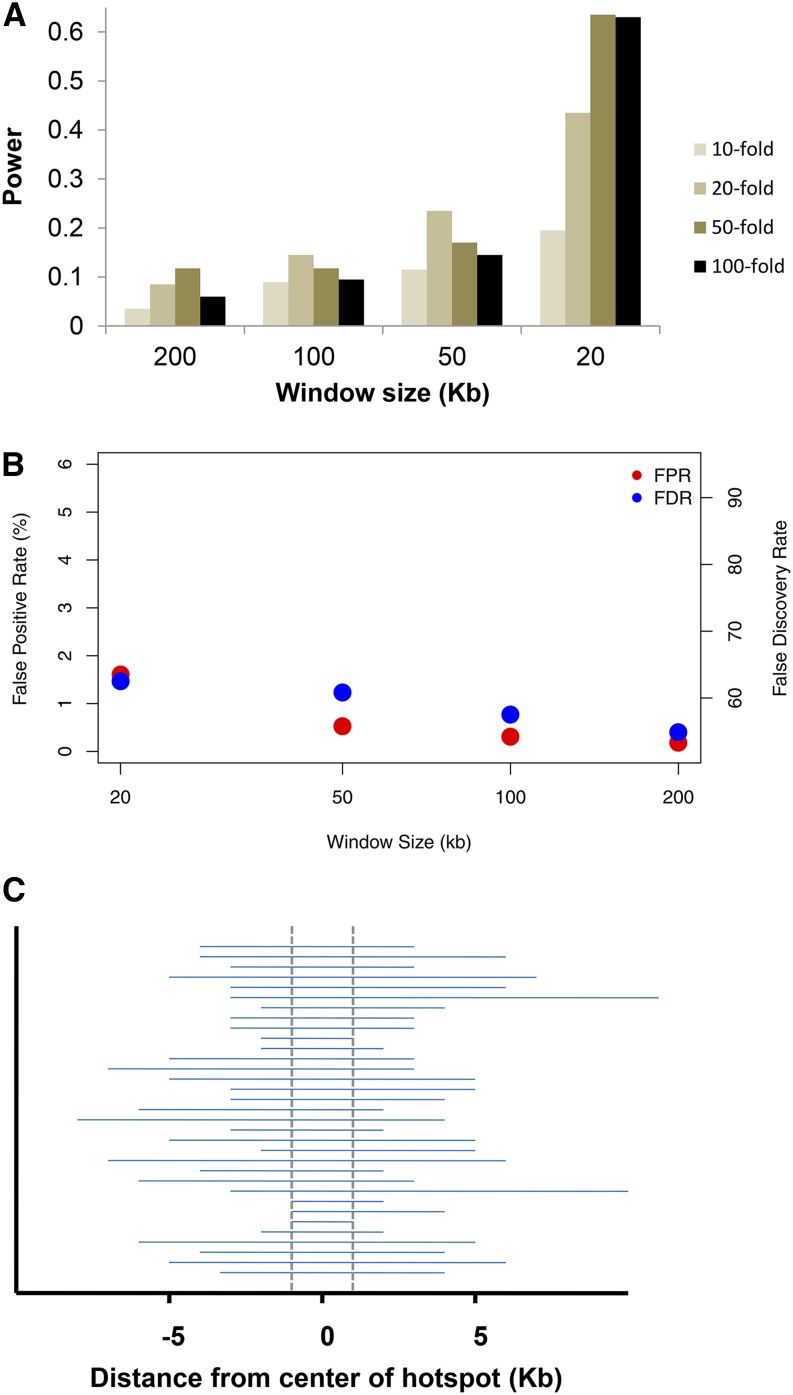
The power results are shown in Figure 1A while the false positive rates and false discovery rates (FDR) are shown in Figure 1B. Two trends are easily apparent. First, the power increases as the window size decreases, with an ∼fivefold increase in power for a 20 kb window size compared with a 200 kb window size. Second, stronger hotspots are not always easier to detect, and the power to detect recombination hotspots under any scenario is quite modest, topping out at 63% for a 20 kb window size and a 50-fold increase in the recombination rate and with a low of 3% power for a 200 kb window size and a 10-fold increase in the recombination rate. The FDR is relatively high at 55–63%. This, plus the lower power, suggests that existing methods might have substantial room for improvement.
Figure 1.
(A) Power to detect a hotspot as a function of the window size (x-axis) and the strength of the hotspot using the protocol of Auton et al. (2012). Background ρ is 0.5/kb. See text for further details. (B) False positive rate for estimated hotspots, defined as the proportion of estimated hotspot sequence that was not actually a simulated hotspot. (C) Estimated hotspot locations without the length limitation for a 100 kb window and a 50-fold hotspot. Dashed vertical lines show location of actual hotspot. Results are similar for other parameter combinations.
The general trends described above are a consequence of the composite likelihood approach used by LDhot, and the protocol used to manage the false positive rate. Since all pairs of sites within a window are used in the likelihood calculations, most of the pairs in large (e.g., ≥100 kb) windows will be uninformative about the precise location of a hotspot. So, if a hotspot is called for a region, it tends to be large (i.e., >5 kb in length), and these large regions are subsequently excluded. Similarly, when the recombination hotspot is strong (50-fold or 100-fold increase), LDhot has more trouble with hotspot localization, leading to large hotspot regions that are also excluded. To illustrate this, Figure 1C shows the estimated hotspot locations relative to the true hotspot location for a 100 kb window size and a 50-fold hotspot. While 85% of the simulated hotspots were identified using a P < 0.01 cutoff, the vast majority of these were excluded from Figure 1A due to their size. This leads to decreasing power for increasing window size.
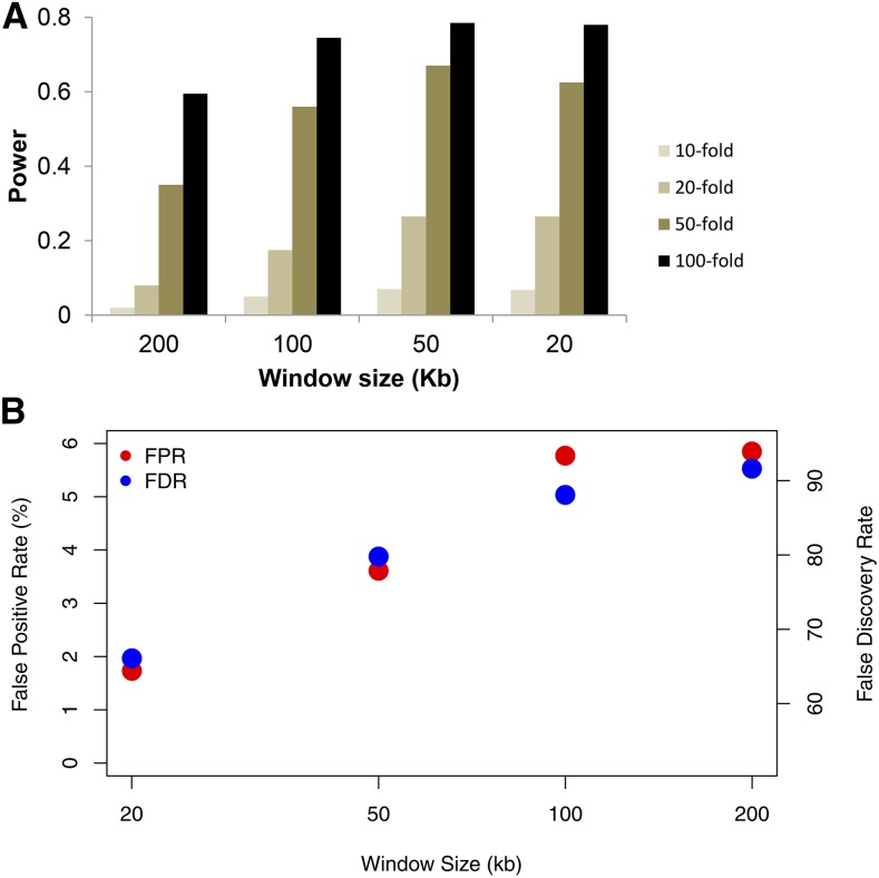
If we drop the 5 kb length restriction, then the power (and false positive rate) increase substantially. Figure 2 shows the power and false positive rates if we use the Auton et al. (2014) criteria for calling hotspots instead. This approach adopts a stricter P < 0.001 cutoff for identifying a candidate region as a hotspot, but does not have any length restriction on the size of the region identified (see Materials and Methods for a more precise description). While the power increases substantially when comparing Figure 1A to Figure 2A, this is achieved at a cost of having a false positive rate that ranges from 1.7 to 5.8% and a FDR of 66–92% (Figure 2B). For these simulations, the average size of the identified hotspot regions varies from 5.1 kb (20 kb window size) to 23.3 kb (200 kb window size). Clearly, a 20 kb “hotspot region” is not very informative, even if it does contain a true recombination hotspot.
Figure 2.
(A) Power to detect a hotspot as a function of the window size and strength of the hotspot using the protocol of Auton et al. (2014). Results are directly comparable to those of Figure 1A and Figure 3A. (B) False positive rate for estimated hotspots, defined as the proportion of estimated hotspot sequence that was not actually a simulated hotspot.
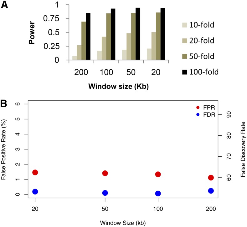
We also tried another approach for hotspot calling, by analyzing each 1 kb region separately, and requiring both P < 0.01 (using LDhot) and ρ ≥ 5/kb (using LDhat) for a region to be called a hotspot. Under this protocol, the power to detect hotspots is much higher than for the previously proposed methods (Figure 3A). For a 20 kb window size, the power ranges from 21% for a 10-fold hotspot to 94% for a 100-fold hotspot, compared with 17% and 40% for the Auton et al. (2012) approach and 7% and 78% for the Auton et al. (2014) approach. The false positive rate (Figure 3B) is intermediate between the results of Figure 1B and Figure 2B, while the FDR ranges from 52 to 54%, lower than for the previous methods used for calling recombination hotspots. We conclude that our new method for calling hotspots using LDhot is better than the previous approaches, and that smaller window sizes (e.g., 20 kb) should be used when analyzing dense SNP or resequencing data.
Figure 3.
(A) Power to detect a hotspot as a function of the window size and strength of the hotspot using our new protocol. Results are directly comparable to those of Figure 1A and Figure 2A. (B) False positive rate for estimated hotspots, defined as the proportion of estimated hotspot sequence that was not actually a simulated hotspot.
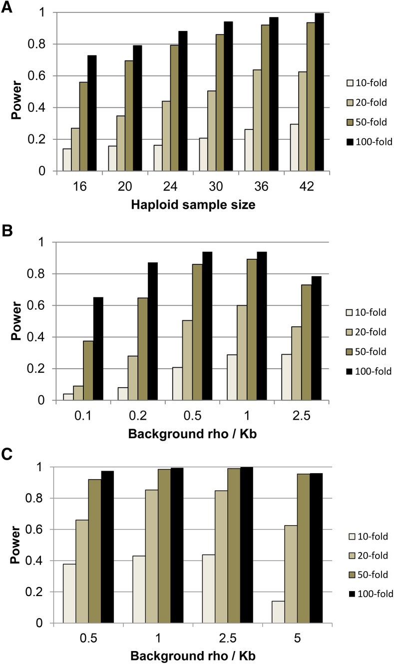
To explore how sensitive these results are to the particular parameter values used, we also estimated the power to detect recombination hotspots for a range of haploid sample sizes and different background recombination rates. In almost all simulations, our new hotspot calling protocol has substantially higher power and lower false discovery rate than the previously described protocols (Auton et al. 2012, 2014), even when we use the same window size for each. For brevity, we include only the results of the new hotspot calling protocol on 20 kb windows. As expected, power increases with increasing sample size (Figure 4A), with some leveling off once n > 30. Additionally, we find that hotspots are easier to detect when the background recombination rate is intermediate (ρ = 0.2–1/kb, cf. Figure 4B), presumably because for low background rates the levels of LD are high even in recombination hotspots and for high background rates the levels of LD are somewhat low even for background regions. Additional simulations suggest that power is also increased (and FDR decreased) when the baseline levels of genetic variation (e.g., θ = 4 Nμ) are higher. Figure 4C shows simulation results with a fivefold higher amount of polymorphism (θ = 5/kb). The increased information content with the higher density of SNPs leads to the increase in power, though this breaks down at the highest recombination rates (presumably due to the low levels of LD even in nonhotspot regions). These results suggest that the ease with which recombination hotspots can be identified in a given species from patterns of LD depends strongly on the values of fundamental biological parameters such as the effective population size, mutation rate, and recombination rate.
Figure 4.
Power to detect hotspots using our new protocol over (A) different haploid sample sizes, (B) different background recombination rates, and (C) higher mutation and (background) recombination rates.
Finally, to see whether the composite likelihood approach of LDhot reduces power compared with computationally intensive full-likelihood approaches, we compared a subset of the results shown in Figure 3 with comparable results obtained from Inferrho (Wang and Rannala 2009). The results, shown in Table 2, are striking. Compared with LDhot, Inferrho has much smaller power, false positive rate, and false discovery rate. Inferrho essentially never finds weak hotspots (e.g., 10- or 20-fold increase in recombination rate) and is very conservative even for the strongest simulated hotspots. On the other hand, when Inferrho does call a region a hotspot, there is a very high likelihood that it really is one (FDR ∼4%). If we use a more liberal definition of power, where any overlap between a called hotspot and an actual hotspot is counted as a hit, Inferrho has moderate power ranging from 2.5% for a 10-fold hotspot to 62.5% for a 100-fold hotspot.
Table 2. Comparison between our implementation of LDhot and Inferrho over 20 different 1 Mb regions.
| Power (%) | LDhot | Inferrho |
|---|---|---|
| 10-fold hotspot | 17.5 | 0.9 |
| 20-fold hotspot | 48.8 | 2.8 |
| 50-fold hotspot | 87.5 | 15.0 |
| 100-fold hotspot | 95.0 | 26.5 |
| False positive rate (%) | 1.59 | <0.2 |
| False discovery rate (%) | 55.6 | ≥4.1 |
Due to computational limitations, we could not calculate exact values of the false positive and false discovery rates for Inferrho.
Discussion
Our simulation results suggest that the efficacy of LDhot in detecting recombination hotspots is quite sensitive to the particular implementation used in the analyses. Specifically, the larger window sizes used in previous studies (e.g., 200 kb in Auton et al. 2012) lead to greatly reduced power and a higher false positive rate when compared to smaller window sizes (e.g., 20 kb in Figure 1, Figure 2, and Figure 3). Qualitatively, this is because of the nature of the underlying composite likelihood used by LDhot – all pairs of sites are used within a window, including ones that are uninformative due to their distance from the central test region. LDhot’s original formulation (McVean et al. 2004) was optimized for human SNP data where the density of markers was relatively low. Now that full resequencing data are available, much smaller window sizes are needed for accurately estimating background vs. putative hotspot recombination rates. While our simulations are not exhaustive, they suggest that the optimal setup is to have the smallest window size that can accurately estimate background recombination rates. For species with human-like evolutionary parameters, this involves a window size of 20 kb (or perhaps slightly smaller). For species with a much higher level of diversity, a 10 kb or smaller window size would be appropriate (results not shown).
Our simulations also found that even for a fixed window size, the protocols used for identifying recombination hotspots have a strong influence on LDhot’s power and false positive rate. While the new protocol proposed here seems superior to previous implementations (Auton et al. 2012, 2014), we caution that our approach is ad hoc and that even better protocols are likely available. We also note that using a suboptimal version of LDhot can have real consequences when analyzing actual data. Auton and colleagues found no evidence of an association between predicted PRDM9 binding sites and recombination hotspots in chimpanzees (Auton et al. 2012). The low power of their LDhot implementation led to fewer called hotspots (<5000), which in turn limited their power to detect any association between sequence motifs and hotspots. Our analysis with an improved LDhot identified twice as many hotspots, and found sequence motifs (corresponding to predicted PRDM9 binding sites) that are overrepresented in recombination hotspots across several great ape species, including chimpanzees, bonobos, and gorillas (Stevison et al. 2016).
Finally, we found that even though LDhot has some serious drawbacks from a statistical standpoint, the computationally intensive full-likelihood approach of Wang and Rannala (2009) has much lower power. These results were unexpected, in part because of the much higher power (74–92%) reported by the authors (Table 1, Wang and Rannala 2009). While part of the difference can be ascribed to the particular parameter values used (e.g., we used a background recombination rate of ρ = 0.5/kb while they used ρ = 0.06/kb), the performance of Inferrho was still surprising. To examine this further, we obtained the 100 simulated data sets analyzed by the authors (with hotspot ρ = 10/kb) and reanalyzed them using both the currently distributed version of Inferrho (IRv1) and the original version (INFERrho) used in Wang and Rannala (2009). Using the same simulation parameters and hotspot calling criteria as their original paper, we were not able to recreate their results (Table 3). These results were unchanged even when we increased the burnin and MCMC chain length by an order of magnitude (see Materials and Methods; results not shown). Consistent with the qualitative results of our previous simulations, we found both versions of Inferrho to have lower power and lower false positive rate than what was originally reported.
Table 3. Comparison between the results published in Wang and Rannala (2009, Table 1) and our computations of Inferrho using the same data sets.
| Wang and Rannal (2009)a | IRv1 | INFERrho | |
|---|---|---|---|
| Power (%) | –a | 19.0 | 28.3 |
| False positive (%)b | 4 | 0 | 0 |
| Overlap (%)c | 74 | 33 | 39 |
False positive refers to the Wang and Rannala (2009) definition – called hotspots that do not overlap at all with true hotspots.
Cannot be determined from the original study.
This refers to the Wang and Rannala (2009) definition – called hotspots that do not overlap with all true hotspots.
This is Wang and Rannala’s (2009) definition of power – the proportion of true hotspots that overlap by at least 1 bp with a called hotspot.
We conclude that methods for identifying recombination hotspots should be tested thoroughly on simulated data and compared with each other across a wide range of parameter values to assess the efficacy of each and to determine which ones are the best to use. We hope that this study can be one step toward this goal.
Acknowledgments
This work was funded in part by National Institutes of Health (NIH) R01 grants HG005226 and GM115433 to J.D.W.
Footnotes
Communicating editor: J. Ross-Ibarra
Literature Cited
- Auton A., Fledel-Alon A., Pfeifer S., Venn O., Segurel L., et al. , 2012. A fine-scale chimpanzee genetic map from population sequencing. Science 336: 193–198. [DOI] [PMC free article] [PubMed] [Google Scholar]
- Auton A., Li Y. R., Kidd J., Oliveira K., Nadel J., et al. , 2013. Genetic recombination is targeted towards gene promoter regions in dogs. PLoS Genet. 9: e1003984. [DOI] [PMC free article] [PubMed] [Google Scholar]
- Auton, A., S. Myers, and G. McVean, 2014 Identifying recombination hotspots using population genetic data. arXiv 1403.4264.
- Axelsson E., Webster M. T., Ratnakumar A., Ponting C. P., Lindblad-Toh K., 2012. Death of PRDM9 coincides with stabilization of the recombination landscape in the dog genome. Genome Res. 22: 51–63. [DOI] [PMC free article] [PubMed] [Google Scholar]
- Baudat F., Buard J., Grey C., Fledel-Alon A., Ober C., et al. , 2010. PRDM9 is a major determinant of meiotic recombination hotspots in humans and mice. Science 327: 836–840. [DOI] [PMC free article] [PubMed] [Google Scholar]
- Billings T., Parvanov E. D., Baker C. L., Walker M., Paigen K., et al. , 2013. DNA binding specificities of the long zinc finger recombination protein PRDM9. Genome Biol. 14: R35. [DOI] [PMC free article] [PubMed] [Google Scholar]
- Borde V., Robine N., Lin W., Bonfils S., Geli V., et al. , 2009. Histone H3 lysine 4 trimethylation marks meiotic recombination initiation sites. EMBO J. 28: 99–111. [DOI] [PMC free article] [PubMed] [Google Scholar]
- Boulton A., Myers R. S., Redfield R. J., 1997. The hotspot conversion paradox and the evolution of meiotic recombination. Proc. Natl. Acad. Sci. USA 94: 8058–8063. [DOI] [PMC free article] [PubMed] [Google Scholar]
- Brick K., Smagulova F., Khil P., Camerini-Otero R. D., Petukhova G. V., 2012. Genetic recombination is directed away from functional genomic elements in mice. Nature 485: 642–645. [DOI] [PMC free article] [PubMed] [Google Scholar]
- Broman K. W., Murray J. C., Sheffield V. C., White R. L., Weber J. L., 1998. Comprehensive human genetic maps: individual and sex-specific variation in recombination. Am. J. Hum. Genet. 63: 861–869. [DOI] [PMC free article] [PubMed] [Google Scholar]
- Brunschwig H., Levi L., Ben-David E., Williams R. W., Yakir B., et al. , 2012. Fine-scale maps of recombination rates and hotspots in the mouse genome. Genetics 191: 757–764. [DOI] [PMC free article] [PubMed] [Google Scholar]
- Buard J., de Massy B., 2007. Playing hide and seek with mammalian meiotic crossover hotspots. Trends Genet. 23: 301–309. [DOI] [PubMed] [Google Scholar]
- Buard J., Barthes P., Grey C., de Massy B., 2009. Distinct histone modifications define initiation and repair of meiotic recombination in the mouse. EMBO J. 28: 2616–2624. [DOI] [PMC free article] [PubMed] [Google Scholar]
- Chan A. H., Jenkins P. A., Song Y. S., 2012. Genome-wide fine-scale recombination rate variation in Drosophila melanogaster. PLoS Genet. 8: e1003090. [DOI] [PMC free article] [PubMed] [Google Scholar]
- Comeron J. M., Ratnappan R., Bailin S., 2012. The many landscapes of recombination in Drosophila melanogaster. PLoS Genet. 8: e1002905. [DOI] [PMC free article] [PubMed] [Google Scholar]
- Coop G., Myers S. R., 2007. Live hot, die young: transmission distortion in recombination hotspots. PLoS Genet. 3: e35. [DOI] [PMC free article] [PubMed] [Google Scholar]
- Crawford D. C., Bhangale T., Li N., Hellenthal G., Rieder M. J., et al. , 2004. Evidence for substantial fine-scale variation in recombination rates across the human genome. Nat. Genet. 36: 700–706. [DOI] [PubMed] [Google Scholar]
- Fearnhead P., 2006. SequenceLDhot: detecting recombination hotspots. Bioinformatics 22: 3061–3066. [DOI] [PubMed] [Google Scholar]
- Hayashi K., Yoshida K., Matsui Y., 2005. A histone H3 methyltransferase controls epigenetic events required for meiotic prophase. Nature 438: 374–378. [DOI] [PubMed] [Google Scholar]
- Horton M. W., Hancock A. M., Huang Y. S., Toomajian C., Atwell S., et al. , 2012. Genome-wide patterns of genetic variation in worldwide Arabidopsis thaliana accessions from the RegMap panel. Nat. Genet. 44: 212–216. [DOI] [PMC free article] [PubMed] [Google Scholar]
- Hudson R. R., 1993. The how and why of generating gene genealogies, pp. 23-36 in Mechanisms of molecular evolution, edited by N. Takahata and A. G. Clark, eds. Sinauer, Sunderland, MA. [Google Scholar]
- Hudson R. R., 2001. Two-locus sampling distributions and their application. Genetics 159: 1805–1817. [DOI] [PMC free article] [PubMed] [Google Scholar]
- Hudson R. R., 2002. Generating samples under a Wright-Fisher neutral model of genetic variation. Bioinformatics 18: 337–338. [DOI] [PubMed] [Google Scholar]
- Jeffreys A. J., Kauppi L., Neumann R., 2001. Intensely punctate meiotic recombination in the class II region of the major histocompatibility complex. Nat. Genet. 29: 217–222. [DOI] [PubMed] [Google Scholar]
- Jeffreys A. J., Neumann R., Panayi M., Myers S., Donnelly P., 2005. Human recombination hot spots hidden in regions of strong marker association. Nat. Genet. 37: 601–606. [DOI] [PubMed] [Google Scholar]
- Johnston H. R., Cutler D. J., 2012. Population demographic history can cause the appearance of recombination hotspots. Am. J. Hum. Genet. 90: 774–783. [DOI] [PMC free article] [PubMed] [Google Scholar]
- Kong A., Gudbjartsson D. F., Sainz J., Jonsdottir G. M., Gudjonsson S. A., et al. , 2002. A high-resolution recombination map of the human genome. Nat. Genet. 31: 241–247. [DOI] [PubMed] [Google Scholar]
- Kong A., Thorleifsson G., Gudbjartsson D. F., Masson G., Sigurdsson A., et al. , 2010. Fine-scale recombination rate differences between sexes, populations and individuals. Nature 467: 1099–1103. [DOI] [PubMed] [Google Scholar]
- Li J., Zhang M. Q., Zhang X., 2006. A new method for detecting human recombination hotspots and its applications to the HapMap ENCODE data. Am. J. Hum. Genet. 79: 628–639. [DOI] [PMC free article] [PubMed] [Google Scholar]
- Mancera E., Bourgon R., Brozzi A., Huber W., Steinmetz L. M., 2008. High-resolution mapping of meiotic crossovers and non-crossovers in yeast. Nature 454: 479–485. [DOI] [PMC free article] [PubMed] [Google Scholar]
- McVean G., Awadalla P., Fearnhead P., 2002. A coalescent-based method for detecting and estimating recombination from gene sequences. Genetics 160: 1231–1241. [DOI] [PMC free article] [PubMed] [Google Scholar]
- McVean G. A., Myers S. R., Hunt S., Deloukas P., Bentley D. R., et al. , 2004. The fine-scale structure of recombination rate variation in the human genome. Science 304: 581–584. [DOI] [PubMed] [Google Scholar]
- Myers S., Bottolo L., Freeman C., McVean G., Donnelly P., 2005. A fine-scale map of recombination rates and hotspots across the human genome. Science 310: 321–324. [DOI] [PubMed] [Google Scholar]
- Myers S., Freeman C., Auton A., Donnelly P., McVean G., 2008. A common sequence motif associated with recombination hot spots and genome instability in humans. Nat. Genet. 40: 1124–1129. [DOI] [PubMed] [Google Scholar]
- Myers S., Bowden R., Tumian A., Bontrop R. E., Freeman C., et al. , 2010. Drive against hotspot motifs in primates implicates the PRDM9 gene in meiotic recombination. Science 327: 876–879. [DOI] [PMC free article] [PubMed] [Google Scholar]
- Oliver P. L., L. Goodstadt, J. J. Bayes, Z. Birtle, K. C. Roach et al, 2009. Accelerated evolution of the Prdm9 speciation gene across diverse metazoan taxa. PLoS Genet. 5: e1000753. [DOI] [PMC free article] [PubMed] [Google Scholar]
- Paape T., Zhou P., Branca A., Briskine R., Young N., et al. , 2012. Fine-scale population recombination rates, hotspots, and correlates of recombination in the Medicago truncatula genome. Genome Biol. Evol. 4: 726–737. [DOI] [PMC free article] [PubMed] [Google Scholar]
- Petes T. D., 2001. Meiotic recombination hot spots and cold spots. Nat. Rev. Genet. 2: 360–369. [DOI] [PubMed] [Google Scholar]
- Ptak S. E., Roeder A. D., Stephens M., Gilad Y., Paabo S., et al. , 2004. Absence of the TAP2 human recombination hotspot in chimpanzees. PLoS Biol. 2: e155. [DOI] [PMC free article] [PubMed] [Google Scholar]
- Sarbajna S., Denniff M., Jeffreys A. J., Neumann R., Soler Artigas M., et al. , 2012. A major recombination hotspot in the XqYq pseudoautosomal region gives new insight into processing of human gene conversion events. Hum. Mol. Genet. 21: 2029–2038. [DOI] [PubMed] [Google Scholar]
- Schwartz J. J., Roach D. J., Thomas J. H., Shendure J., 2014. Primate evolution of the recombination regulator PRDM9. Nature Comm. 5: 4370. [DOI] [PMC free article] [PubMed] [Google Scholar]
- Stevison L. S., Woerner A. E., Kidd J. M., Kelley J. L., Veeramah K. R., et al. , 2016. The time-scale of recombination rate evolution in great apes. Mol. Biol. Evol. 33: 928–945. [DOI] [PMC free article] [PubMed] [Google Scholar]
- The 1000 Genomes Project Consortium , 2010. A map of human genome variation from population-scale sequencing. Nature 467: 1061–1073. [DOI] [PMC free article] [PubMed] [Google Scholar]
- Tsai I. J., Burt A., Koufopanou V., 2010. Conservation of recombination hotspots in yeast. Proc. Natl. Acad. Sci. USA 107: 7847–7852. [DOI] [PMC free article] [PubMed] [Google Scholar]
- Wall J. D., Hudson R. R., 2001. Coalescent simulations and statistical tests of neutrality. Mol. Biol. Evol. 18: 1134–1135. [DOI] [PubMed] [Google Scholar]
- Wall J. D., Frisse L. A., Hudson R. R., Di Rienzo A., 2003. Comparative linkage-disequilibrium analysis of the beta-globin hotspot in primates. Am. J. Hum. Genet. 73: 1330–1340. [DOI] [PMC free article] [PubMed] [Google Scholar]
- Wang Y., Rannala B., 2009. Population genomic inference of recombination rates and hotspots. Proc. Natl. Acad. Sci. USA 106: 6215–6219. [DOI] [PMC free article] [PubMed] [Google Scholar]
- Wang Y., Rannala B., 2014. Bayesian inference of shared recombination hotspots between humans and chimpanzees. Genetics 198: 1621–1628. [DOI] [PMC free article] [PubMed] [Google Scholar]
- Winckler W., Myers S. R., Richter D. J., Onofrio R. C., McDonald G. J., et al. , 2005. Comparison of fine-scale recombination rates in humans and chimpanzees. Science 308: 107–111. [DOI] [PubMed] [Google Scholar]
Associated Data
This section collects any data citations, data availability statements, or supplementary materials included in this article.
Data Availability Statement
The authors state that all data necessary for confirming the conclusions presented in the article are represented fully within the article.


