

Abstract
The purpose of this manuscript is to add some more information in the present scientific literature on these nearly forgotten triangles of surgical importance. The neck is an area that lends itself to anatomical geometry, such as triangles. Many triangles of the neck have been described, and some are well-known, yet, some have been nearly forgotten, i.e., Lesser's triangle, Farabeuf triangle, Pirogoff's triangle, and Beclard's triangle. From the anatomic and surgical point of view, the neck is an amazingly interesting place. It is like a connection where crucial functional units meet and pass. Added surgical landmarks are always helpful to the surgeon while dealing with the neck. Described triangles of neck in this article are always reliable and constant landmarks for head and neck surgeons
Keywords: Beclard's triangle, carotid triangle, Farabeuf triangle, Lesser's triangle, Pirogoff's triangle
INTRODUCTION
From the anatomic and surgical point of view, the neck is an amazingly attractive place. It is like a connection where vital functional units meet and transit. The operating field is on a favorable scale for the surgeon's hands: Not so undersized that it can be explored only with a microscope, nor so huge as to require ample movements of the arms.[1,2,3] Many anatomical triangles of the neck have been described in scientific literature, and some triangles of the neck are very distinguished, on the other hand, some have been nearly over and done.[4,5,6] The purpose of this manuscript is to add some more information in the present scientific literature on these nearly forgotten triangles of surgical importance. Acquaintance of these triangles, their contents, and relationships to other structures of neck could be imperative for craniofacial surgeons, general and neurosurgeons that operate within this field.[6]
ANATOMIC LAYOUT
The neck serves to connect the head with the rest of the body, and it is the most mobile part of the trunk. It is cylindrical in shape with constant length but diameter of the neck is uneven. The well-known landmarks in the neck are the thyroid cartilage, the hyoid bone, the trachea, and the sternocleidomastoid muscle. Many localizing triangles of the neck have been described, and some are renowned, however, some have been nearly forgotten. Each side of the neck is divided into two major well-known triangles, i.e., anterior triangle and posterior triangle.
Boundaries of anterior triangle
Laterally, by anterior border of sternocleidomastoid muscle and medially by midline, and superiorly by lower border of mandible.
Boundaries of posterior triangle
Anteromedially by posterior border of sternocleidomastoid muscle, posterolaterally by anterior border of trapezius muscle and inferiorly by clavicle.
Both these triangles are covered by deep fascia of neck and only after the removal of this fascia the secondary triangles of each neck comes into view.[3] Anterior triangle is further subdivided into three paired triangles known as carotid (superior carotid) triangle, submandibular (submaxillary or digastric) triangle, muscular (inferior carotid) triangle, and an unpaired triangle known as submental triangle which is present inferior to chin between anterior belly of digastric of both sides. Posterior triangle is subdivided into occipital triangle and subclavian (omoclavicular triangle).[2,3]
The carotid triangle is bounded below and anterior-inferiorly by superior belly of omohyoid, above by stylohyoid and posterior belly of digastric, and posteriorly by sternocleidomastoid muscle.[3] The carotid triangle contains triangle of Farabeuf.
FARABEUF's TRIANGLE
It is named after the French surgeon Louis Hubert Farabeuf (1841–1910). This triangle is bounded posteriorly by internal jugular vein, common facial vein anteroinferiorly, and hypoglossal nerve forms base for this triangle superiorly [Figure 1]. Contents of this triangle are one branch of common carotid artery or carotid bifurcation, jugulodiagastric lymph node.[4,5,6] According to Deaver[7] this triangle is another useful and neglected feature of carotid anatomy which forms an admirable rallying point of the neck. This triangle is a well-known sign for identification of common carotid artery/carotid bifurcation.[7]
Figure 1.
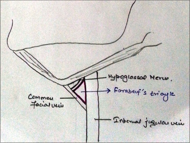
Schematic drawing of Farabeuf's triangle
Submandibular triangle is bordered superiorly by inferior border of the body of the mandible and lies between anterior and posterior belly of digastric muscle. Submandibular triangle is subdivided into an anterior and posterior part by stylomandibular ligament submandibular gland is the main content of this triangle and present in anterior part of this triangle, superficial to which is anterior facial vein and beneath the gland, on the surface of mylohyoid muscle, are submental artery and mylohyoid nerve and vessels. In the posterior part of this triangle is external carotid artery, ascending deep into the substance of parotid gland.[3] Beclard's, Lesser's and Pirogoff's triangles which are also known as forgotten/smaller triangles of neck are housed within submandibular triangle.
LESSER's TRIANGLE
It is a triangle contained within the submandibular triangle. Its boundaries are the hypoglossal nerve, and the anterior and posterior belly of the digastric muscle [Figure 2]. This triangle was named after a German surgeon named Ladislaus Leon Lesser, who lived from 1846 to 1925. Anterior belly of digastric and intermediate tendon of digastric forms the inferior boundary, superior border by hypoglossal nerve and posterior border is formed by posterior margin of mylohyoid muscle at the intermediate tendon of digastric muscle. Floor of this is formed by hyoglossus and mylohyoid muscles.[4]
Figure 2.
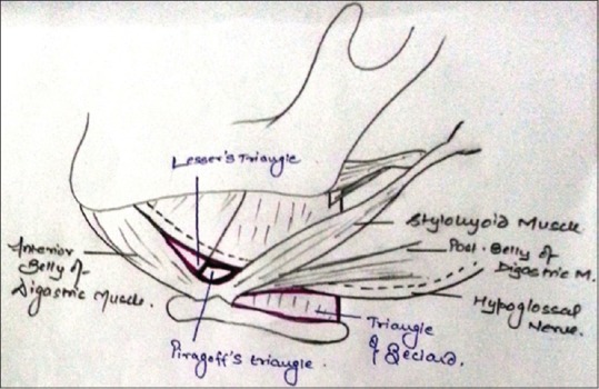
Schematic drawing of Lesser's, Beclard's and Pirogoff's Triangle adopted from Tubbset al[4]
PIROGOFF's TRIANGLE
It was named after Russian surgeon and scientist Nikolai I Pirogoff (1810–1881), who performed the first description of this anatomic area of the neck. Following are the boundaries of this triangle: Superior boundary is formed by hypoglossal nerve, inferior boundary is formed by intermediate tendon of digastric muscle and posterior border is formed by posterior border of mylohyoid muscle [Figure 2]. It is also considered that this triangle is posterior continuation of Lesser's triangle.[4]
Deaver in his book titled “Surgical anatomy of human body” referred this triangle as both Pinaud's and hypoglossohyoid triangle.[7] Sometimes, lingual artery can be found in the Pirogoff triangle underneath the fibers of the hyoglossus muscle.
BECLARD's TRIANGLE
It is named after French anatomist Pierre A. Beclard (1785–1825). Greater cornu of hyoid bone forms the inferior boundary, posterior belly of digastric muscle forms superior boundary, and posterior border of hyoglossus muscle forms posterior boundary and its base [Figure 2].
It is also known as posterior triangle of lingual artery and contents of this triangle are lingual artery and hypoglossal nerve. Sometimes, arcus raninus, i.e., anastomosis between right and left end branch of deep lingual artery is also present in this triangle. This triangle also helps in ligating external carotid artery.[4]
CONCLUSION
Added surgical landmarks are always helpful to the surgeon while dealing with the neck. These above-described triangles of the neck are always reliable and constant landmarks for head and neck surgeons in the identification of common carotid artery, ligation of lingual artery, in the identification of hypoglossal nerve for certain measures, and for recognition of the common facial vein.[6]
Financial support and sponsorship
Nil.
Conflicts of interest
There are no conflicts of interest.
REFERENCES
- 1.Lucioni M. Practical Guide to Neck Dissection. Berlin, Heidelberg, Germany: Springer; 2007. [Google Scholar]
- 2.Hollinshead WH. Anatomy for Surgeons: The Head and Neck. 3rd ed. Vol. I. Philadelphia: Harper & Row Publishers; 1982. [Google Scholar]
- 3.Harold E. Clinical Anatomy, A Revision and Applied Anatomy for Clinical Students. 11th ed. Newjersey: Blackwell Publishing Limited; 2006. [Google Scholar]
- 4.Tubbs RS, Rasmussen M, Loukas M, Shoja MM, Cohen-Gadol AA. Three nearly forgotten anatomical triangles of the neck: Triangles of Beclard, Lesser and Pirogoff and their potential applications in surgical dissection of the neck. Surg Radiol Anat. 2011;33:53–7. doi: 10.1007/s00276-010-0697-2. [DOI] [PubMed] [Google Scholar]
- 5.Beclard PA. Elements of General Anatomy. Philadelphia: Carey and Lea; 1830. [Google Scholar]
- 6.Tubbs RS, Rasmussen M, Loukas M, Shoja MM, Mortazavi M, Cohen-Gadol AA. Use of the triangle of Farabeuf for neurovascular procedures of the neck. Biomed Int. 2011;2:39–42. [Google Scholar]
- 7.Deaver JB. Surgical Anatomy of the Human Body. Philadelphia: The Blakiston co.; 1926. [Google Scholar]