Abstract
We have measured muscle fibre diameters using two methods of interactive computer-aided microscopy. They are simple to perform, reproducible and more convenient than manual methods of measurement. The technique is of general application to histological measurement.
Full text
PDF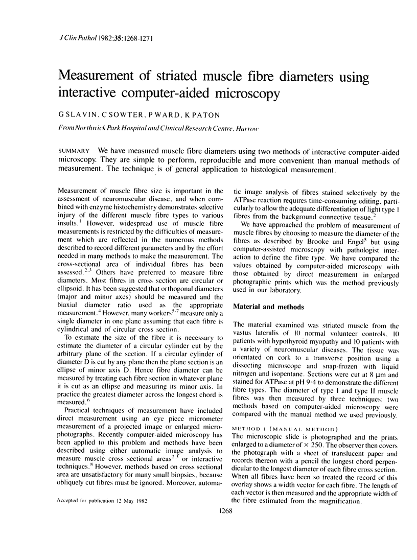
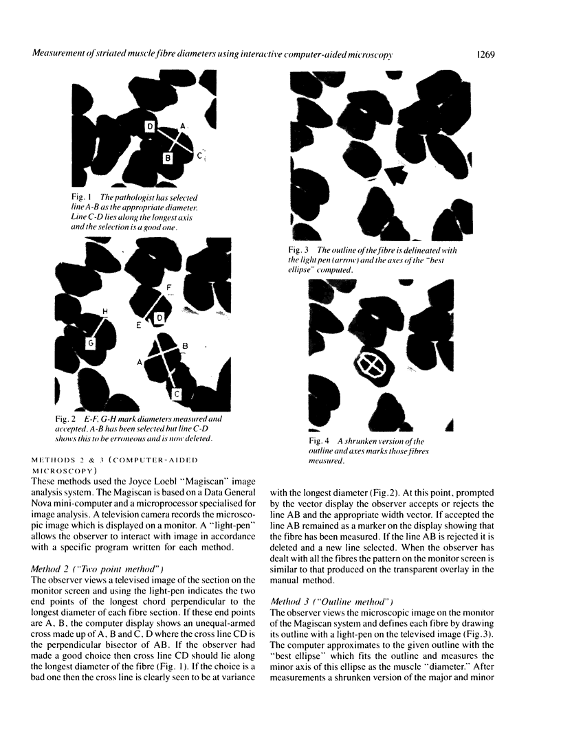
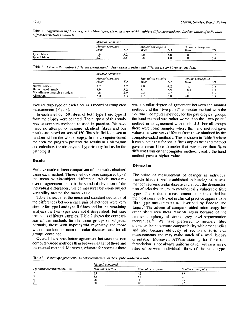
Images in this article
Selected References
These references are in PubMed. This may not be the complete list of references from this article.
- Brooke M. H., Engel W. K. The histographic analysis of human muscle biopsies with regard to fiber types. 1. Adult male and female. Neurology. 1969 Mar;19(3):221–233. doi: 10.1212/wnl.19.3.221. [DOI] [PubMed] [Google Scholar]
- Jaffe D. M., Terry R. D., Spiro A. J. Disuse atrophy of skeletal muscle. A morphometric study using image analysis. J Neurol Sci. 1978 Feb;35(2-3):189–200. doi: 10.1016/0022-510x(78)90002-3. [DOI] [PubMed] [Google Scholar]
- McKeran R. O., Slavin G., Andrews T. M., Ward P., Mair W. G. Muscle fibre type changes in hypothyroid myopathy. J Clin Pathol. 1975 Aug;28(8):659–663. doi: 10.1136/jcp.28.8.659. [DOI] [PMC free article] [PubMed] [Google Scholar]
- Round J. M., Jones D. A., Edwards R. H. A flexible microprocessor system for the measurement of cell size. J Clin Pathol. 1982 Jun;35(6):620–624. doi: 10.1136/jcp.35.6.620. [DOI] [PMC free article] [PubMed] [Google Scholar]
- SPRAGG S. P. USE OF A DIGITAL COMPUTER FOR EVALUATING ULTRACENTRIFUGE DATA. Nature. 1963 Dec 21;200:1200–1201. doi: 10.1038/2001200a0. [DOI] [PubMed] [Google Scholar]
- Scott K. W., Hoy J. The cross sectional area of diaphragmatic muscle fibres in emphysema, measured by an automated image analysis system. J Pathol. 1976 Oct;120(2):121–128. doi: 10.1002/path.1711200208. [DOI] [PubMed] [Google Scholar]


