Abstract
Background Despite its seldom occurrence, fetal tachycardia can lead to poor fetal outcomes including hydrops and fetal death. Management can be challenging and result in maternal adverse effects secondary to high serum drug levels required to achieve effective transplacental antiarrhythmic drug therapy.
Case A 33-year-old woman at 33 weeks of gestation with a diagnosis of a fetal sustained superior ventricular tachycardia developed chest pain, shortness of breath, and bigeminy on electrocardiogram secondary to digoxin toxicity despite subtherapeutic serum drug levels. She required supportive care with repletion of corresponding electrolyte abnormalities. After resolution of cardiac manifestations of digoxin toxicity, the patient was discharged home. The newborn was discharged at day 9 of life on maintenance amiodarone.
Conclusion We describe an interesting case of digoxin toxicity with cardiac manifestations of digoxin toxicity despite subtherapeutic serum drug levels. This case report emphasizes the significance of instituting an early diagnosis of digoxin toxicity during pregnancy, based not only on serum drug levels but also on clinical presentation. In cases of refractory supportive care, digoxin Fab fragment antibody administration should be considered. With timely diagnosis and treatment, excellent maternal and perinatal outcomes can be achieved.
Keywords: pregnancy, fetal SVT, digoxin, toxicity
Pathologic fetal tachycardia occurs in 1 in 25,000 pregnancies.1 It is defined as a fetal heart rate (HR) above 160 beats per minute (bpm) between 20 and 40 weeks gestational age. It is further classified as nonsustained and sustained tachycardias. Superior ventricular tachycardia (SVT) or paroxysmal atrioventricular (AV) re-entry tachycardia make up more than 50% of all fetal pathologic tachycardia.2 Primary atrial tachycardia (i.e., atrial flutter) and sinus tachycardia are responsible for the remaining cases.3 Known predisposing factors include congenital heart deformities, congenital diaphragm defects, illicit drug abuse, caffeine or nicotine exposure, and hyperthyroidism. Although infrequent, sustained fetal tachycardia can lead to high cardiac output heart failure and ultimately to fetal hydrops. The latter is a serious condition associated with poor fetal outcomes and mortality risk up to 30%. Diagnosis of in utero arrhythmias is primarily made by M mode and Doppler fetal echocardiogram. These tools are also useful in identifying the type of abnormal fetal heart rhythm and in evaluating for structural heart deformities. Management is primary based on transplacental antiarrhythmics such as digoxin, sotalol, flecainide, amiodarone, or propranolol with the goal to slow ventricular rate and avoid hydrops. Success of single drug therapy varies between 50 and 70%.4 Fetal response to therapy may take several days after achieving maintenance dose. Digoxin remains the first drug of choice used in the management of fetal tachycardia due to its safety profile and therapeutic success. Initiation of management usually requires in-house maternal and fetal monitoring and plasma drug level monitoring to avoid maternal and fetal toxicity. Clinical manifestations of digoxin toxicity include multiple arrhythmias such as AV blocks, atria and ventricular fibrillation, and bigeminy or premature beats, among others. The latter emphasizes the need for daily electrocardiogram (EKG) monitoring while achieving effective therapeutic digoxin levels. Patients may also report nausea, vomiting, abdominal pain, confusion, and weakness. Renal dysfunction and electrolyte disturbances (hypomagnesemia, hypercalcemia, and hypokalemia) can precipitate digoxin toxicity. Due to limitations of serum drug level testing or radioimmunoassay, detecting toxicity should be based mainly on clinical signs and symptoms rather than laboratory testing alone.
Case
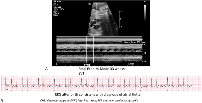
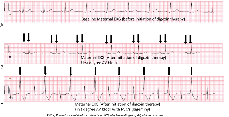
A 33-year-old primigravida at 33 weeks of gestation presented to our labor and delivery unit for evaluation of fetal tachycardia and leakage of fluid. Her prenatal course was unremarkable. She had no past surgical history or past medical history. Her vital signs were as follows: blood pressure 115/68 mm Hg, HR 73 bpm, respiratory rate 17 breaths per minute, and temperature 36.7°C. Her physical exam was unremarkable. After being ruled out for rupture of membranes, fetal ultrasound showed grossly normal fetus with an estimated fetal weight of 2,383 g and amniotic fluid index 15.6 cm. No signs of hydrops fetalis were noted. Electronic fetal HR monitoring showed a sustained baseline fetal heart of 210 bpm with minimal to moderate variability, with absent accelerations or decelerations. Middle cerebral artery Doppler ultrasonography was within normal. Fetal echo on M mode revealed a 1:1 atrial ventricular rate of 205 bpm consistent with SVT (Fig. 1A). Maternal baseline EKG was obtained and was normal (Fig. 2A). Baseline electrolytes and maternal thyroid panel were within normal limits. Plan was made for administration of betamethasone series for lung maturity and transplacental digoxin therapy for fetal rate control. Digoxin was administered with a 1.5 mg intravenous (IV) load over first 24 hours, followed by per os (PO) maintenance of 0.25 mg daily. Digoxin levels along with electrolytes were checked daily. Doses were adjusted targeting serum therapeutic levels between 1.5 and 2 ng/mL for maximum benefit for arrhythmia treatment. Despite adjusting the patient's digoxin dose for 6 days, serum digoxin levels remained low therapeutic (1.2 ng/mL) and the fetal arrhythmia persisted. Her last dose was 0.5 mg PO BID with an additional 0.25 mg given IV in a 24-hour period. On hospital day 7, a decision was made to move toward delivery for fetal benefits secondary to sustained fetal SVT. After regional anesthesia, the patient complained of chest discomfort and developed significant bradycardia (30 bpm) and telemetry revealed first-degree heart block (Fig. 2B). Ephedrine and glycopyrrolate were administered with resolution of symptoms and bradycardia. The patient was transferred to the postanesthesia care unit after completion of cesarean delivery. In the recovery room, she developed again significant bradycardia with bigeminy (Fig. 2C). At that time, laboratory workup revealed a potassium of 4.0 mmol/L (normal range: 3.5–5.0 mmol/L), magnesium of 1.5 mg/dL (normal range: 1.7–2.4 mg/dL), digoxin level 1.1 ng/mL (therapeutic 0.8–2.0 ng/mL), and ionized calcium of 4.7 mg/dL (normal range: 4.5–5.3 mg/dL). After IV infusion of magnesium, the patient became asymptomatic and the EKG anomalies resolved within 1 hour. The patient was discharged home on postoperative day 2. After delivery, the newborn was evaluated by pediatric cardiology and was found to have atrial flutter with a 1:1 conduction rate (Fig. 1B). Fetal echocardiogram results were normal. Despite vasovagal maneuvers, synchronized cardioversion attempts x3, adenosine, and procainamide administration, the flutter persisted. The arrhythmia later resolved after amiodarone administration. The newborn was discharged on day 9 of life on amiodarone and is currently off any medications and doing well.
Fig. 1.

(A) M mode fetal echo showing atrial ventricular relationship consistent with supraventricular tachycardia. (B) Newborn electrocardiogram showing atrial flutter.
Fig. 2.

Electrocardiogram of pregnant patient. (A) Baseline electrocardiogram before initiation of digoxin therapy. (B) Electrocardiogram after initiation of digoxin therapy (day 7). Arrows denote prolonged PR interval (first degree block: > 200 milliseconds). (C) Electrocardiogram after initiation of digoxin therapy (day 7). Arrows denote bigeminy.
Conclusion
The initial evaluation of a fetus with arrhythmia should include a detailed history and physical examination. The fetal HR tracing should be analyzed and a detailed fetal heart echocardiography should be performed to rule out congenital malformations. Similarly, maternal history of arrhythmias such as Wolf–Parkinson–White and prolonged QT syndrome should be investigated. Fetal heart evaluation should include M mode and pulsed Doppler ultrasonography to assess the AV relationship/atrial and ventricular rates.
The management of fetal SVT should be individualized. According to the American Heart Association, in utero SVT management should be based on gestational age, presence and degree of fetal compromise, maternal condition, degree of tachycardia, intermittent versus sustained tachycardia, and whether hydrops is present or not.5 When counseling patients, the risk of early delivery with potential preterm complications must be weighed against the therapies available and their effectiveness. Unless near term, all pregnant women with sustained fetal tachycardia should receive pharmacological treatment. Sustained fetal tachycardia is defined as fetal HR greater than 160 bpm present more than 50% of the time on fetal heart monitoring. Intermittent SVT does not usually require treatment unless there is cardiac compromise or findings consisting with hydrops. The mainstay of management involves transplacental administration of digoxin, flecainide, or sotalol (Table 1). Amiodarone is seldom used due to its higher toxicity profile. Digoxin, being the oldest and most consistent drug in pregnancy, is the initial drug of choice. Classically, in the presence of fetal hydrops, decreased transplacental drug transfer occurs secondary to placental enlargement and edema. In this case, double drug therapy or direct fetal treatment should be considered with intramuscular fetal administration of digoxin. Other indications to consider adding a second agent include refractory response despite maximum therapeutic dose for 5 days, unable to reach maximal dose due to maternal side effects, or need for faster conversion (e.g., premature hydrops fetus).
Table 1. Common antiarrhythmic drugs used for treatment of fetal arrhythmia.
| Drug | Administration | Potential adverse effects | Therapeutic level | Contraindications | FDA drug class |
|---|---|---|---|---|---|
| Digoxin | Total digitalizing dose (loading dose): 1–1.5 mg IV over 24 h (0.5 mg IV every 4–8 h) Initial maintenance dose: PO or IV 0.25–0.375 mg/d (will likely require higher doses during pregnancy due to increased renal clearance) |
Fatigue, visual changes, gastrointestinal distress, and cardiac arrhythmias: ventricular ectopy (earliest sign), atrial tachyarrhythmias, and high degree heart block | 0.8–2.0 ng/mL | Wolff–Parkinson–White syndrome (enhance antegrade conduction) AV block or sinus node dysfunction (HR < 50 bpm) History of ventricular fibrillation |
C |
| Flecainide | PO: 50–100 mg PO every12 h, increasing to a maximum of 300 mg/d | Dizziness, headache, visual disturbances, paresthesias, tremors, flushing, nausea, vomiting | 0.2–1.0 µg/mL | Sick sinus syndrome Atrial flutter (increased ventricular rate response) AV or bifasicular block (RBBB and left hemiblock) |
C |
| Sotalol | PO: 80 mg PO every 12 h, increasing to a maximum of 320 mg/d | Fatigue, dizziness, dyspnea, chest pain, palpitations 20–30% QTc prolongation |
Levels not available | AV block or sinus node dysfunction (HR < 50 bpm) QTc ≥ 480 ms Congenital or acquired long QT syndrome |
B |
Abbreviations: AV, atrioventricular; bpm, beats per minute; FDA, Food and Drug Administration; HR, heart rate; IV, intravenous; PO, per os; QTc, corrected QT interval; RBBB, right bundle branch block.
The diagnosis of digoxin toxicity remains challenging due to its nonspecific presentation and findings. Moreover, radioimmunoassay techniques are used to quantify serum digoxin levels in most medical centers.6 This assay has serious limitations making it difficult to correlate drug levels to clinical toxicity. First of all, the assay itself is subject to interference or result variability. This occurs secondary to critical methodical steps, such as plate washing, plate incubation times, and antibody affinity to bound versus free digoxin. Second, drug level results depend on the type of analyzer used. Moreover, published evidence shows a great variability in serum levels among patients with or without toxicity.7 Other factors that confound this relationship include the time the test is performed relative to the occurrence of the overdose and patient's characteristics including age, electrolyte abnormalities, cardiac disease, or thyroid disorders.
Electrolyte abnormalities may potentiate digoxin toxicity. Hypokalemia, hypomagnesemia, and hypercalcemia are all associated with digoxin toxicity and should be avoided at all times while treatment is in effect. As with any other pharmacological agent, timing of sampling is crucial in the diagnosis of toxicity. Levels obtained shortly after administering a dose may not reflect actual total levels, as further absorption and distribution of the agent may not be completed yet. Age is a critical factor with the use of digoxin; compared with adults at same serum levels, the pediatric population has been shown to be resistant to digoxin cardiotoxicity.8 Similar to the nonobstetrical population, pregnant women with hypothyroidism, electrolyte imbalances, and profound myocardial disease are more sensitive to digoxin and are more prone to develop toxicity at normal serum levels.9 Finally, endogenous digoxin-like substances may be responsible for incorrect high digoxin level results from the immunoassay, specially, in the newborn population, in patients with liver or kidney impairment, and in the pregnant population.8
Due to all the earlier limitations, we recommend physicians no to base their clinical decision solely on one laboratory value but rather on the whole clinical picture to manage digoxin toxicity in a timely fashion to achieve better maternal and fetal outcomes. Extracardiac manifestations of digoxin toxicity include fatigue, nausea, vomiting, anorexia, abdominal pain, weakness, and ocular symptoms affecting yellow and green color vision. Cardiac manifestations are potentially life threatening and require quick recognition and treatment. Second and third degree AV blocks are the result of significant depression of the AV node, while sinus bradycardia, sinus arrest, and sinus node block are related to interference with sinus node function. Enhanced impulse formation is seen with atrial, junctional, or ventricular arrhythmias. A combination of blockade of conduction and enhanced ectopic impulse formation is seen in atrial tachycardias with heart block.
Treatment of digoxin toxicity is mainly supportive, replacing, and treating any electrolyte abnormalities (potassium and magnesium with levels should be kept above 4.0 mmol/L and 2.0 mg/dL, respectively, at all times). Digoxin-specific Fab antibodies should be considered for refractory cases. Each vial of DigiFab (Protherics, Blaenwaun, Wales) (40 mg of Fab) binds 0.5 mg digoxin. The number of vials to be administered in patients with known steady-state serum level is calculated by multiplying the patient's weight in kilograms times the serum digoxin level in ng/mL divided by 100:
Number of vials to administer: (kg × serum level in ng/mL)/100
If unknown serum levels, it is recommended to administer 6 to 10 vials of DigiFab IV over 30 minutes.
When Digoxin Fab antibodies are administered, total digoxin levels rise significantly and should not be used to guide therapy unless free digoxin levels are obtained. It is also important to note that hypokalemia may be caused with the treatment of Digoxin Fab antibodies. This is especially important in patients who are hyperkalemic and treatment (glucose, insulin, and bicarbonate) has been already instituted in addition to Fab which can further exacerbate the hypokalemia.
The case presented in this report emphasizes the significance of instituting an early diagnosis of digoxin toxicity during pregnancy, based not only on serum drug levels but also on clinical presentation. Toxicity can occur even with subtherapeutic or therapeutic serum drug levels. In cases refractory to supportive care, digoxin Fab antibodies administration should be considered. With timely diagnosis and treatment, excellent maternal and perinatal outcomes can be achieved.
Précis
When considering transplacental digoxin therapy for fetal SVT, physicians should rely not only on serum drug levels but also on clinical manifestations of maternal drug toxicity.
Footnotes
Conflict of Interest The authors did not report any potential conflict of interest.
References
- 1.Owen P, Cameron A. Fetal tachyarrhythmias. Br J Hosp Med. 1997;58(4):142–144. [PubMed] [Google Scholar]
- 2.Vergani P, Mariani E, Ciriello E. et al. Fetal arrhythmias: natural history and management. Ultrasound Med Biol. 2005;31(1):1–6. doi: 10.1016/j.ultrasmedbio.2004.10.001. [DOI] [PubMed] [Google Scholar]
- 3.Jaeggi E. New York: Informa Healthcare London; 2009. Electrophysiology for the perinatologist; pp. 435–447. [Google Scholar]
- 4.Jaeggi E, Tulzer G. Philadelphia: Elsevier; 2009. Pharmacological and interventional fetal cardiovascular treatment; p. 199. [Google Scholar]
- 5.Donofrio M T, Moon-Grady A J, Hornberger L K. et al. Diagnosis and treatment of fetal cardiac disease: a scientific statement from the American Heart Association. Circulation. 2014;129(21):2183–2242. doi: 10.1161/01.cir.0000437597.44550.5d. [DOI] [PubMed] [Google Scholar]
- 6.Smith T W, Antman E M, Friedman P L, Blatt C M, Marsh J D. Digitalis glycosides: mechanisms and manifestations of toxicity. Part I. Prog Cardiovasc Dis. 1984;26(5):413–458. doi: 10.1016/0033-0620(84)90012-4. [DOI] [PubMed] [Google Scholar]
- 7.Lewander W J, Gaudreault P, Einhorn A, Henretig F M, Lacouture P G, Lovejoy F H Jr. Acute pediatric digoxin ingestion. A ten-year experience. Am J Dis Child. 1986;140(8):770–773. doi: 10.1001/archpedi.1986.02140220052031. [DOI] [PubMed] [Google Scholar]
- 8.Graves S W, Brown B, Valdes R Jr. An endogenous digoxin-like substance in patients with renal impairment. Ann Intern Med. 1983;99(5):604–608. doi: 10.7326/0003-4819-99-5-604. [DOI] [PubMed] [Google Scholar]
- 9.Bayer M J Recognition and management of digitalis intoxication: implications for emergency medicine Am J Emerg Med 1991920129–32., discussion 33–34 [DOI] [PubMed] [Google Scholar]


