

Abstract
Bedside pleuroscopy can be used in daily practice by medical pulmonologists if a patient cannot tolerate either general anesthesia or being moved to an operating or endoscopy room due to their critical condition. It is a simple and safe technique that rarely has complications. The aim of this review is to summarize recent literatures about bedside pleuroscopy and share our experiences with using it in Taiwan.
Keywords: Bedside, Intensive Care Unit (ICU), Pleuroscopy, Taiwan
1. Introduction
Pleuroscopy is an indispensable tool for the diagnosis and treatment of pleural disease. The instrument is easy to manipulate because the endoscope is of a similar design as a standard flexible bronchoscope. Previous studies have shown the efficacy of pleuroscopy, particularly in cases with pleural effusion [1-3]. Yet it is a procedure seldom used on critically-ill patients and in intensive care unit (ICU) bedsides, so here in we have reviewed bedside pleuroscopy and share our experiences with using it in Taiwan.
2. The development of pleuroscopy in Taiwan- Flexible pleuroscopy
The earliest use of a fexible bronchoscope as a fiberoptic pleuroscope was done in 1975 in America [4, 5]. At the time pulmonologists dubbed the technique “pleuroscopy”. Since then, flexible bronchoscopes have been introduced to hospitals around the world, especially where no suitable tools were available for the diagnosis or treatment of pleural disease [6, 7]. Recently, pleuroscopies performed under local anesthesia using a chest tube with a flexible fiberoptic bronchoscope have been reported [8, 9]. This technique has been used in many countries included developed and developing ones (Japan, China, Egypt, and Taiwan, for example) [8-10]. This so-called flexible fiberoptic pleuroscopy may be able to provide a diagnosis of exudative pleural effusions when other less invasive procedures fail to do so [11, 12].
This flexible pleuroscopy under local analgesia using a flexible bronchoscope is a simple procedure performed at the bedside and suitable for those critically-ill patients who cannot be moved to an operating or endoscopic room [13, 14]. At our institution, respiratory physicians have been performing pleuroscopies with a flexible bronchoscopies for over 5 years (since 2010) in the ICU [8,9]; however, there are still some limitations: They are as follows:
A pleuroscopy with a flexible bronchoscopy is more difficult to manipulate within the pleural cavity than within the bronchi and does not provide a good orientation within the pleural space. This procedure also has a long learning curve and needs supervision from an experienced endoscopist as well as a lot of practice [15, 16].
A flexible bronchoscope has a small specimen in comparison with a rigid thoracoscope. Thus, we have increased the biopsy site and during the procedure take more than 10 specimens [17, 18].
Damage done to the rubber shirt of the bronchoscope: we use a plastic trocar and a chest tube to protect the shirt of the bronchoscope [19, 20]. This helps to diminish the damage done to the tool. However, it must be considered that there are different facilities available in different countries or just different hospitals [21-23]. In our department, we did not have a semiflexible pleuroscope from 2010 to 2014, so we had to make do with using a flexible bronchoscope during that time for pleuroscopies at critically-ill patients’ bedsides once they were determined to require pleuroscopies [6-10].
However, the advantages of a flexible pleuroscopy are that at least it does not require extra- money to buy an additional instrument and that there is no need to worry about whether there is a suitable tool in the hospital to perform pleuroscopies [26, 27].
2.1. Semi-flexible pleuroscopy
A semi-flexible pleuroscope with rigid shafts and flexible tips was developed in 1978 in Japan by Takeno [28]. Today, the most commonly used semi-flexible pleuroscope was developed by Olympus Corporation in 2002 [29], with a working channel of 2.8 mm and incorporated video imaging. This semi-felxible pleuroscope was introduced into Taiwan only in 2014 after the various efforts of flexible pleursocopy performed at Taichung’s Tzu Chi Hospital [6-10]. This pleuroscope consists of a handle that is similar to a standard flexible bronchoscope and a shaft that measures 7 mm in outer diameter and 27 cm in length. The shaft is made up of two sections, a 22 cm proximal rigid portion and a 5 cm flexible distal end [30, 31]. The flexible tip is movable by a lever on the handle, which allows two-way angulation capability of 160° upward and 130° downward. It also has a 2.8-mm working channel that can accommodate biopsy forceps, needles, and other accessories and is compatible with various electrosurgical and laser procedures. The other advantage of the semi-flexible pleuroscope is that it interfaces easily with existing processors and light sources manufactured for flexible video bronchoscopy [32, 33].
3. Bedside pleuroscopy for critically-ill patients
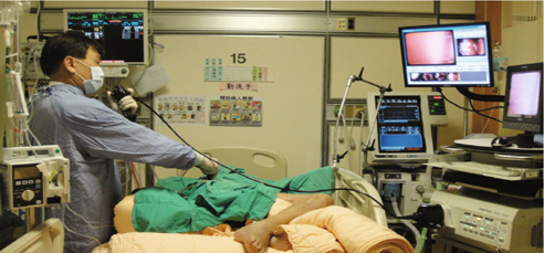
The main indication for the necessity of performing a pleuroscopy is an exudative pleural effusion with an unknown etiology [30-33]. For critically-ill patients with acute respiratory failure due to an unresolved exudative pleural effusion were challengeable [9, 10]. It is not always possible to move critically-ill patients to the operating or endoscopy room for a pleuroscopy due to their critical condition; and there is the uncertainty of the waiting time for the operation to consider too. Because the crude mortality rates are higher for intensive care unit (ICU) patients with pleural effusions than for those without pleural effusions, in 2010 we began using pleuroscopy at the bedside in the ICU to diagnose pleural effusions in patients with acute respiratory failure [Fig. 1]. This was when there were a large number of critically-ill patients that needed their pleural problems solved in the ICU in order to decrease their mortality rate [34, 35].
It is known that a standard rigid or semi-rigid thoracoscopy has several advantages over a pleuroscopy under local analgesia [35, 37], such as the ability to obtain larger biopsy specimens and better control of the bleeding. However, a thoracoscopy is a more invasive technique than a pleuroscopy that requires general anesthesia with a double-lumen endotracheal tube and selective lung ventilation [38, 39]. Therefore, a thoracoscopy has to be carried out by surgeons with a large number of operative instruments, and anesthesiologists are also needed in the surgical suite [40]. Pleuroscopy with local anesthesia is a less invasive and less expensive approach to thoracoscopy. Many studies have reported that pleuroscopy performed by pulmonologists is a safe and effective modality for the diagnosis of pleural effusions [36-40].
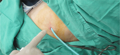
In our case in Taiwan, because of the small size of the trocar insertion wound and the small diameter of the instruments, we were able to use a pigtail 16 Fr catheter as the drainage catheter without any suture stitches. Standard chest tube insertion requires sutures and also uses tubes of a bigger size like 32 Fr [41]. To the best of our knowledge, this is the first use ever of bedside pleuroscopy in an ICU using a pigtail 16 Fr catheter [Fig. 2] for drainage without any suture stitches [6-9].
4. The etiology and pleural finding in ICU bedsides
The causes of most pleural effusions in critically-ill patients are secondary to malignancy or infections. The presences of malignancies (60%) and infections (36%) also have been noted in our study. We performed adhesiolysis at the time of pleuroscopy at ICU bedsides [42], as well as ensuring adequate drainage with a pigtail catheter to allow for the re-expansion of the lungs. The list of etiologies of pleural effusions is extensive; however, a bloody effusion with malignancy is still the main cause of an undiagnosed pleural effusion [43]. The endoscopic appearance of pleural lesions suggestive of a malignancy includes nodules, polypoid lesions, masses, and localized “candle wax drops” in the literature [44, 45].
Actually, most of the pleural lesions in critically-ill patients appear to be infiltrating (40%) or a combination of nodules and infiltrating (40%), but not all are nodular in appearance (20% are not) as found in previous studies [22, 23, 24]. Clinicians should be alert to this when performing a pleuroscopy for an undiagnosed pleural effusion in patients with acute respiratory failure [6-9].
5. Complications of bedside pleuroscopy
Major complications resulting from pleuroscopy have been reported in 0.0001 to 0.24% of patients [46], the most serious being bleeding or death. Major complications are not seen frequently,however. In our institution we had one patient who experienced CO2 narcosis during these periods, so protecting the patient’s airway and the equipped monitors was very important during the procedure. Minor complications of the procedure include subcutaneous emphysema, insignificant pneumothorax, wound pain, and postoperative fever and infection [47, 48]. All of these conditions have easily been controlled and have been self-limited [49, 50].
6. A great vision for critically-ill patients and intensivists
Rapid response or early management for critically-ill patients with undiagnosed exudative pleural effusion is very important. A quick clinical practice in the ICU at a patient’s bedside requires little to no waiting, and the easy to manipulated pleuroscopy has few complications. Indeed, bedside pleuroscopy is a simple and well-tolerated procedure with local analgesia. It can be used as a routine by medical pulmonologists or ICU physicians if their patients are not able to undergo general anesthesia or moving them to the operating or endoscopic room is unwise due to their critical condition. It is a great vision for all critically-ill patients and also their intensivists.
References
- 1.Wang B, Ge SJ. Nonintubated anesthesia for thoracic surgery. J Thorac Dis. 2014;6:1868–74. doi: 10.3978/j.issn.2072-1439.2014.11.39. [DOI] [PMC free article] [PubMed] [Google Scholar]
- 2.Chen JS. New directions and technologies for minimal invasive thoracic surgery. J Thorac Dis. 2014;6:1. doi: 10.3978/j.issn.2072-1439.2014.01.08. [DOI] [PMC free article] [PubMed] [Google Scholar]
- 3.Wang W, Peng GL, Guo ZH, Liang LX, Dong QL, He JX, et al. Radical resection of right upper lung cancer using uniportal videoassisted thoracic surgery with non-intubated anesthesia. J Thorac Dis. 2015;7:2362–65. doi: 10.3978/j.issn.2072-1439.2015.12.51. [DOI] [PMC free article] [PubMed] [Google Scholar]
- 4.Huang YK, Chou C, Li CL, Chiu HG, Chang YT. Minimally invasive thoracic surgery in pediatric patients: the Taiwan experience. Biomed Res Int 2013; 2013 [DOI] [PMC free article] [PubMed]
- 5.Chou YP, Kuo LC, Soo KM, Tarng YW, Chiang HI, Huang FD, et al. The role of repairing lung lacerations during video-assisted thoracoscopic surgery evacuations for retained haemothorax caused by blunt chest trauma. Eur J Cardiothorac Surg. 2014;46:107–11. doi: 10.1093/ejcts/ezt523. [DOI] [PMC free article] [PubMed] [Google Scholar]
- 6.Ooi H. A new modified autofluorescence pleuroscopy in the undiagnosed lung cancer with pleural effusion. Eur J Cancer. 2011;47:S215. doi: 10.1016/S0959-8049(11)71048-6. [DOI] [Google Scholar]
- 7.Ooi H, Chang SM, Liu CM, Chiu KL, Wu JY, Hsu NY. The bedside autofluorescence pleuroscopy for the undiagnosed lung cancer with pleural effusion in a intensive care unit. Eur Resp J. 2012;40:S56. [Google Scholar]
- 8.Ooi H, Liu MC, Chiu KL. Bedside Medical Thoracoscopy in Management of the Undiagnosed Refractory Exudative Pleural Effusion by Using a Flexible Bronchoscope and Pigtailed Catheter. Chest. 2010;138:347. doi: 10.1378/chest.10470. [DOI] [Google Scholar]
- 9.Ooi H, Chang SM, Liu CM, Chiu KL, W JY, Hsu NY, et al. Bedside pleuroscopy in the management of undiagnosed exudative pleural effusion with acute respiratory failure. Anaesth Intensive Care. 2013;41:473–5. doi: 10.1177/0310057X1304100406. [DOI] [PubMed] [Google Scholar]
- 10.Oldenburg FA., Jr A safe, accurate diagnostic procedure using the rigid thoracoscope and local anesthesia. Chest. 1979;75:45–50. doi: 10.1378/chest.75.1.45. [DOI] [PubMed] [Google Scholar]
- 11.Davidson AC, George RJ, Sheldon CD, Sinha G, Corrin B, Geddes DM. Thoracoscopy: assessment of a physician service and comparison of a flexible bronchoscope used as a thoracoscope with a rigid thoracoscope. Thorax. 1988;43:327–32. doi: 10.1136/thx.43.4.327. [DOI] [PMC free article] [PubMed] [Google Scholar]
- 12.Takeno Y. Thoracoscopic treatment of spontaneous pneumothorax. Ann Thorac Surg. 1993;56:688–90. doi: 10.1016/0003-4975(93)90953-F. [DOI] [PubMed] [Google Scholar]
- 13.Janssen JP, Thunissen FBJM, Visser FJ. Comparison of the 2.0 mm and 3.5 mm minithoracoscopy set to standard equipment for medical thoracoscopy. Eur Respir J. 2003;22(45):S541. [Google Scholar]
- 14.Casal RF, Eapen GA, Morice RC, Jimenez CA. Medical thoracoscopy. Curr Opin Pulm Med. 2009;15:313–20. doi: 10.1097/MCP.0b013e32832b8b2d. [DOI] [PubMed] [Google Scholar]
- 15.Lee P, Mathur PN, Colt HG. Advances in thoracoscopy: 100 years since Jacobaeus. Respiration. 2010;79:177–86. doi: 10.1159/000268617. [DOI] [PubMed] [Google Scholar]
- 16.Janssen JP. Why you do or do not need thoracoscopy. Eur Respir Rev. 2010;19:213–16. doi: 10.1183/09059180.00005410. [DOI] [PMC free article] [PubMed] [Google Scholar]
- 17.Antony VB, Loddenkemper R, Astoul P, Boutin C, Goldstraw P, Hott J, et al. Management of malignant pleural effusions. (ATS/ERS Statement) Eur Respir J. 2001;18:402–19. doi: 10.1183/09031936.01.00225601. [DOI] [PubMed] [Google Scholar]
- 18.Rahman NM, Ali NJ, Brown G, Chapman SJ, Davies RJ, et al. Local anaesthetic thoracoscopy: British Thoracic Society pleural disease guideline 2010. Thorax 2010. 2010;65(2):ii54–ii60. doi: 10.1136/thx.2010.137018. [DOI] [PubMed] [Google Scholar]
- 19.Tassi G, Marchetti G. Minithoracoscopy: a less invasive approach to thoracoscopy. Chest. 2003;124:1975–77. doi: 10.1378/chest.124.5.1975. [DOI] [PubMed] [Google Scholar]
- 20.Tassi GF, Marchetti GP, Pinelli V. Minithoracoscopy: a complementary technique for medical thoracoscopy. Respiration. 2011;82:204–06. doi: 10.1159/000324072. [DOI] [PubMed] [Google Scholar]
- 21.McLean AN, Bicknell SR, McAlpine LG, Peacock AJ. Investigation of pleural effusion: an evaluation of the new Olympus LTF semiflexible thoracofiberscope and comparison with Abram’s needle biopsy. Chest. 1998;114:150–53. doi: 10.1378/chest.114.1.150. [DOI] [PubMed] [Google Scholar]
- 22.Harris RJ, Kavuru MS, Rice TW, Kirby TJ. The diagnostic and therapeutic utility of thoracoscopy. A review. Chest. 1995;108:828–41. doi: 10.1378/chest.108.3.828. [DOI] [PubMed] [Google Scholar]
- 23.Loddenkemper R. Thoracoscopy—state of the art. Eur Respir J. 1998;11:213–21. doi: 10.1183/09031936.98.11010213. [DOI] [PubMed] [Google Scholar]
- 24.Munavvar M, Khan MA, Edwards J, Waqaruddin Z, Mills J. The autoclavable semirigid thoracoscope: the way forward in pleural disease? Eur Respir J. 2007;29:571–74. doi: 10.1183/09031936.00101706. [DOI] [PubMed] [Google Scholar]
- 25.Lee P, Colt HG. Rigid and semirigid pleuroscopy: the future is bright. Respirology. 2005;10:418–25. doi: 10.1111/j.1440-1843.2005.00737.x. [DOI] [PubMed] [Google Scholar]
- 26.Loddenkemper R. Medical Thoracoscopy/Pleuroscopy: step by step. Breathe. 2011;8:156–68. doi: 10.1183/20734735.011611. [DOI] [Google Scholar]
- 27.Medford AR. Theoretical cost benefits of medical thoracoscopy (MT) Respir Med. 2010;104:1075–76. doi: 10.1016/j.rmed.2010.03.005. [DOI] [PubMed] [Google Scholar]
- 28.Boutin C, Viallat JR, Cargnino P, Farisse P. Thoracoscopy in malignant pleural effusions. Am Rev Respir Dis. 1981;124:588–92. doi: 10.1164/arrd.1981.124.5.588. [DOI] [PubMed] [Google Scholar]
- 29.Schwarz C, Lübbert H, Rahn W, Schönfeld N, Serke M, Loddenkemper R. Medical thoracoscopy: hormone receptor content in pleural metastases due to breast cancer. Eur Respir J. 2004;24:728–30. doi: 10.1183/09031936.04.00069104. [DOI] [PubMed] [Google Scholar]
- 30.Colt HG. Thoracoscopic management of malignant pleural effusions. Clin Chest Med. 1995;16:505–18. [PubMed] [Google Scholar]
- 31.Liu YJ, Hung MH, Hsu HH, Chen JS, Cheng YJ. Effects on respiration of nonintubated anesthesia in thoracoscopic surgery under spontaneous ventilation. Ann Transl Med. 2015;3:107. doi: 10.3978/j.issn.2305-5839.2015.04.15. [DOI] [PMC free article] [PubMed] [Google Scholar]
- 32.Steiropoulos P, Kouliatsis G, Karpathiou G, Popidou M, Froudarakis ME. Rare cases of primary pleural Hodgkin and nonHodgkin lymphomas. Respiration. 2009;77:459–63. doi: 10.1159/000135255. [DOI] [PubMed] [Google Scholar]
- 33.Boutin C, Rey F. Thoracoscopy in pleural malignant mesothelioma: a prospective study of 188 consecutive patients. Part 1: Diagnosis. Cancer. 1993;72:389–93. doi: 10.1002/1097-0142(19930715)72:2<389::AID-CNCR2820720213>3.0.CO;2-V. [DOI] [PubMed] [Google Scholar]
- 34.Scherpereel A, Astoul P, Baas P, Berghmans T, Clayson H, de Vuyst P, et al. Guidelines of the European Respiratory Society and the European Society of Thoracic Surgeons for the management of malignant pleural mesothelioma. Eur Respir J. 2010;35:479–95. doi: 10.1183/09031936.00063109. [DOI] [PubMed] [Google Scholar]
- 35.Boutin C, Viallat JR, Aelony Y. Practical thoracoscopy. Berlin Heidelberg New York, Springer, 1991. 56. Loddenkemper R, Boutin C. Thoracoscopy: present diagnostic and therapeutic indications. Eur Respir J. 1993;6:1544–55. [PubMed] [Google Scholar]
- 36.Ernst A, Hersh CP, Herth F, Thurer R, LoCicero J, 3rd, Beamis J, et al. A novel instrument for the evaluation of the pleural space: an experience in 34 patients. Chest. 2002;122:1530–34. doi: 10.1378/chest.122.5.1530. [DOI] [PubMed] [Google Scholar]
- 37.Froudarakis ME. New challenges in medical thoracoscopy. Respiration. 2011;82:197–200. doi: 10.1159/000324266. [DOI] [PubMed] [Google Scholar]
- 38.Rodriguez-Panadero F, Antony VB. Pleurodesis: State of the art. Eur Respir J. 1997;10:1648–54. doi: 10.1183/09031936.97.10071648. [DOI] [PubMed] [Google Scholar]
- 39.Diacon AH, Van de Wal BW, Wyser C, Smedema JP, Bezuidenhout J, Bolliger CT, et al. Diagnostic tools in tuberculous pleurisy: a direct comparative study. Eur Respir J. 2003;22:589–591. doi: 10.1183/09031936.03.00017103a. [DOI] [PubMed] [Google Scholar]
- 40.Hooper C, Lee YCG, Maskell N. Investigation of a unilateral pleural effusion in adults: British Thoracic Society pleural disease guidelines 2010. Thorax 2010 [DOI] [PubMed]
- 41.Chung CL, Chen CH, Yeh CY. Early effective drainage in the treatment of loculated tuberculous pleurisy. Eur Respir J. 2008;31:1261–7. doi: 10.1183/09031936.00122207. [DOI] [PubMed] [Google Scholar]
- 42.Hung MH, Hsu HH, Cheng YJ, Chen JS. Nonintubated thoracoscopic surgery: state of the art and future directions. J Thorac Dis. 2014;6:2–9. doi: 10.3978/j.issn.2072-1439.2014.01.16. [DOI] [PMC free article] [PubMed] [Google Scholar]
- 43.Hung WH, Hsu HH, Hung MH, Hsieh PY, Cheng YJ, Chen JS. Nonintubated uniportal thoracoscopic surgery for resection of lung lesions. J Thorac Dis. 2016;8:S242–S50. doi: 10.3978/j.issn.2072-1439.2016.02.09. [DOI] [PMC free article] [PubMed] [Google Scholar]
- 44.Luh SP, Liu HP. Video-assisted thoracic surgery―the past, present status and the future. J Zhejiang Univ Sci B. 2006;7:118–28. doi: 10.1631/jzus.2006.B0118. [DOI] [PMC free article] [PubMed] [Google Scholar]
- 45.Luh SP, Liu HP. Video-assisted thoracoscopic surgery (VATS) for the treatment of hepatic hydrothorax: report of twelve cases. J Zhejiang Univ Sci B. 2009;10:547–51. doi: 10.1631/jzus.B0820374. [DOI] [PMC free article] [PubMed] [Google Scholar]
- 46.Chou SH, Cheng Y J, Kao EL, Chai CY. Spontaneous haemothorax: an unusual presentation of primary lung cancer. Thorax. 1993;48:1185–86. doi: 10.1136/thx.48.11.1185. [DOI] [PMC free article] [PubMed] [Google Scholar]
- 47.Sole`r M, Wyser C, Bolliger CT, Perruchoud AP. Treatment of early parapneumonic empyema by ‘‘medical’’ thoracoscopy. Schweiz Med Wochenschr. 1997;127:1748–1753. [PubMed] [Google Scholar]
- 48.Loddenkemper R, Kaiser D, Frank W. Treatment of parapneumonic pleural effusion and empyema—conservative view. Eur Respir Mon. 2004;29:199–207. [Google Scholar]
- 49.Brutsche MH, Tassi GF, Gyo ¨rik S, Gökcimen M, Renard C, Marchetti GP, et al. Treatment of sonographically stratified multiloculated thoracic empyema by medical thoracoscopy. Chest. 2005;128:3303–09. doi: 10.1378/chest.128.5.3303. [DOI] [PubMed] [Google Scholar]
- 50.Kern L, Robert J, Brutsche M. Management of parapneumonic effusion and empyema: medical thoracoscopy and surgical approach. Respiration. 2011;82:193–96. doi: 10.1159/000326337. [DOI] [PubMed] [Google Scholar]