

Abstract
Objective:
Aim of this study is to evaluate antiurolithiatic potential of whole plant hydro-alcoholic (30:70) extract of Vernonia cinerea Less. in accordance to its claims made in ancient literature and also being one of the ingredients of cystone, a marketed formulation widely used in the management of urolithiasis.
Materials and Methods:
To induce urolithiasis, 0.75% v/v ethylene glycol was administered orally for 14 days. The curative dose of 400 mg/kg b.w. and preventive doses of 100, 200, and 400 mg/kg b.w. were administered from 15th to 28th and 1st to 28 days, respectively. Cystone 750 mg/kg b.w. was selected as the reference standard for both curative and preventive doses. On 28th day, urinate of 24 h was collected and subjected for estimation of calcium, oxalate, and phosphates. Serum biochemical and kidney homogenate analysis was done for determination of renal oxalate contents.
Results:
The diseased Group II showed marked increase (P < 0.001 vs. normal Group I) in levels of urine calcium, oxalate, and phosphate. Serum creatinine, urea, and uric acid levels were also increased. Histopathological studies of kidney sections revealed significant changes. Treatment with hydro-alcoholic extract of V. cinerea showed significant (P < 0.01 vs. calculi-induced Group II) dose-dependent activity. A progressive increase in urine output, body weight, and decline in concentrations of stone-forming components such as calcium, oxalates, and phosphates was observed.
Conclusion:
It can be inferred that V. cinerea Less. is effective in ethylene glycol-induced urolithiasis and may have a potential in preventing and curing urolithiasis.
Keywords: Antiurolithiatic, cystone, ethylene glycol-induced urolithiasis, Vernonia cinerea Less
The most consistent feature of urolithiasis is the formation and retention of stone (s) in various parts of urinary tract, the kidney, ureter, and bladder wherein the size and type of calculi vary significantly. As per the statistical data reported 12% of world population is affected with urolithiasis. The prevalence is more in the working class between the age group of 25–50. It occurs more in men as compared to women, may be because of enhancing testosterone and inhibitory property of estrogen in the pathogenesis of uroliths.[1]
Urolithiasis is a comprehensive disease condition wherein various endogenous and/or exogenous etiological factors, as well as multivariate pathogenesis, are involved. Endogenous factors include improper metabolism of calcium and phosphorous, metabolism of oxalic acid, uric acid, and cystine. Furthermore, hyperthyroidism, difficulties in excretion of nitrogenous waste products may also be a major contributing factor in stone formation. Exogenous factors comprise food habits, dehydration, less fluid intake, hot climatic conditions, excessive consumption of some vitamins like Vitamin A and D, unsafe or unnecessary use of certain drug substances and hard water usage for drinking purpose.[2]
Calcium oxalate stones are most common neproliths, accounting for more than 80% of stones, whereas 5–10% of uric acid stones are present. The other types are cystine, struvite, and urate stones, have very less percentage. Urolithiasis is the outcome of various physicochemical changes such as supersaturation of urine, nucleation, growth of crystal and aggregation. Urine is invariably saturated with the stone-forming components such as calcium, oxalate, urate, cystine, xanthenes, and phosphate. However, the natural tendency to inhibit crystallization prevents stone formation, whereas this natural inhibition capacity varies person to person and which is poor in stone formers.[3] Revolutionary developments in surgical procedures and extracorporeal shock wave lithotripsy (ESWL) in the treatment of kidney stone has taken place, but the expenses, and recurrence or relapse rate still remains the challenge. Moreover, renal damage and decreased kidney function becomes the major concern in these procedures.[4] Diverse therapeutic approaches to treat urolithiasis were adopted such as diuretics, chelating agents (magnesium citrate, magnesium, and citrates), and diet therapy (Increased water intake, reduced intake of Vitamin C, guidance regarding calcium intake, etc).[5]
Despite these advancements in treatment of kidney stone, no satisfactory medication is available. The evidence-based information suggests that the drugs and formulations derived from herbal source are widely preferred as a promising therapy in treating urolithiasis. The hypothetical underlying mechanism of these medicinal plant materials is believed to be, the effect on kidney and urinary blood circulation, diuresis, changes the pH of urine and also exerts spasmolytic and analgesic effect. These are interrelated with the liver, pancreas, and intestines through metabolism and removal of stone-forming components.[2]
Emphasis was given by the WHO (2002) on the development and standardization of herbal medicine due to its cost effectiveness and less or no side effects. As the advancement took place, the gap between the modern and traditional medicine widened due to lack of scientific evidence and its less convincing therapeutic effects, where safety remains a major concern.
Vernonia cinerea L. root is used as diuretic[6] and is one of the major ingredients of well-known marketed preparation Cystone (Himalaya Herbal Healthcare – Bengaluru, Karnataka, India) used for treating urolithiasis. Phytochemical investigation revealed the presence of alkaloids, saponins, triterpenoids, steroids, and flavonoids.[7] It has been screened for its analgesic, antipyretic, anti-inflammatory effects.[8] Flower extract is used in arthritis[9] whereas leaf extract showed larvicidal activity.[10] Although claims are made, no scientific data are available supporting its diuretic activity. Nevertheless, diuretic activity alone cannot be considered responsible for preventing and curing urolithiasis. The aim of this study undertaken is to evaluate and validate the antiurolithiatic potential of hydro-alcoholic extract of V. cinerea Less against ethylene glycol-induced urolithiasis in rats.
Materials and Methods
Chemicals
The chemicals and solvents used were of analytical grade procured from Sigma-Aldrich Co., USA. The kits used for urine estimation and serum analysis purchased from Erba Mannheim, Transasia Biomedicals Ltd., Solan. Ethlyene glycol was obtained from S D Fine Chemicals. The standard marketed preparation – Cystone tablets (Himalaya Herbal Healthcare – Bengaluru, Karnataka, India) were purchased from the market.
Collection and Authentication of Plant Material
V. cinerea Less. plant was collected from botanical garden of Shri B M Kankanwadi Ayurveda Mahavidyalaya, Belagavi and nearby areas of Belagavi. The plant material was identified and authenticated by Scientist B, RMRC, Belagavi. The herbarium was prepared and stored at RMRC, Belagavi, Karnataka, India, bearing specimen number RMRC-913.
Preparation of Plant Extract
The collected whole plant V. cinerea was cleaned properly, dried in shade and further pulverized to obtain a coarse powder. The powder, thus, obtained was extracted with hydro-alcohol ([30:70] water and 95% ethanol) using soxhlet apparatus. The extract was concentrated using rotary evaporator (IKA RV 10 Digital) under reduced pressure at 40°C to obtain (Yield - 9.6% w/w) a semisolid mass. It was labeled and stored in a glass bottle for further studies. A suspension of the extract was formulated using 5% Tween-80 for oral administration.[3] The extract was analyzed for various phytochemical constituents (flavonoids, alkaloids, tannins, phytosterols, glycosides, terpenoids, and saponins) using standard procedures.[11,12,13,14]
Antiurolithiatic Activity
Considering various parameters, the hydro-alcoholic extract of V. cinerea Less was screened for its antiurolithiatic potential.
Animal selection
Wistar albino rats weighing about 150–200 g, of either sex, were selected for acute toxicity study and male adult rats were used for screening of antiurolithiatic activity. The experimental protocol was approved by Institutional Animal Ethical Committee vide Resolution No. KLECOP/IAEC/Res. 16-20/04/2013. The animals were kept in transparent polypropylene cages at a temperature 25°C ± 2°C and 12–12-day night cycle. The animals were provided with standard pellets and drinking water ad libitium. They were acclimatized to the laboratory conditions and randomly divided into eight groups.
Acute oral toxicity study
The acute oral toxicity study was performed as per OECD No. 425 guidelines. For the hydro-alcoholic extract of V. cinerea, a starting dose of 2000 mg/kg b.w. was selected. After overnight fasting, the animals were administered with extract orally with gastric intubation method. The animals were observed for any physiological and behavioral changes for first 4 h continuously, then at every 4 h for 12 h, followed by for first 14 days daily once. The LD50 cut off dose was determined, and 1/10th of it was considered for antiurolithiatic activity.[15]
Experimental protocol
Animals were divided into eight groups containing six animals in each group. During the experimental protocol, animals were allowed free access to food and water.
Group I: Normal control
Group II: Calculi-induced, received 0.75% ethylene glycol in drinking water from 1st to 14th day and were left untreated
Group III: Standard curative-received 400 mg/kg b.w. standard drug cystone from day 15th to 28th
Group IV: Standard preventive-received standard drug cystone from day 1st to 28th
Group V: Curative– received 400 mg/kg b.w. hydro-alcoholic extracts from day 15th to 28th
Group VI: Preventive I-received 100 mg/kg b.w. hydro-alcoholic extracts from day 1st to 28th
Group VII: Preventive II-received 200 mg/kg b.w. hydro-alcoholic extracts from day 1st to 28th
Group VIII: Preventive III-received 400 mg/kg b.w. hydro-alcoholic extracts from day 1st to 28th.
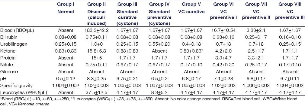
Collection and Analysis of Urine
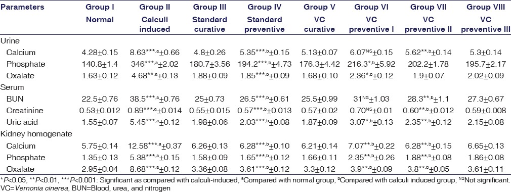
The animals were kept in polypropylene cages separately, and 24 h urine was collected on the 28th day. The volume of urine collected from each animal of all the groups was measured. During the study, the animals were having free access to drinking water. The collected urine was stored at 4°C by adding a drop of concentrated. HCl. Using UroColor 10 strips (SD Bio Standard Diagnostics) routine urine analysis was done for the presence of protein, glucose, blood, nitrite, etc., Furthermore, quantitative determination of specific gravity and pH was done. Urine was analyzed for calcium, phosphate, and oxalate using semi-auto analyzer[16,17] [Table 1].
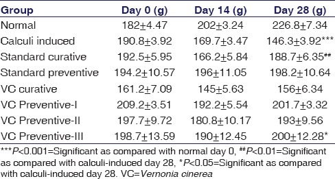
Table 1.
Effect of Vernonia cinerea on urine and serum parameters in experimental animals-curative/preventive dose
Urine Volume
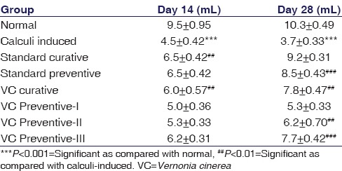
On 14th and 28th day, 24 h urine was collected of all the eight groups, measured the urine volume and recorded.
Change in Body Weight
The difference in initial body weight (day 0) and final body weight (day 14 and day 28) was noted, and further comparative analysis was done among different groups.
Urine Microscopy
Urine microscopy was done on a 28th day to observe the presence of crystals of CaOX and phosphates at ×40, with characteristic size and shape. For this, an electron microscope (Labomed TCM 400) fitted with a Canon digital camera (12 megapixel) was used, and photographs were taken.
Serum Analysis
From the anesthetized animals, the blood sample was withdrawn from the retro-orbital sac. It was centrifuged at 14,500 rpm (14,100 ×g) for 10 min in a centrifuge machine (Eppendorf, Minispin plus); serum was separated and analyzed for blood, urea and nitrogen (BUN), creatinine and uric acid.
Kidney Homogenate Analysis
After the animals were sacrificed, the abdomen was cut open to remove both the kidneys from each animal. The kidneys were cleaned and preserved in formalin (10%). A sample of one kidney (100 g) was boiled for ½ h in 10 mL of 1 N HCl and homogenized. It was further centrifuged at 2000 rpm (1930 ×g) for 10 min; supernatant was analyzed for calcium, oxalate, and phosphate content.
Histopathological Analysis of Kidney
Examination of histopathological changes such as tubular congestion, tubular necrosis, glomerular congestion, peritubular inflammation, hemorrhage, and presence of calculi was done. The remaining kidney was embedded in liquid paraffin, 5 µm sections were taken, stained with hematoxylin and eosin and mounted in diphenyl xylene. The microscopic examination of thus prepared slides was done using compound microscope (×50). To determine the nephritic damage and recovery, photographs were taken and scored in the format as, no damage (−), Mild (+), Moderate (++), and Severe (+++) damage. For this, at least 10 fields were analyzed and marked.[18]
Statistical Analysis
Results were expressed as mean ± standard error of the mean. One-way analysis of variance, followed by Dunnett's multiple comparison test (GraphPad Prism software for Windows, Version 5.01, GraphPad Software, Inc. 7825 Fay Avenue, Suite 230 La Jolla, CA 92037 USA) was used to analyze the statistical significance among different groups. The P < 0.05 was considered to be statistically significant.
Results
The phytochemical study of V. cinerea Less. confirmed the presence of alkaloids, saponins, triterpenoids, glycosides, steroids, and flavonoids.
In acute toxicity study, the extract does not show any adverse clinical signs and mortality as well. Therefore, 2000 mg/kg b.w. was considered as LD50 cut off dose and 1/10th of it was taken as therapeutic dose for the study, i.e. 200 mg/kg b.w.
The preventive and curative antilithiatic effects of hydro-alcoholic extract of V. cinerea were evaluated considering various parameters. In this study, ethylene glycol (0.75% v/v) in drinking water was administered to induce calculi except normal control Group I. The preliminary confirmation was done by doing urine microscopy where the presence of various stones was observed [Figure 1]. As per the data reported, urine calcium, oxalate, and phosphate excretion significantly increased (P < 0.001) in calculi-induced (Group II) as compared to normal control (Group I). However, in both the preventive and curative dose regimen, there was an increased degree (P < 0.001–0.05) of reduction in the levels of urine calcium, oxalate, and phosphate, relevant to the dose of hydro-alcoholic extract administered, and highly significant decrease was observed in cystone treated group [Table 1, Group III to VIII]. Increase in dissolution and significant reduction (P < 0.001 vs. Group II) in the concentration of calcium, phosphate, and oxalates results in better prevention of stone formation.
Figure 1.
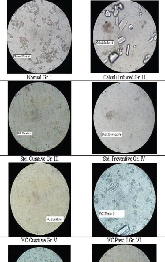
Microscopic study (×40) of urine collected of Group I to Group VIII
In the analysis of other routine urine parameters, it was observed that there was increase in urinary pH of 8.3 in calculi-induced Group II, but it was brought down gradually toward normal value in the groups treated with hydro-alcoholic extract of V. cinerea and cystone. In other groups, the parameters such as ketone bodies, nitrites, bilirubin, urobilinogen, and leukocytes were statistically insignificant. All the groups showed absence of glucose in urine. In calculi-induced group, the severe nephritic damage was confirmed by the presence of protein in urine. There was a considerable recovery recorded with a decrease in protein in the groups treated with both preventive and curative dose regimen of cystone and hydro-alcoholic extract of V. cinerea [Table 2].
Table 2.
Effect of Vernonia cinerea hydro-alcoholic extracts on urine analysis parameters in rats in curative/preventive dose regimen
In diseased Group II, urine output was drastically reduced (P < 0.001 vs. Group I). There was increased (P < 0.001) urine output observed in the groups treated with cystone and hydro-alcoholic extract of V. cinerea [Table 3]. The increase in urine volume was appreciable on day 28 when compared with day 14.
Table 3.
Effect of hydro-alcoholic extract of Vernonia cinerea on urine volume in urolithiatic animals-curative/preventive dose
A consistent growth in body weight was recorded [Table 4] in normal control (Group I) animals, whereas significant (P < 0.001) weight loss was observed in calculi-induced Group II. Cystone treated animals (Group III and IV) gained body weight significantly (P < 0.01). The progressive increase in body weight was recorded on day 14 and day 28, in animals treated with extract (Group V, VI, VII, and VIII).
Table 4.
Effect of hydro-alcoholic extract of Vernonia cinerea on change in body weight in urolithiatic animals curative/preventive dose
Microscopic examination of urine of normal control animal's (Group I) showed the absence of renal calculi, whereas in calculi-induced Group II showed rectangular, larger calcium oxalate crystals [Figure 1]. Cystone treated animals (Group III and IV) showed absence or stones were almost dissolved whereas hydro-alcoholic extract of V. cinerea curative dose showed equally less amount of crystals in urine. In preventive treatment groups, an increased absence of small crystal and similar structures were observed in accordance with the dose given (Group VI < VII < VIII). However, small fragments of crystals were observed in lower dose (Group VI).
In serum analysis, increased values of BUN, creatinine, and uric acid were reported in calculi-induced animals (Group II) as compared to normal control animals (Group I). This results in severe nephritic injury and impaired renal function. The animals treated with cystone, and hydro-alcoholic extracts of V. cinerea showed significant decrease in corresponding values thus protecting renal injury and improved kidney function.
Kidney homogenate analysis revealed increased amount (P < 0.001) of stone-forming components calcium, phosphate, and oxalate in diseased animals (Group II vs. Group I). The animals treated with cystone (Group III and IV) showed decreased levels of stone-forming components. Whereas animals treated with hydro-alcoholic extract of V. cinerea curative (Group V) and preventive (Group VI, VII, and VIII) dosage regimen showed reduced content of calcium, phosphate, and oxalate.
Histopathology of kidney, confirms the absence of performed stones [Figure 2a] and associated relevant abnormalities in normal control animals (Group I). A significant (P < 0.001) CaOX crystal deposition was observed in calculi-induced group II. The associated abnormalities such as peritubular congestion, glomerular congestion, epithelial desquamation, blood vessel congestion, and inflammation of cells were noted [Figure 2b]. Both, curative [Figure 2c] and preventive [Figure 2d] treatment with cystone and extract [Curative - Figure 2e and Preventive I (2f), II (2g), III (2h) resp.] decreased CaOX depositions and associated abnormalities significantly.
Figure 2.
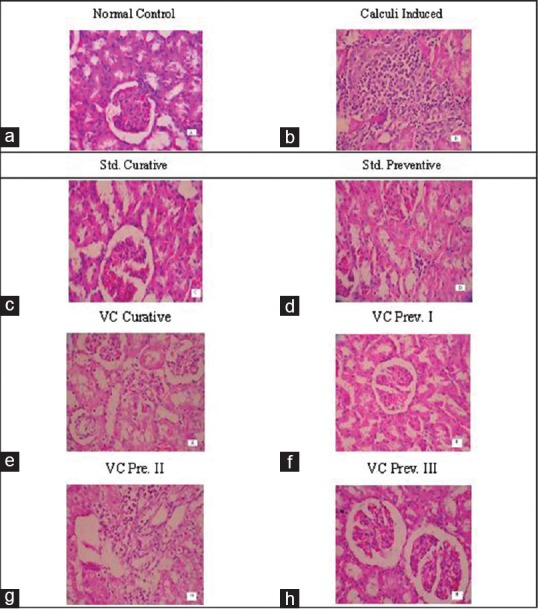
Histopathological examination of kidney section of. (a) Normal control, (b) calculi-induced, (c) standard curative, (d) standard preventive (e) Vernonia cinerea curative, (f) Vernonia cinerea Prev. I, (g) Vernonia cinerea Prev. II (h) Vernonia cinerea Prev. III
Discussion
Kidney stone formation is an aggravating disease condition for human being. The patients seeking treatments in present days will be prescribed with alkalizers, diuretics, anti-inflammatory, analgesics, and antispasmodic drugs. Not only this, the technological advancements provides the treatments like ESWL, surgery, percutaneous nephrolithotripsy to get rid of uroliths.[19] However, these techniques are associated with various side effects such as hemorrhage, hypertension, tubular necrosis, and cell injury. Moreover, these techniques are not capable of avoiding the stone formation, as recurrences of stones were observed.
In traditional system of medicine, many herbs, and herbal products are used to prevent and treat kidney stone. The reports suggest that the alternative system of medicine, i.e., plant-based drugs have significant effect in preventing and treating urolithiasis. It is essential to standardize these phytotherapeutic substances and establish a rationale behind their use.[2]
In the present study, evaluation of antiurolithiatic property of hydro-alcoholic extract of V. cinerea was carried out. Animal selection was done based on the physiological resemblance with human system; male Wister albino rats were used for the study. As per the earlier reports, male rats are more susceptible for stone formation than female due to the inhibitory action of female sex hormones.
In the present study, ethylene glycol-induced urolithiasis model was used. From the evidence of earlier reports, the metabolic pathway involved in stone formation is ready absorption and metabolism of ethylene glycol into glycolic acid in liver through alcohol dehydrogenase or aldehyde dehydrogenase. Oxidation of glycolic acid leads to the formation of glyoxylic acid, which is oxidized to oxalic acid by lactate dehydrogenase or glycolate oxidase.[5]
The major risk factors in urolithiasis are decreased urine output, elevation in pH, hyperoxalurea, and hypercalciurea. To determine the characteristic types of kidney stones, urine analysis plays an important role.
As compared with vehicle-treated Group I, in Group II, there was a remarkable decrease in urine output which is indicative of the performed stones. Whereas, a progressive improvement in the animals treated with Cystone (curative Group III and preventive Group IV) and hydro-alcoholic extract of V. cinerea (curative Group V and preventive Group VI, VII, and VIII) was recorded. The decreased glomerular filtration rate (GFR) is the result of an obstruction in urine output in urolithiatic condition. This accounts for supersaturation of urine with calcium, oxalate, and phosphates. The improvement in GFR suggests preventive as well as curative therapeutic efficacy of hydro-alcoholic extract.
In urolithiasis, nucleation, and aggregation of various types of stones is pH dependent. Usually, at pH <5.5 uric acid stones are predominant whereas calcium oxalate and calcium phosphate stones occur at pH >7.2. In normal control Group I, the pH of urine was found to be 6.5 ± 0.12.
The significant alteration in pH (8.3 ± 0.25) in calculi-induced Group II confirms the presence of kidney stones. While the animals treated with hydro-alcoholic extract, do not show any statistically significant change in pH, except Group VI where a slight increase was observed which may be attributed to lower dose. The stone formation was further supported with the presence of abnormal constituents such as protein and blood in urine, which is the result of nephritic injury caused by stones. The proportion of protein and red blood cells (RBC's) content is dose dependent in case of extract treated group while it was highest in calculi-induced Group II (15 ± 5 and 183.3 ± 42.2, respectively). This dose-dependent decrease in protein and RBC content may be due to the inhibitory effect of the hydro-alcoholic extract.
Kidney stone is the major factor to create mental and physiological imbalance. This can be identified by significant decrease (P < 0.001) in body weight in calculi-induced Group II. This weight loss is due to decreased food intake, which may be due to the physiological imbalance and mental stress developed. In contrary, treatment with standard drug cystone and extracts brought down the undesired physiological changes and mental stress developed, toward normal condition. Cystone and hydro-alcoholic extract showed better diuretic and/or antiurolithiatic effect which in turn avoids stone formation. This results in increased diet and subsequent increase in body weight.
The characteristic shape, size observed in microscopic study, and earlier observation confirms the predominant CaOX crystals present in calculi-induced Group II. It was evident from the observation that the groups treated with cystone and extract (higher dose) show the absence of stones. This absence and gradual decreased amount of crystals was in accordance with the dose administered. The effect observed may be due to the prevention of urine saturation with stone-forming components or dissolution of performed crystals. This observation was suggestive of promising lithotriptic effect.
The decreased urine excretion was due to the crystal deposition resulting in decreased GFR and subsequent obstructive urine output. This reduction in urine output causes accumulation of nitrogenous waste products such as creatinine, urea, and uric acid in blood which was observed in serum analysis. In Group II (diseased), a remarkable increase in serum BUN, creatinine, and uric acid was observed. This suggests the damage to glomerulus and kidney tubule.[20] However, the groups treated with standard cystone and hydro-alcoholic extract; statistically significant increase in the concentration of nitrogenous waste products was not observed. The probable underlying cause for this may be due to diuresis or dissolution of performed crystals or prevention of nucleation and aggregation of stones.
It is well-defined that the major cause for urolithiasis is believed to be hyperoxalurea. There was a significant rise (P < 0.001) in kidney homogenate contents of oxalates and phosphates in calculi-induced Group II. This accelerates the process of stone formation. Increased oxalate concentration is more crucial factor than calcium in urolithiasis. The enhanced stone formation is the result of increased urinary calcium, causing aggregation, and crystal growth. However, this process of stone formation was hindered by decreased oxalate levels, reduced calcium excretion, and maintained phosphate levels in urine, which was observed in the animals treated with hydro-alcoholic extract.
Accumulation of irregular shaped polymorphic crystals in the tubules was observed in histopathological examination of kidney sections. The increased oxalate and calcium concentrations cause dilation of proximal tubules and interstitial inflammation in kidney as observed in calculi-induced Group II [Figure 2b]. A remarkable prevention as well recovery in renal damage was observed in the animals treated with hydro-alcoholic extracts of V. cinerea.. [Preventive dose - Group VI < Group VII < Group VIII (Figure 2f < 2g < 2h respectively) and Curative dose Group V [Figure 2e].
Conclusion
The antiurolithiatic effect reported in the data supports the claims and its inclusion as one of the major ingredients of well-known marketed preparation cystone. Although the exact underlying mechanism is not known, a hypothetical conclusion may be drawn. The hydro-alcoholic extract of V. cinerea exerted its efficacy by decreasing the concentration of stone-forming components in turn inhibiting lithogenesis. The therapeutic effect of plant extract may also be due to diuretic property or dissolution of kidney stones formed or may be synergistic effect of all.
Financial Support and Sponsorship
Nil.
Conflicts of Interest
There are no conflicts of interest.
Acknowledgments
The authors are thankful to K. L. E. University College of Pharmacy, Belagavi, India, for providing facilities to carry out this research work. The authors are grateful to Scientist B, RMRC, Belagavi, for authenticating the plant material.
References
- 1.Joy JM, Prathyusha S, Mohanalakshmi S, Praveen Kumar AV, Ashokkumar CK. Potent herbal wealth with litholytic activity: A review. Int J Innov Drug Discov. 2012;2:66–75. [Google Scholar]
- 2.Smyslova OA, Markaryan AA, Evdokimova OV, Glazkova IU, Yaroshenko MA. Characteristics of the new comprehensive herbal medicine for the treatment and prevention of urolithiasis. Biol Med. 2015;7:4. [Google Scholar]
- 3.Rathod NR, Biswas D, Chitme HR, Ratna S, Muchandi IS, Chandra R. Anti-urolithiatic effects of Punica granatum in male rats. J Ethnopharmacol. 2012;140:234–8. doi: 10.1016/j.jep.2012.01.003. [DOI] [PubMed] [Google Scholar]
- 4.Butterweck V, Khan SR. Herbal medicines in the management of urolithiasis: Alternative or complementary? Planta Med. 2009;75:1095–103. doi: 10.1055/s-0029-1185719. [DOI] [PMC free article] [PubMed] [Google Scholar]
- 5.Chavada K, Fadadu KN, Patel KV, Patel KG, Gandhi TR. Effect of flavanoid rich fraction of Citrus medica Linn. on ethylene glycol induced urolithiasis in rats. J Drug Deliv Ther. 2012;2:109–16. [Google Scholar]
- 6.Pandey G. Materia Medica-Vegetable Drugs. III. Chowkhamba Krishnadas Academy; 2004. Dravyaguna Vijnana; p. 270. [Google Scholar]
- 7.Youn UJ, Park EJ, Kondratyuk TP, Simmons CJ, Borris RP, Tanamatayarat P, et al. Anti-inflammatory sesquiterpene lactones from the flower of Vernonia cinerea. Bioorg Med Chem Lett. 2012;22:5559–62. doi: 10.1016/j.bmcl.2012.07.010. [DOI] [PubMed] [Google Scholar]
- 8.Iwalewa EO, Iwalewa OJ, Adeboye JO. Analgesic, antipyretic, anti-inflammatory effects of methanol, chloroform and ether extracts of Vernonia cinerea less leaf. J Ethnopharmacol. 2003;86:229–34. doi: 10.1016/s0378-8741(03)00081-3. [DOI] [PubMed] [Google Scholar]
- 9.Latha RM, Geetha T, Varalakshmi P. Effect of Vernonia cinerea Less flower extract in adjuvant-induced arthritis. Gen Pharmacol. 1998;31:601–6. doi: 10.1016/s0306-3623(98)00049-4. [DOI] [PubMed] [Google Scholar]
- 10.Arivoli S, Tennyson S, Jesudoss Martin J. Larvicidal efficacy of Vernonia cinerea (L.) (asteraceae) leaf extracts against the filarial vector culex quinquefasciatus say (diptera: Culicidae) J Biopesticides. 2011;4:37–42. [Google Scholar]
- 11.Kokate CK. 9th ed. Pune: Nirali Prakashan; 2000. Practical Pharmacognosy – Evaluation of Crude Drugs; pp. 122–4. [Google Scholar]
- 12.Kokate CK. 9th ed. Pune: Nirali Prakashan; 2002. Practical Pharmacognosy – Techniques and Experiments; pp. 149–53. [Google Scholar]
- 13.Geneva 27: Switzerland: 2007. WHO. WHO Guidelines for Assessing Quality of Herbal Medicines with Reference to Contaminants and Residues. WHO Press, World Health Organization; p. 118. [Google Scholar]
- 14.Khandelwal KR. 22nd ed. Pune: Nirali Prakashan; 2012. Practical Pharmacognosy, Techniques and Experiments; pp. 231–259. [Google Scholar]
- 15.Vol. 425. Paris: OECD Publishing; 2008. OECD/OCDE. Acute oral toxicity – Up-and-down-procedure (UDP). OECD Guidelines for the Testing of Chemicals; pp. 1–27. DOI: http://dx.doi.org/101787/9789264071049-en. [Google Scholar]
- 16.Atmani F, Slimani Y, Mimouni M, Hacht B. Prophylaxis of calcium oxalate stones by Herniaria hirsuta on experimentally induced nephrolithiasis in rats. BJU Int. 2003;92:137–40. doi: 10.1046/j.1464-410x.2003.04289.x. [DOI] [PubMed] [Google Scholar]
- 17.Gadge NB, Jalalpure SS. Curative treatment with extracts of Bombax ceiba fruit reduces risk of calcium oxalate urolithiasis in rats. Pharm Biol. 2012;50:310–7. doi: 10.3109/13880209.2011.604332. [DOI] [PubMed] [Google Scholar]
- 18.Singh D, Chopra K. The effect of naringin, a bioflavonoid on ischemia-reperfusion induced renal injury in rats. Pharmacol Res. 2004;50:187–93. doi: 10.1016/j.phrs.2004.01.007. [DOI] [PubMed] [Google Scholar]
- 19.Selvam R, Kalaiselvi P, Govindaraj A, Bala Murugan V, Sathish Kumar AS. Effect of A. lanata leaf extract and vediuppu chunnam on the urinary risk factors of calcium oxalate urolithiasis during experimental hyperoxaluria. Pharmacol Res. 2001;43:89–93. doi: 10.1006/phrs.2000.0745. [DOI] [PubMed] [Google Scholar]
- 20.Ghodkar PB. Mumbai: Bhalani Publishing House; 1994. Chemical tests in kidney disease. Textbook of Medical Laboratory Technology; pp. 118–32. [Google Scholar]