Figure 3.
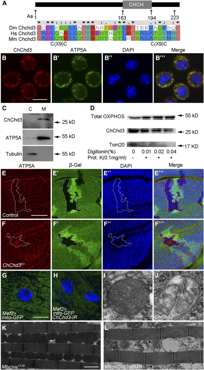
ChChd3 localizes to the inner mitochondrial membrane and is required for mitochondrial fusion and crista formation. (A) Schematic diagram of Drosophila ChChd3 protein domain structure and sequence comparison of Drosophila ChChd3 with human ChChd3 and mouse ChChd3 within the CHCH domain. (B–B′′′) S2 cells showing colocalization of ChChd3 and ATP5A. Samples were stained with anti-ChChd3 and anti-ATP5A antibodies. (C) Western blot analysis of cytosolic and mitochondrial fractions separated by centrifugation. Samples were probed with anti-ChChd3, anti-ATP5A (inner membrane), and anti-tubulin antibodies. ChChd3 is enriched in the mitochondrial fraction. (D) Western blot analysis of ATP5A (detected by anti-Total OXPHOS), ChChd3, and Tom20 proteins from S2 cell mitochondria. Isolated mitochondria were treated with the indicated concentrations of digitonin followed by Proteinase K digestion. (E–F′′′) Wing imaginal discs containing control (E–E′′′) or ChChd3D1 homozygous mutant (F–F′′′) clones stained with anti-β-Gal and anti-ATP5A antibodies. Clones are marked by the absence of β-Gal and the dashed white line. ChChd3 mutant clone cells display punctate ATP5A staining. (G) Larval body wall cells from the control larvae expressing UAS-mito-GFP with Mef2-Gal4 and showing mitochondria with tubular morphology. (H) Knockdown of ChChd3 in the larval body wall cells results in shorter mitochondria. (I and J) TEM images of mitochondria from wild-type (I) or ChChd3D1 mutant larvae. Crista content was reduced in ChChd3D1 mutant mitochondria. (K and L) TEM images of adult indirect flight muscle from the Mhc-Gal4 control (K) and ChChd3 knockdown flies (L). Knockdown of ChChd3 leads to fragmented mitochondria and reduced crista content. Bars, 10 μm in B; 40 μm in E; 10 μm in G; 0.1 μm in I; and 2 μm in K.

