Abstract
SmeYZ efflux pump is a critical pump responsible for aminoglycosides resistance, virulence-related characteristics (oxidative stress susceptibility, motility, and secreted protease activity), and virulence in Stenotrophomonas maltophilia. However, the regulatory circuit involved in SmeYZ expression is little known. A two-component regulatory system (TCS), smeRySy, transcribed divergently from the smeYZ operon is the first candidate to be considered. To assess the role of SmeRySy in smeYZ expression, the smeRySy isogenic deletion mutant, KJΔRSy, was constructed by gene replacement strategy. Inactivation of smeSyRy correlated with a higher susceptibility to aminoglycosides concomitant with an increased resistance to chloramphenicol, ciprofloxacin, tetracycline, and macrolides. To elucidate the underlying mechanism responsible for the antimicrobials susceptibility profiles, the SmeRySy regulon was firstly revealed by transcriptome analysis and further confirmed by quantitative real-time polymerase chain reaction (qRT-PCR) and promoter transcription fusion constructs assay. The results demonstrate that inactivation of smeRySy decreased the expression of SmeYZ pump and increased the expression of SmeDEF pump, which underlies the ΔsmeSyRy-mediated antimicrobials susceptibility profile. To elucidate the cognate relationship between SmeSy and SmeRy, a single mutant, KJΔRy, was constructed and the complementation assay of KJΔRSy with smeRy were performed. The results support that SmeSy-SmeRy TCS is responsible for the regulation of smeYZ operon; whereas SmeSy may be cognate with another unidentified response regulator for the regulation of smeDEF operon. The impact of inverse expression of SmeYZ and SmeDEF pumps on physiological functions was evaluated by mutants construction, H2O2 susceptibility test, swimming, and secreted protease activity assay. The increased expression of SmeDEF pump in KJΔRSy may compensate, to some extents, the SmeYZ downexpression-mediated compromise with respect to its role in secreted protease activity.
Introduction
To get rid of toxic substances and waste products, bacteria are equipped with efficient efflux systems. The efflux systems contributing to multidrug resistance have been extensively reported in several bacterial pathogens [1]. On the basis of structural characteristics, the multidrug efflux systems are classified into five families: resistance nodulation cell division (RND), major facilitator superfamily (MFS), small multidrug resistance (SMR), multidrug and toxic compound extrusion (MATE), and ATP-binding cassette (ABC) [2]. Among them, RND transporter is an effective mediator of multi-drug resistance in Gram-negative bacteria. The RND efflux pump forms a tripartite transporter system comprised of an inner membrane protein (IMP), an outer membrane protein (OMP), and a membrane fusion protein (MFP) to link IMP and OMP [3].
The known determinants involved in the regulation of RND efflux pumps include local regulator and global regulator [4]. The local regulators genes generally locate next to one of the regulated genes and can be easily recognized; however, the genes encoding the global regulators situate chromosomally elsewhere. In general, there are two kinds of regulatory systems involved in the expression of the multidrug efflux systems; one is the transcriptional regulator and the other is the two-component regulatory system (TCS). TCSs classically consist of an inner membrane-spanning sensor kinase (SK) and a cytoplasmic response regulator (RR) [5]. The SK and RR genes are often encoded adjacent to one another in the genome, forming an operon. Typically, a specific signal triggers the SK, which undergoes autophosphorylation at a specific histidine residue. This phosphoryl group is then transferred to an aspartate residue of the cognate RR, resulting in its activation. The activated RR usually acts as a transcriptional regulator to alter the genes expression.
Stenotrophomonas maltophilia, an opportunistic human pathogen, is well known for its intrinsic resistance to a wide range of antibiotics, including β-lactam, quinolone, and aminoglycoside [6]. Multi-drug resistances of S. maltophilia have been attributed to expression of antibiotic hydrolyzing or modifying enzymes [7–8] and multidrug efflux systems [9–14]. Eight putative RND-type efflux systems, SmeABC, SmeDEF, SmeGH, SmeIJK, SmeMN, SmeOP, SmeVWX, and SmeYZ, have been revealed in the sequenced genome of S. maltophilia K279a [15]. Of them, six pumps, SmeABC, SmeDEF, SmeIJK, SmeOP, SmeVWX, and SmeYZ, have been characterized [9–14]. The genome organization of the six pump operons and their individually contiguous regulatory genes are summarized in S1 Fig. The SmeABC pump is intrinsically quiescent [9]. The smeRS TCS divergently located upstream of smeABC is involved in the smeABC overexpression. The smeDEF and smeOP operons are negatively regulated by the products of the smeT and smeRo genes, which encode the TetR-type transcriptional repressors. SmeT and smeRo are located upstream of the smeDEF and smeOP operons and are divergently transcribed [12, 16]. SmeDEF and smeOP operons are expressed and can be further overexpressed by smeT and smeRo inactivation, respectively [12, 16]. The substrates extruded by SmeDEF pump are mainly chloramphenicol, quinolone, tetracycline, and macrolide [10]. SmeOP pump can extrude nalidixic acid, doxycycline, aminoglycosides, and macrolides [12]. SmeVWX pump is intrinsically quiescent and its expression is positively regulated by the divergently transcribed smeRv, which encodes a LysR-type transcription regulator [13]. SmIJK- and SmeYZ-overexpression strains contribute the resistance to aminoglycosides/leucomycin and aminoglycosides/trimethoprim-sulfamethoxazole, respectively [11, 17]. There is no recognizable regulatory gene flanking the smeIJK operon. However, we noticed that a TCS (designated as smeSy and smeRy), located upstream of smeYZ operon. This genomic organization highly suggests that SmeRySy TCS is responsible for the expression of smeYZ operon. In this study, we assessed the regulatory role of the SmeRySy TCS in the expression of SmeYZ. Unexpectedly, we found that the SmeDEF pump was upexpressed in the case of SmeRySy TCS inactivation, and the underlying regulatory circuit was further elucidated.
Materials and Methods
Bacterial strains, primers, and growth condition
S1 Table summarizes bacterial strains, plasmids, and primers used in this study. The strains used were derivatives of S. maltophilia KJ [8]. All primers used in this study were designed based on the S. maltophilia K279a genome sequence [15]. For the general purpose, strains were grown aerobically in Luria-Bertani (LB) medium except special note.
Construction of deletion mutants
To construct ΔsmeT, ΔsmeDEF, ΔsmeRy, and ΔsmeRSy mutants, the expected 454-, 406-, 820-, 503-, 390-, and 580-bp products were amplified from the genome of S. maltophilia KJ using primers SmeT3-F/R, SmeD5-F/R, SmeF3-F/R, SmeSy3-F/R, SmeRy3-F/R, and SmeRy5-F/R (S2 Fig). The 454-bp and 406-bp PCR products were subsequently cloned into the vector pEX18Tc, yielding plasmid pΔT, similarly, the amplicons of 406-bp and 820-bp for the construct of pΔDEF, the amplicons of 503-bp and 580-bp for pΔRSy, and the amplicons of 390-bp and 580-bp for pΔRy. The plasmids pΔT, pΔDEF, pΔRSy, and pΔRy were introduced into E. coli S17-1 by transformation and mobilized into S. maltophilia KJ via conjugation [18]. Transconjugants carrying deleted smeT, smeDEF, smeRSy, and smeRy in the chromosome were obtained by two-step selection on LB agar containing tetracycline (30 μg/ml)/norfloxacin (2.5 μg/ml) and then LB agar containing 10% (wt/vol) sucrose, yielding the deletion mutants KJΔT, KJΔDEF, KJΔRSy, and KJΔRy respectively (S2 Fig). The correctness of mutants was confirmed by colony PCR [19].
Construction of promoter-xylE transcription fusions
The aforementioned 406-bp PCR amplicon (primered by SmeD5-F and SmeD5-R) was treated with SmaI. The resulting 359-bp SmaI-SmaI DNA fragment, encompassing the 224-bp smeT-smeD intergenic region, was cloned into the pRK415 at both directions respectively. A xylE gene retrieved from pTxylE [8] was subsequently cloned behind the 224-bp DNA fragment, to generate smeT and smeDEF promoter transcription fusions constructs, pSmeTxylE and pSmeDxylE (Figure A in S2 Fig). The orientation of xylE gene inserted was opposite to that of the lacZ’ promoter of pRK415. A 343-bp DNA fragment spanning nucleotide -310 to +34 relative to the smeY start codon was obtained by PCR using primer sets SmeY5-F and SmeY5-R. We cloned this amplicon in front of the xylE reporter gene in a pRKXylE vector [11], yielding a smeYZ promoter transcription fusion construct, pSmeYxylE (Figure B in S2 Fig).
Transcriptome assay by RNA sequencing
Overnight-cultured bacteria cultures were inoculated into fresh LB broth with an initial OD450nm of 0.15. All cultures were grown at 37°C for 5 h and DNA-free RNA was extracted as described previously [11]. Ribosomal RNA (rRNA) was depleted from 5 μg of total RNA using the Ribo-Zero rRNA Removal Kit for bacteria (Epicentre, USA). The sequencing library for mRNA-seq was prepared according to the protocol for the "TruSeq RNA sample preparation” (Illumina Inc., USA). Briefly, the rRNA depleted mRNA was fragmented, and first-strand cDNA was synthesized using random hexamers following by second-strand cDNA synthesis, end repair, addition of a single A base and adapter ligation. The adapter-ligated cDNA library was amplified by PCR for 6 cycles using KAPA HiFi DNA polymerase (Kapa Biosystems). The enriched cDNA library was sequenced on a MiSeq (Illumina Inc., USA) using 250 bp paired-end reads. After trimming of low quality of bases (< Q30), the first 12 bases and adapters, the trimmed Reads were mapped to the Stenotrophomonas maltophilia K279a genome (GenBank acc. no. NC_010943.1) and run RNA-seq analysis by CLC Genomics Workbench v 6.0 (CLC Bio).
Antimicrobial susceptibility test
The susceptibilities of S. maltophilia strains to a number of antibiotics were tested by serial twofold dilutions in Mueller-Hinton agar according to the guidelines of Clinical Laboratory Standards Institute (CLSI) [20]. After overnight incubation at 37°C, cell growth was examined visually. The MIC was defined as the lowest concentration of antimicrobial agent that inhibited visible growth. All antibiotics were purchased from Sigma.
Catechol 2,3-dioxygenase (C23O) activity assay
The catechol-2,3-dioxygenase activity was measured as described previously [21]. The rate of hydrolysis was calculated by using 44,000 M-1cm-1 as the extinction coefficient. One unit of enzyme activity (Uc) was defined as the amount of enzyme that converts 1 nmole substrate per minute. The specific activity was expressed as Uc/OD450nm.
Quantitative Real-Time PCR (qRT-PCR)
DNA-free cellular RNA was prepared [11] and its purity was checked by qPCR without the additive of reverse transcriptase. The RNA was converted into cDNA which was then used directly as a template for qRT-PCR [11]. The primers used for qRT-PCR are listed in S1 Table. Amplification and detection of specific products were performed in the ABI Prism 7000 Sequence Detection System (Applied Biosystems) using the Smart Quant Green Master Mix (Protech Technology Enterprise Co., Ltd.) according to the manufacturer’s protocols. The mRNA of 16S rDNA was chosen as the internal control to normalize the relative expression level. Relative quantities of mRNA from each gene of interest were determined by the comparative cycle threshold method [22]. Each experiment was performed in triplicate.
H2O2 susceptibility test, swimming, and secreted protease activity assay
The physiological functions, including H2O2 susceptibility test, swimming, and secreted protease activity assay, were determined following the established protocols [17]. Each experiment was performed with at least 3 replicates. Results shown are mean with standard deviations. Statistical significance was assessed by Student t test.
Results and Discussion
Sequence analysis of the smeRySy-smeYZ cluster
The genetic organization surrounding the smeYZ genes (Smlt2198 and Smlt2197) in the genome of S. maltophilia K279a was surveyed. Smlt2200 and Smlt2199, transcribed divergently from the smeYZ, were found immediately upstream of smeYZ (Figure B in S2 Fig). The coding regions of Smlt2200 and Smlt2199 overlapped, signifying that they comprise a co-transcribed operon. Reverse transcriptase-PCR (RT-PCR) was carried out to verify the presence of Smlt2200-Smlt2199 operon (S3 Fig). The regulatory regions and relevant consensus sequences of Smlt2200 and Smlt2199 proteins were analyzed. The relevant domains, linked to the functions of sensor kinase and response regulator of a TCS, were identified in SmeSy and SmeRy, respectively (S4 Fig). In addition, based on the comparisons with other sensor kinases and response regulators, the His148 residue of SmeSy and the Asp81 residue of SmeRy likely represent the autophosphorylated histidine and the phosphoaceptor aspartate, respectively (S4 Fig). Herein, we designated the Smlt2200 and Smlt2199 as SmeRy and SmeSy, respectively.
Inactivation of SmeRySy system decreases the expression of smeYZ operon
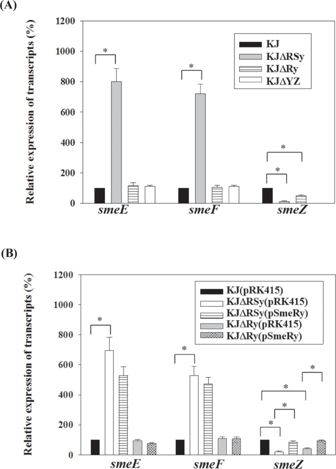
SmeRySy TCS is divergently transcribed from the smeYZ operon. To test the regulatory role of SmeRySy TCS in the expression of smeYZ operon, smeRySy was deleted from the chromosome of wild-type strain KJ and the impacts on antimicrobial susceptibility and smeYZ expression were examined. The resulting strain, KJΔRSy, was more susceptible to all of the SmeYZ substrates tested (Table 1) [17], suggesting that SmeRySy inactivation decreases the expression of smeYZ operon. The smeYZ expression in KJΔRSy was further checked by qRT-PCR. Compared with those in wild-type KJ, the smeZ transcript in KJΔRSy was decreased (Fig 1). Decreased transcription of the smeYZ operon in KJΔRSy versus KJ was also confirmed by the promoter transcription fusion assay. Fig 2 demonstrates that the promoter activity of smeYZ operon was drastically decreased in KJΔRSy (pSmeYxylE in KJ and KJΔRSy), further confirming the positive regulatory role of SmeRySy TCS in the expression of smeYZ operon. However, it is worthily mentioned that the expression of SmeYZ pump was not totally abolished in the case of SmeRySy inactivation. The MICs reduction in KJΔRSy did not reach the level displayed in KJΔYZ (Table 1), and there was detectable smeZ transcript in KJΔRSy (Fig 1) and detectable C23O activity in the strain KJΔRSy(pSmeYxylE) (Fig 2), indicating that a remnant expression of smeYZ pump exists in KJΔRSy.
Table 1. Antimicrobial susceptibilities of S. maltophilia KJ and its derived constructs.
| MIC (μg/ml) | ||||||||
|---|---|---|---|---|---|---|---|---|
| CHL | CIP | TET | ERY | LM | KAN | GEN | TOB | |
| KJ | 8 | 1 | 16 | 64 | 256 | 256 | 1024 | 512 |
| KJΔRSy | 16 | 4 | 32 | 256 | 512 | 32 | 32 | 32 |
| KJΔRSyΔDEF | 4 | 0.5 | 8 | 32 | 128 | 32 | 32 | 32 |
| KJΔDEF | 4 | 0.5 | 8 | 32 | 128 | 256 | 1024 | 512 |
| KJΔYZ | 8 | 1 | 8 | 128 | 128 | 8 | 8 | 32 |
| KJΔRy | 8 | 1 | 16 | 64 | 256 | 32 | 32 | 32 |
| KJΔT | 32 | 8 | 32 | 512 | 2048 | 64 | 128 | 64 |
| KJΔRSyΔT | 32 | 8 | 32 | 512 | 1024 | 4 | 8 | 16 |
| KJΔRSy(pSmeRy) | 16 | 4 | -a | 256 | 512 | 64 | 256 | 512 |
| KJΔ Ry(pSmeRy) | 8 | 1 | -a | 65 | 256 | 256 | 1024 | 512 |
CHL, chloramphenicol; CIP, ciprofloxacin; TET, tetracycline; ERY, erythromycin; LM, leucomycin; KAN, kanamycin; GEN, gentamicin; TOB, tobramycin.
aThe complementation plasmid carries the tetracycline-resistant gene
Fig 1. Quantitative RT-PCR (qRT-PCR) analysis of smeE, smeF, and smeZ transcripts.
The RNA was prepared from the exponentially growing cells, converted to cDNA by RT-PCR, and used as the template for quantitative reverse-transcriptase PCR (qRT-PCR). Data are the means of three independent experiments. Error bars indicate the standard deviation for three triplicate samples. *, p≤0.05 significance calculated by a Student’s t-test. (A) qRT-PCR analysis of smeE, smeF, and smeZ transcripts of strains KJ, KJΔRSy, KJΔRy, and KJΔYZ. (B) qRT-PCR analysis of smeE, smeF, and smeZ transcripts in the smeRy-complemented strains of KJΔRSy and KJΔRy.
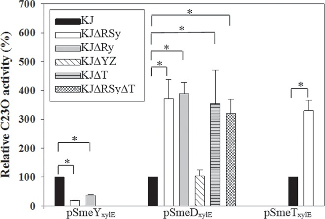
Fig 2. The C23O activities expressed by the promoter transcription fusion constructs of pSmeYxylE, pSmeDxylE, and pSmeYxylE in wild-type KJ and its derived mutants.
The overnight-cultured bacteria assayed were inoculated into fresh LB with an initial OD450nm of 0.15, cultured for 5 h, and the C23O activities were determined. The data represent means of three repetitions. Error bars indicate the standard deviation for three triplicate samples. *, p≤0.05 significance calculated by a Student’s t-test.
Inactivation of SmeRySy system increases the expression of smeDEF operon
In the results of susceptibility test of KJΔRSy, it was surprisingly noticed that the MICs of KJΔRSy to chloramphenicol, ciprofloxacin, tetracycline, and macrolides were increased (Table 1), and these antimicrobials are not the substrates of the SmeYZ pump [17]. It is thus likely that in addition to smeYZ operon, the SmeRySy system regulates other antimicrobials resistance-relevant determinants, which are responsible for the increased MICs of chloramphenicol, ciprofloxacin, tetracycline, and macrolides. We sought to determine genes that comprise the SmeRySy regulon by comparing transcription in wild-type KJ and smeRySy mutant, KJΔRSy. The transcriptomic data showed that the difference in gene expression caused by the smeRySy deletion was small, with only 21 genes showing greater than a 3-fold change (up or down) in their transcript abundance (Table 2; S2 Table). Interestingly, the greatest dysregulation was displayed by two RND-type efflux pump operons, smeYZ and smeDEF. The expression of smeDEF and smeYZ was inversely changed in response to the smeRySy inactivation (Table 2). To validate the expression profiles obtained by the transcriptome analysis, the smeE, smeF, and smeZ transcripts in KJ and KJΔRSy were quantified by qRT-PCR. Fig 1 shows that smeDEF and smeYZ operons were upregulated and downregulated, respectively, in response to smeSyRy inactivation, which is consistent with the RNA-seq data. It has been known the substrates extruded by the SmeDEF pump include chloramphenicol, ciprofloxacin, tetracycline, and macrolide [10]. Therefore, it is highly suggested that the smeRySy-mediated smeDEF upregulation is responsible for the increased MICs observed in KJΔRSy (Table 1).
Table 2. Transcriptomic analysis of genes differentially expressed in the smeRySy mutant compared to the wild-type S. maltophilia KJ.
| Gene ID | Description / gene name | Fold changea |
|---|---|---|
| Smlt4071 | SmeE (RND-type multidrug efflux pump) | 11.036 |
| Smlt4070 | SmeF (RND-type multidrug efflux pump) | 10.410 |
| Smlt4072 | SmeD (RND-type multidrug efflux pump) | 9.576 |
| Smlt3594 | transmembrane protein | 6.115 |
| Smlt4069 | transmembrane protein | 3.655 |
| Smlt0271 | hypothetical protein | 3.057 |
| Smlt2932 | glutamine amidotransferase class-I | 3.019 |
| Smlt4073 | SmeT | 2.718 |
| Smlt2201 | SmeY(RND-type multidrug efflux pump) | -19.440 |
| Smlt2202 | SmeZ (RND-type multidrug efflux pump) | -9.273 |
| Smlt1290 | conjugal transfer protein / trbC | -5.738 |
| Smlt1284 | conjugal transfer protein /trbG | -5.734 |
| Smlt2200 | SmeRy | -5.072 |
| Smlt2203 | Hypothetical protein | -4.641 |
| Smlt2531 | hypothetical protein | -4.568 |
| Smlt1419 | transmembrane protein | -4.055 |
| Smlt0593 | methionine sulfoxide reductase | -3.891 |
| Smlt2283 | flagellarbasal body-associated protein /fliL | -3.634 |
| Smlt2290 | flagellar MS-ring protein / fliE | -3.577 |
| Smlt1293 | conjugal transfer coupling protein TraG/traG | -3.478 |
| Smlt2312 | flagellar basal body rod protein FlgG/ flgG | -3.418 |
| Smlt2314 | flagellar hook protein FlgE/ flgE | -3.371 |
aA positive value signifies upregulation and a negative value signifies downregulation in smeRySy mutant.
To validate the SmeRySy TCS regulatory role in smeDEF and smeYZ operons, the promoter transcription fusions of pSmeDxylE and pSmeYxylE were prepared. The C23O activities expressed by pSmeDxylE and pSmeYxylE in strains KJ and KJΔRSy were comparatively determined. KJΔRSy(pSmeDxylE) had a ca. 3.72-fold increase in C23O activity relative to KJ(pSmeDxylE); and the C23O activity expressed by KJΔRSy(pSmeYxylE) was 82% lower than that by KJ(pSmeYxylE) (Fig 2), in consistent with the transcriptome result.
To determine whether upregulation of smeDEF in KJΔRSy may account for the increased antimicrobials resistance (Table 1), the smeDEF operon was deleted from the chromosome of strain KJΔRSy, yielding KJΔRSyΔDEF. The MICs of KJΔRSyΔDEF to chloramphenicol, ciprofloxacin, tetracycline, and macrolides were reverted to the comparable level as those of KJΔDEF (Table 1); but the MICs of KJΔRSyΔDEF to aminoglycosides were still as low as those of KJΔRSy. Taken together, we concluded that inactivation of the smeRySy system results in the upexpression of smeDEF operon, which then confers to the phenotype of increased resistance to chloramphenicol, ciprofloxacin, tetracycline, and macrolides.
SmeYZ is irrelevant to the ΔsmeRySy-mediated upexpression of smeDEF
It has been demonstrated that altering the expression of a single RND pump may have downstream effect on any number of other RND efflux systems [23]. In view of the regulatory role of SmeRySy TCS in smeYZ operon, we wondered whether the increased smeDEF transcript in strain KJΔRSy results from the coordinated regulation between the SmeYZ pump and SmeDEF pump, other than from the direct regulation of the SmeRySy system. To solve the question, the C23O activity expressed by pSmeDxylE was comparatively assessed in KJ and KJΔYZ cells. The promoter activity of smeDEF operon was little affected in the case of smeYZ inactivation (Fig 2). Furthermore, Table 1 also shows that KJΔYZ exhibited a similar degree of resistance to chloramphenicol, ciprofloxacin, tetracycline and macrolides, compared with wild-type KJ, further supporting that the elevated resistance in KJΔRSy is not simply compensatory upregulation in response to decrease expression of SmeYZ.
SmeRy is not involved in the ΔsmeRySy-mediated increment of smeDEF transcript and antibiotic resistance
To further elucidate the SmeSy and SmeRy cognate relationship responsible for the ΔsmeRySy-mediated phenotype, we deleted the smeRy gene in wild-type KJ, yielding KJΔRy. The antibiotic susceptibility and the expression of smeDEF and smeYZ operons in KJΔRy were performed. KJΔRy increased the susceptibility toward aminoglycosides, which are the substrates of SmeYZ, but did not alter the susceptibility toward the antibiotics, which are the known substrates of SmeDEF pump (Table 1). The smeZ transcript and the promoter activity assay (PsmeYZ) of KJΔRy were decreased relative to those of wild-type KJ (Fig 1A & 2). The smeE and smeF transcripts of KJΔRy were comparable to those of wild-type KJ (Fig 1), in despite of an increment in the PsmeDEF promoter activity of KJΔRy (Fig 2). These observations imply the irrelevance of SmeRy to ΔsmeRySy-mediated increment of smeDEF transcript and antibiotic resistance. To further verify this assumption, complementation assay was performed. Complementation of KJΔRSy or KJΔRy with a smeRy gene restored the resistance toward aminoglycosides (Table 1) and the smeZ transcript (Fig 1B), but little affected the susceptibility toward chloramphenicol, ciprofloxacin, tetracycline, and macrolides (Table 1) as well as the smeE and smeF transcripts (Fig 1B).
Based on these observations, we propose that SmeSy may cooperate with two different response regulators to individually regulate smeDEF and smeYZ operons. When SmeRy acts as a cognate response regulator of SmeSy, it can activate the expression of smeYZ and thus contribute to the SmeYZ pump-mediated resistance. Nevertheless, there should be another unidentified response regulator that can work with SmeSy to regulate smeDEF operon. In view of the increased PsmeD promoter activity (Fig 2) and the decreased smeDEF transcript (Fig 1A) in KJΔRy, SmeSy-mediated regulation in KJΔRy may link to the stability of smeDEF transcript. When smeSy and smeRy are simultaneously inactivated, the phenotype of SmeDEF pump upexpression and SmeYZ pump downexpression can coexist.
The regulatory roles of SmeT and SmeRySy TCS in the expression of smeDEF
Since the expression of the smeDEF operon is under the negative control of the TetR-type transcriptional regulator SmeT, which is divergently transcribed from the smeDEF operon (S1 Fig) [16], we wondered whether SmeRySy TCS affects the expression of smeT and consequently alters the expression of smeDEF operon. Based on the transcriptome data, the smeT transcript had a 2.71-fold upregulation in smeRSy mutant compared with that in wild-type KJ (Table 2, S2 Table). To confirm this, the smeT promoter transcription fusion construct, pSmeTxylE, was transferred into wild-type KJ and KJΔRSy. As seen in Fig 2, the C23O activity expressed by KJΔRSy(pSmeTxylE) was increased compared to that by KJ(pSmeTxylE).
In view of upexpression of smeDEF operon in strains KJΔT and KJΔRSy, we further assessed whether simultaneous inactivation of smeSyRy and smeT has an additive effect on smeDEF upexpression. To assess this, the expressions of pSmeDxylE in wild-type KJ, KJΔRSy, KJΔT, and KJΔRSyΔT were comparatively determined. The C23O activities determined from KJΔRSy(pSmeDxylE), KJΔT(pSmeDxylE), and KJΔRSyΔT(pSmeDxylE) were comparable (Fig 2). As a result, simultaneous inactivation of smeRySy and smeT did not further upregulate the expression of smeDEF operon. The antimicrobial susceptibilities of strains KJΔT and KJΔRSyΔT displayed a consistent result (Table 1).
In this article, we found that SmeRySy inactivation simultaneously upregulates the expression of smeDEF and smeT (Fig 2). This observation seems to contradict the previous report that the repression extent of smeDEF operon has a positive correlation with the amount of repressor SmeT [16]. Herein, we proposed two explanations for this phenotype. (i) The binding activity of transcriptions repressor toward DNA affects the repressor function [24]. Therefore, the smeDEF upexpression observed in SmeRySy inactivation may result from the compromise of SmeT-operator interaction and is less relevant to the amount of SmeT. It is highly possible that a modulator, whose expression is altered in the case of smeRySy inactivation, compromises the affinity between SmeT and operator, and thus simultaneously derepresses the expression of smeDEF and smeT. (ii) Both SmeT and the unidentified regulator have a direct effect and are needed for the repression of smeDEF operon, in which case the absence of one or another will produce the same effect on the smeDEF upregulated.
SmeDEF upexpression in ΔsmeRySy compensates the SmeYZ downexpression-mediated alterations with respect to secreted protease activity
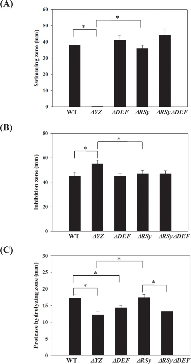
The overall expression of MDR pumps is closely monitored, and whenever the levels of one of these systems are altered, compensatory changes in the levels of the other MDR pumps may follow. For example, there is an inverse correlation between the MexAB-OprM and MexEF-OprN expression in P. aeruginosa [23]. Inactivation of SmeRySy TCS inversely regulates the expression of SmeDEF and SmeYZ pumps, implying that there are some overlapped functions between SmeDEF and SmeYZ, and the inverse expression between both pumps may have a partially compensatory effect benefiting bacterial survival. Given the distinct difference in substrate profiles of SmeDEF and SmeYZ pumps (Table 1), the compensatory effect is less relevant to antibiotics extrusion. In our recent study, we have demonstrated that the SmeYZ pump contributes to an array of physiological functions, including oxidative stress susceptibility, swimming, and secreted protease activity [17]. Therefore, we were interested in assessing whether SmeDEF upexpression in ΔsmeRySy compensates the SmeYZ downexpression-mediated compromise of physiological functions. The physiological functions for oxidative stress susceptibility, swimming, and secreted protease activity in strains KJ, KJΔYZ, KJΔDEF, KJΔRSy, and KJΔRSyΔDEF were comparatively evaluated. Consistent with the previous reports [17], KJΔYZ displayed a compromise in oxidative stress susceptibility, swimming, and secreted protease activity; however, these compromises in KJΔRSy were not as severe as those in KJΔYZ, as compared with wild-type KJ (Fig 3). This observation suggested two possibilities: (i) Small level of SmeYZ expression could still mediate certain functions, since there are remnant expression of SmeYZ pump in KJΔRSy (Fig 1 & 2); (ii) some compensatory mechanisms for the physiological functions of SmeYZ pump may occur in KJΔRSy. As shown in Fig 3A, mutant KJΔYZ cannot swim but the mutants KJΔRSy and KJΔRSyΔDEF can. This suggests that downregulation of SmeYZ is not sufficient to abolish swimming but total loss of SmeYZ is. Since smeDEF inactivation in KJΔRSy did not change this, then either small level of SmeYZ expression can support swimming phenotype, or there is a compensatory mechanism occurring in KJΔRSy (but it is not SmeDEF). The similar phenomenon can be noticed in wild-type KJ and mutants KJΔYZ, KJΔRSy, and KJΔRSyΔDEF with respect to oxidative stress susceptibility (Fig 3B). Nevertheless, compared with wild-type KJ, KJΔDEF decreased the secreted protease activity, but hardly affected oxidative stress susceptibility and swimming (Fig 3), supporting that SmeDEF and SmeYZ have an overlapped physiological function in protease secretion, but not in oxidative stress tolerance and swimming. In an attempt to determine the linkage of SmeDEF upregulation to the compensatory circuit, ΔsmeDEF allele was introduced into KJΔRSy and the physiological functions of the resultant mutant KJΔRSyΔDEF were assessed. Inactivation of smeDEF conferred KJΔRSy a decreased secreted protease activity (Fig 3C), signifying that upregulation of SmeDEF in KJΔRSy compensates the SmeYZ downexpression-mediated compromise in secreted protease activity.
Fig 3. The physiological functions evaluation between wild-type KJ and its derived mutants.
The data represent means of three repetitions. Error bars indicate the standard deviation for three triplicate samples. *, p≤0.05 significance calculated by a Student’s t-test. (A) Motility ability. Five microliter bacterial cell suspension was inoculated at the swimming agar (1% tryptone, 0.5% NaCl and 0.15% agar), and the diameter (mm) of swimming zone was measured after 48 h incubation at 37°C. (B) H2O2 susceptibility test. Sterile filter paper with 20 μl of 10% H2O2 was placed onto MH agar, which was uniformly spread with bacterial cell suspension. The diameter of a zone of growth inhibition was measured after 24 h incubation at 37°C. (C) Secreted protease activity assay. Forty microliter bacterial cell suspension was dipped on LB agar containing 1% skin milk. The proteolytic activity of bacteria was assessed by measuring the transparent zones around the bacteria after incubation at 37°C for 72 h.
Microorganism contains a variety of TCSs that regulate complex antibiotic resistance. Based on the regulatory mechanisms of those TCSs already characterized, several regulatory models have been proposed. Firstly, the TCSs definitely regulate the antibiotics resistance determinants (either operons or genes), for instance, AdeABC by the AdeRS system of Acintobacter baumannii, CzcCBA by the CzcRS system of Pseudomonas aeruginosa and SmeABC by the SmeRS system of S. maltophilia [9, 25–26]. Secondly, the different response regulators from individually TCS act on the same antibiotics resistance determinants. The PhoP-PhoQ and PmrA-PmrB systems of P. aeruginosa provide such an example. The phosphorylated response regulators, PhoP and PmrA, can positively upregulate the transcription of arnBCADTEF operon and result in the resistance to polymyxin B and cationic antimicrobials peptides [27]. Thirdly, the activated TCSs can simultaneously affect the expression of a variety of targets genes which involved in the different resistance mechanisms. The CzcRS system of P. aeruginosa regulates the expression of czcCAB operon and oprD gene [28]. The BaeSR TCS is responsible for the regulation of MdtABC and AcrD [29]. The EvgSA system activates the expression of MdeEF and EmrKY [30–31]. The present results provide another kind of example that a sensor kinase can partner with different response regulators to inversely modulate two RND-type efflux pumps and the outcome can benefit the maintenance of bacterial physiologic functions.
Supporting Information
(DOCX)
(DOCX)
(DOCX)
(DOCX)
(DOCX)
(DOCX)
Acknowledgments
The authors acknowledge the High-throughput Genome Analysis Core Facility of National Core Facility Program for Biotechnology, Taiwan (NSC-102-2319-B-010-001), for Illumina sequencing.
Data Availability
All relevant data are within the paper and its Supporting Information files.
Funding Statement
This work was supported by grant MOST 104-2320-B-010-023-MY3 from Ministry of Science and Technology of Taiwan and grant 30219003 from Professor Tsuei-Chu Mong Merit Scholarship. The funders had no role in study design, data collection and analysis, decision to publish, or preparation of the manuscript.
References
- 1.Poole K. Efflux-mediated antimicrobial resistance. J Antimicrob Chemother. 2005; 56:20–51. [DOI] [PubMed] [Google Scholar]
- 2.Bolhuis H, van Veen HW, Poolman B, Driessen AJ, Konings WN. Mechanisms of multidrug transporters. FEMS Microbiol Rev. 1997; 21:55–84. [DOI] [PubMed] [Google Scholar]
- 3.Misra R, Bavro VN. Assembly and transport mechanism of tripartite drug efflux systems. Biochim Biophys Acta.2009; 1794:817–825. 10.1016/j.bbapap.2009.02.017 [DOI] [PMC free article] [PubMed] [Google Scholar]
- 4.Grkovic S, Brown MH, Skurray RA. Regulation of bacterial drug export systems. Microbiol. Mol Biol Rev. 2002: 66:671–701. [DOI] [PMC free article] [PubMed] [Google Scholar]
- 5.Stock AM, Robinson VL, Goudreau PN. Two-component signal transduction. Annu Rev Biochem. 2000; 69:183–215. [DOI] [PubMed] [Google Scholar]
- 6.Brooke JS. Stenotrophomonas maltophilia: an emerging global opportunistic pathogen. Clin Microbiol Rev. 2012; 25:2–41. 10.1128/CMR.00019-11 [DOI] [PMC free article] [PubMed] [Google Scholar]
- 7.Lambert T, Ploy MC, Denis F, Courvalin P. Characterization of the chromosomal aac(6')-Iz gene of Stenotrophomonas maltophilia. Antimicrob Agents Chemother. 1999; 43:2366–2371. [DOI] [PMC free article] [PubMed] [Google Scholar]
- 8.Hu RM, Huang KJ, Wu LT, Hsiao YJ, Yang TC. Induction of L1 and L2 β-lactamases of Stenotrophomonas maltophilia. Antimicrob Agents Chemother. 2008; 52:1198–1200. [DOI] [PMC free article] [PubMed] [Google Scholar]
- 9.Li XZ, Zhang L, Poole K. SmeC, an outer membrane multidrug efflux protein of Stenotrophomonas maltophilia. Antimicrob Agents Chemother. 2002; 46:333–343. [DOI] [PMC free article] [PubMed] [Google Scholar]
- 10.Alonso A, Martinez JL. Cloning and characterization of SmeDEF, a novel multidrug efflux pump from Stenotrophomonas maltophilia. Antimicrob Agents Chemother. 2000; 44:3079–3086. [DOI] [PMC free article] [PubMed] [Google Scholar]
- 11.Huang YW, Liou RS, Lin YT, Huang HH, Yang TC. A linkage between SmeIJK efflux pump, cell envelope integrity, and σE-mediated envelope stress response in Stenotrophomonas maltophilia. PLoS ONE, 2014;9:e111784 10.1371/journal.pone.0111784 [DOI] [PMC free article] [PubMed] [Google Scholar]
- 12.Lin CW, Huang YW, Hu RM, Yang TC. SmeOP-TolCsm efflux pump contributes to the multidrug resistance of Stenotrophomonas maltophilia. Antimicrob Agents Chemother. 2014; 58:2405–2408. 10.1128/AAC.01974-13 [DOI] [PMC free article] [PubMed] [Google Scholar]
- 13.Chen CH, Huang CC, Chung TC, Hu RM, Huang YW, Yang TC. Contribution of resistance-nodulation-division efflux pump operon smeU1-V-W-U2-X to multidrug resistance of Stenotrophomonas maltophilia. Antimicrob Agents Chemother. 2011; 55:5826–5833. 10.1128/AAC.00317-11 [DOI] [PMC free article] [PubMed] [Google Scholar]
- 14.Gould VC, Okazaki A, Avison MB. Coordinate hyperproduction of SmeZ and SmeJK efflux pumps extends drug resistance in Stenotrophomonas maltophilia. Antimicrob Agents Chemother. 2013; 57:655–657. 10.1128/AAC.01020-12 [DOI] [PMC free article] [PubMed] [Google Scholar]
- 15.Crossman LC, Gould VC, Dow JM, Vernikos GS, Okazaki AK, Sebaihia M et al. The complete genome, comparative and functional analysis of Stenotrophomonas maltophilia reveals an organism heavily shielded by drug resistance determinants. Gen Biol. 2008; 9:R74. [DOI] [PMC free article] [PubMed] [Google Scholar]
- 16.Sanchez P, Alonso A, Martinez JL. Cloning and characterization of SmeT, a regulator of the Stenotrophomonas maltophilia multidrug efflux pump SmeDEF. Antimicrob Agents Chemother. 2002; 46:3386–3393. [DOI] [PMC free article] [PubMed] [Google Scholar]
- 17.Lin YT, Huang YW, Chen SJ, Chang CW, Yang TC. The SmeYZ efflux pump of Stenotrophomonas maltophilia contributes to drug resistance, virulence-related characteristics, and virulence in mice. Antimicrob Agents Chemother. 2015; 59:4067–4073. 10.1128/AAC.00372-15 [DOI] [PMC free article] [PubMed] [Google Scholar]
- 18.Yang TC, Huang YW, Hu RM, Huang SC, Lin YT. AmpDI is involved in expression of chromosomal L1 and L2 β-lactamases of Stenotrophomonas maltophilia. Antimicrob Agents Chemother. 2009; 53:2902–2907. 10.1128/AAC.01513-08 [DOI] [PMC free article] [PubMed] [Google Scholar]
- 19.Lin CW, Chiou CS, Chang YC, Yang TC. Comparison of pulsed-field gel electrophoresis and three rep-PCR methods for evaluating the genetic relatedness of Stenotrophomonas maltophilia isolates. Lett Appl Microbiol. 2008; 47:393–398. 10.1111/j.1472-765X.2008.02443.x [DOI] [PubMed] [Google Scholar]
- 20.CLSI. 2012. Performance standards for antimicrobial susceptibility testing. 22nd informational supplement. M100-S22. Clinical and Laboratory Standards Institute, Wayne, PA.
- 21.Lin CW, Huang YW, Hu RM, Chiang KH, Yang TC. The role of AmpR in the regulation of L1 and L2 β-lactamases in Stenotrophomonas maltophilia. ResMicrobiol.2009; 160:152–158. [DOI] [PubMed] [Google Scholar]
- 22.Livak KJ, Schmittgen TD. Analysis of relative gene expression data using real-time quantitative PCR and the 2(-ΔΔC(T)) method. Methods. 2001; 25:402–408. [DOI] [PubMed] [Google Scholar]
- 23.Li XZ, Barre N, K. Poole K. Influence of the MexA-MexB-OprM multidrug efflux system on expression of the MexC-MexD-OprJ and MexE-MexF-OprN multidrug efflux systems in Pseudomonas aeruginosa. J Antimicrob Chemother. 2000; 46:885–893. [DOI] [PubMed] [Google Scholar]
- 24.Saito K, Akama H, Yoshihara E, Nakae T. Mutations affecting DNA-binding activity of the MexR repressor of mexR-mexA-mexB-oprM operon expression. J Bacteriol. 2003; 185: 6195–6198. [DOI] [PMC free article] [PubMed] [Google Scholar]
- 25.Marchand I, Damier-Piolle L, Courvalin P, Lambert T. Expression of the RND-type efflux pump AdeABC in Acinetobacter baumannii is regulated by the AdeRS two-component system. Antimicrob Agents Chemother. 2004; 48:3298–3304. [DOI] [PMC free article] [PubMed] [Google Scholar]
- 26.Gooderham WJ, Hancock REW. Regulation of virulence and antibiotic resistance by two-component regulatory systems in Pseudomonas aeruginosa. FEMS Microbiol Rev. 2009; 33:279–294. 10.1111/j.1574-6976.2008.00135.x [DOI] [PubMed] [Google Scholar]
- 27.McPhee JB, Bains M, Winsor G, Lewenza S, Kwasnicka A, Brazas MD et al. Contribution of the PhoP-PhoQ and PmrA-PmrB two-component regulatory systems to Mg21-induced gene regulation in Pseudomonas aeruginosa. J Bacteriol. 2006; 188:3995–4006. [DOI] [PMC free article] [PubMed] [Google Scholar]
- 28.Perron K, Caille O, Rossier C, Van Delden C, Dumas JL, Kohler T. CzcR-CzcS, a two-component system involved in heavy metal and carbapenem resistance in Pseudomonas aeruginosa. J Biol Chem. 2004; 279:8761–8768. [DOI] [PubMed] [Google Scholar]
- 29.Baranova N, Nikaido H. The baeSR two-component regulatory system activates transcription of the yegMNOB (mdtABCD) transproter gene cluster in Escherichia coli and increases its resistance tonovobiocin and deoxycholate. J Bacteriol. 2002; 184:4168–4176. [DOI] [PMC free article] [PubMed] [Google Scholar]
- 30.Nishino K, Yamaguchi A. Overexpression of the response regulator evgA of the two-component signal transduction system modulates multidrug resistance conferred by multidrug resistance transporters. J Bacteriol. 2001; 183:1455–1458. [DOI] [PMC free article] [PubMed] [Google Scholar]
- 31.Nishino K, Yamaguchi A. EvgA of the two-component signal transduction system modulates production of the YhiUV multidrug transporter in Escherichia coli. J Bacteriol. 2002;184:2319–2323. [DOI] [PMC free article] [PubMed] [Google Scholar]
Associated Data
This section collects any data citations, data availability statements, or supplementary materials included in this article.
Supplementary Materials
(DOCX)
(DOCX)
(DOCX)
(DOCX)
(DOCX)
(DOCX)
Data Availability Statement
All relevant data are within the paper and its Supporting Information files.


