SUMMARY
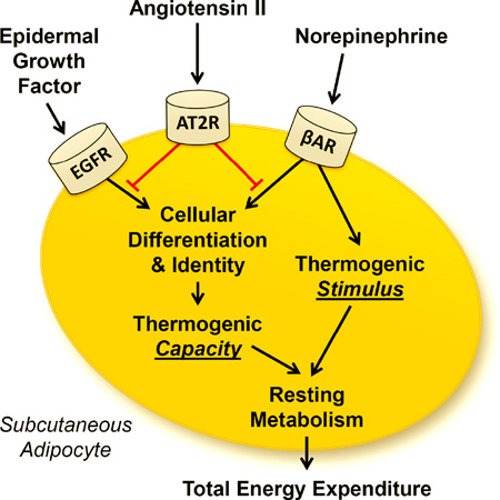
Activation of the brain renin-angiotensin system (RAS) stimulates energy expenditure through increasing resting metabolic rate (RMR), and this effect requires simultaneous suppression of the circulating/adipose RAS. To identify the mechanism by which the peripheral RAS opposes RMR control by the brain RAS, mice with transgenic activation of the brain RAS (sRA mice) were examined. sRA mice exhibit increased RMR through increased energy flux in the inguinal adipose tissue, and this effect is attenuated by angiotensin II type 2 receptor (AT2) activation. AT2 activation in inguinal adipocytes opposes norepinephrine-induced uncoupling protein-1 (UCP1) production and aspects of cellular respiration, but not lipolysis. AT2 activation also opposes inguinal adipocyte function and differentiation responses to epidermal growth factor (EGF). These results highlight a major, multifaceted role for AT2 within inguinal adipocytes in the control of RMR. The AT2 receptor may therefore contribute to body fat distribution and adipose depot-specific effects upon cardio-metabolic health.
Graphical Abstract
INTRODUCTION
The renin-angiotensin system (RAS) is well recognized for its varied roles in cardiovascular physiology. Increasing evidence also supports tissue-specific actions of angiotensin II (ANG) in the control of energy balance. Human obesity is associated with increased activity of the circulating RAS (Engeli et al., 2005), and various studies have documented beneficial effects of RAS inhibition upon glycemic control endpoints in obese humans (Grassi et al., 2003; Hsueh et al., 2010; Lindholm et al., 2003; Shimabukuro et al., 2007). In rodent models, pharmacological or genetic interference with the RAS also generally has beneficial effects upon body mass (as reviewed recently (Claflin and Grobe, 2015; Littlejohn and Grobe, 2015)). It is therefore confusing that RAS blockade does not have an overt effect upon body mass in humans. We hypothesize that the multifaceted contribution of the RAS to energy balance (through opposing effects on intake and activity behaviors, digestive efficiency, and resting metabolic rate (RMR)) may result in a net caloric balance in humans, and thereby no change in body mass. Understanding the tissue-specific molecular mechanisms by which the RAS mediates control of individual components of energy balance, such as RMR, will allow for the development of novel therapeutics for obesity and its sequelae.
While RAS-mediated mechanisms controlling food intake, digestive efficiency and physical activity have been defined, RMR control by the RAS is much more complicated with tissue- and receptor-specific actions of ANG functioning in apparent opposition (Claflin and Grobe, 2015; Grobe et al., 2013). Stimulation of the brain RAS by transgenic activation, ICV infusion of ANG, or deoxycorticosterone acetate (DOCA)-salt treatment increase RMR through brain AT1-dependent mechanisms (de Kloet et al., 2011; de Kloet et al., 2013; Grobe et al., 2011; Grobe et al., 2010; Porter et al., 2003; Porter and Potratz, 2004). Oddly, chronic infusion of ANG in the periphery can reverse these effects of a stimulated brain RAS (Grobe et al., 2010), highlighting the opposing effects of the brain and peripheral versions of the RAS in energy homeostasis (Grobe et al., 2013). Thus, there remains a critical lack of understanding of the mechanisms that mediate control of RMR by the RAS, and in particular, how the brain RAS and peripheral RAS interact in this control. We hypothesize that while ANG acts within the brain to stimulate RMR, actions of ANG in the periphery suppress this mechanism of energy expenditure. The objective of the current project was therefore to clarify the mechanisms by which the peripheral RAS opposes the RMR-stimulating effects of the brain RAS.
RESULTS
The brain RAS stimulates RMR
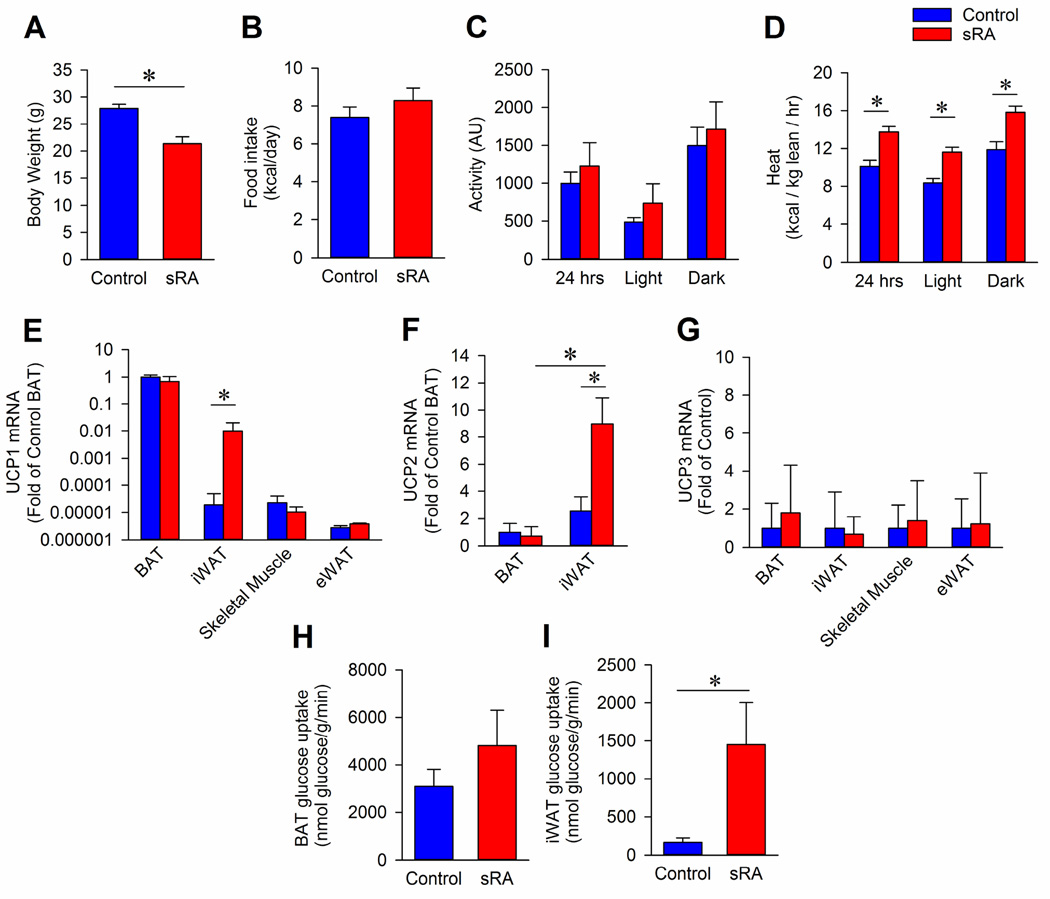
Double-transgenic “sRA” mice exhibit brain-specific elevations in RAS activity due to transgenic expression of both a human renin transgene via the synapsin promoter (sR) and a human angiotensinogen transgene under transcriptional control by its own promoter (A) (Sakai et al., 2007). Under baseline conditions, double-transgenic sRA mice exhibit decreased body weight and proportional fat mass compared to single- and non-transgenic littermate controls (Figure 1A and Table S1). Although these animals display normal food intake (Figure 1B) and physical activity (Figure 1C), sRA mice exhibit an elevated metabolic rate throughout the light-dark cycle (Figure 1D). These data confirm our previous studies of elevated energy expenditure in the sRA mouse model (Grobe et al., 2010; Grobe et al., 2013) and those of our group and others studying pharmacological models of elevated brain RAS activity including mice and rats with ICV infusion of ANG or DOCA-salt treatment (de Kloet et al., 2011; Grobe et al., 2011; Hilzendeger et al., 2013; Porter et al., 2003; Porter and Potratz, 2004). Collectively, these findings support a major stimulatory effect of the brain RAS upon RMR and thereby energy expenditure.
Figure 1. sRA mice exhibit increased RMR mediated by inguinal white adipose tissue.
(AD) Body weight (A), food intake (B), activity (C), and heat production as measured by respirometry in control and sRA mice (N=8 per group; 4 males, 4 females). (E) UCP1 mRNA in BAT, iWAT, Skeletal Muscle (gastrocnemius), and eWAT (N=3). (F) UCP2 mRNA in BAT and iWAT (N=3). (G) UCP3 mRNA in BAT, iWAT, Skeletal Muscle, and eWAT (N=3). (H) BAT glucose uptake in control (N=8; 4 males, 4 females) and sRA (N=7; 3 males, 4 females). (I) iWAT glucose uptake in control (N=8; 4 males, 4 females) and sRA (N=7; 3 males, 4 females). * p<0.05 was considered significant. All data presented as mean ± sem. See also Figure S1.
Selective modulation of inguinal fat function
We previously established that sRA mice exhibit increased thermogenesis as measured by core body temperature, adipose sympathetic nerve activity, and RMR when measured at thermoneutrality (Grobe et al., 2010). To identify the thermogenic tissue responsible for the increased RMR of sRA mice, we measured uncoupling protein-1 (UCP1) mRNA, which mediates non-shivering thermogenesis in a variety of tissues (Shabalina et al., 2013). sRA mice demonstrated a significant (~500-fold) increase in UCP1 mRNA specifically within the inguinal white adipose tissue (iWAT), however there was no change in UCP1 expression between sRA and control mice in interscapular brown adipose tissue (BAT), skeletal muscle, and epididymal fat (eWAT) (Figure 1E). We also examined the expression profile of UCP2, which has low expression in various tissues, and UCP3, which is more predominant in skeletal muscle and BAT (Vidal-Puig et al., 1997). The expression of UCP2 was not different between control and sRA mice in BAT, but was elevated in iWAT of sRA mice (Figure 1F). UCP3 expression was not changed between genotypes in BAT, iWAT, skeletal muscle, or eWAT (Figure 1G). These data point to the inguinal fat as the likely mediator of the elevated RMR in sRA mice.
Acknowledging that UCP1 mRNA or protein levels do not equate to energy flux or heat production (Nedergaard and Cannon, 2013), we next examined rates of glucose uptake in various tissues as a more direct assessment of energy flux. To analyze glucose uptake in tissues from control and sRA mice, we performed a glucose tracer test by injecting radiolabeled glucose ([3H]2-deoxyglucose in 20% glucose solution). Glucose uptake in BAT (Figure 1H), heart (Figure S1A), and quadriceps skeletal muscle (Figure S1B) were not significantly different in sRA mice compared to control mice, though a trend toward a possible increase in BAT was noted. In contrast, and complementing the above UCP1 gene expression data, iWAT exhibited a tremendous, 9-fold, increase in glucose uptake in sRA mice compared to littermate controls (Figure 1I). Thus, the elevated UCP1 expression in the inguinal fat is associated with an increase in energy flux specifically in this fat depot.
AT2 activation suppresses metabolic rate
Previously we determined that the suppressed activity of the circulating RAS in sRA mice is essential for the increased RMR in these animals. Specifically, we found that chronic peripheral infusion of a low, non-pressor dose of ANG via subcutaneous minipump normalized the elevated RMR in these mice without effect in littermate controls (Grobe et al., 2010). The tissue and receptor type mediating this action of the exogenous ANG, however, remained unclear. To examine whether this effect was directly mediated through actions upon adipocytes or indirectly mediated through actions upon other tissues, and to identify the specific receptors which mediate this effect, we next examined the effects of ANG action in cultured adipocytes.
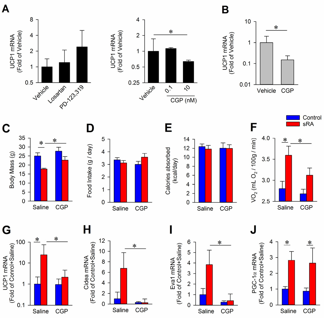
Blockade of the AT1 receptor with losartan in differentiated 3T3-L1 adipocytes failed to alter UCP1 mRNA (Figure 2A). In contrast, antagonism of AT2 with PD-123,319 appeared to increase UCP1 expression and low-dose activation of AT2 with CGP-42112a (“CGP”, at doses where CGP maintains AT2-selectivity (Hines et al., 2001)) significantly suppressed UCP1 expression (Figure 2A). We confirmed the reduction of UCP1 expression following AT2 activation by CGP treatment (10 nM) in differentiated primary adipocytes isolated from iWAT of neonatal wildtype C57BL/6J mice (Figure 2B). Collectively, these data support a direct, suppressive effect of adipocyte AT2 receptors upon UCP1 mRNA. Given our in vivo data demonstrating a suppressive role for circulating ANG upon RMR in sRA mice (Grobe et al., 2010), we therefore hypothesized that selective activation of AT2 receptors in sRA mice should normalize RMR and weight gain in these animals.
Figure 2. Chronic AT2 activation reverses metabolic phenotypes of sRA mice.
(A) UCP1 mRNA from differentiated 3T3-L1 adipocytes treated with vehicle, losartan (10 µM), PD-123,319 (10 µM), or CGP-42112a “CGP” for 6 hrs (N=3 per treatment). (B) UCP1 mRNA in mouse primary adipocytes isolated from iWAT in control mice treated with vehicle (N=7) or CGP (10 nM; N=7) for 6 hrs. (C–F) Body mass (C), daily food intake (D), calories absorbed via the gastrointestinal track (E), and oxygen consumption at thermoneutrality (F) in control and sRA mice treated with saline or CGP. Con+Sal N=5 (3 males, 2 females); sRA+Sal N=5 (2 males, 3 females); Con+CGP N=7 (4 males, 3 females); sRA+CGP N=7 (4 males, 3 females). (G) UCP1 mRNA in the iWAT (N=6). (H) Cidea mRNA in the iWAT (N=6). (E) Eva1 mRNA in the iWAT (N=6). (J) PGC-1a mRNA in the iWAT (N=6). * p<0.05 was considered significant. All data presented as mean ± sem. See also Figure S2 and Table S1 and S2.
Control and sRA mice were chronically infused with saline or low-dose CGP (50 ng/kg/min, 8 wks, s.c.) to test whether AT2 activation is sufficient to normalize metabolic function in sRA mice. CGP infusion significantly increased sRA body mass (Figure 2C) without altering feeding behavior (Figure 2D). Caloric absorption through the gastrointestinal tract, as determined by bomb calorimetry, was also not different between sRA and littermate mice or with CGP treatment (Figure 2E), which was consistent with unaltered pancreatic lipase and colipase expression (Figure S2A). We concluded that AT2 activation did not alter energy intake/uptake, but rather inhibited energy expenditure.
While chronic AT2 activation had no effect on energy intake, this manipulation significantly reduced RMR in sRA mice (Figure 2F), consistent with a suppression of energy expenditure. Despite no change in the iWAT mass with CGP treatment (Table S2), the elevated UCP1 expression in this fat pad was normalized with CGP treatment in sRA mice (Figure 2G). Conversely, despite a reduction in whole-body RMR, CGP increased UCP1 mRNA in BAT (Figure S2B), further supporting a dominant role for inguinal fat in the observed energy balance changes of sRA mice. In addition to UCP1, the expression of brown/beige-adipose markers (Wu et al., 2012) Cidea (Figure 2H) and Eva1 (Figure 2I) were normalized in iWAT from sRA mice treated with CGP supporting further the concept that AT2 activation reversed a ‘browning’ of the iWAT in sRA mice. Interestingly, despite an increase in PGC-1α mRNA in iWAT of sRA mice, CGP did not alter PGC-1α expression (Figure 2J). In addition, analysis of brown/beige-fat markers identified altered Slc27a1, Sp100, Ear2, Tmem26, Cd40, Hspb7, Acot2, and Oplah in the iWAT of sRA mice relative to control littermate mice, whereas Ear2, Tbx1, Acot2, and Pdk4 were significantly changed with CGP (Figure S2C, S2D and Table S3). CGP did not have any significant effect upon any markers in wildtype control mice (Figures 2G–J). Finally, with the exception of dermatopontin expression which was suppressed in sRA mice and stimulated with CGP treatment, white adipose markers were largely unchanged in sRA mice (Table S4). These data support the concept that chronic AT2 activation reduces RMR, likely through modulation of the molecular identity and thereby thermogenic program of the inguinal fat pad. Notably these findings do not exclude the possibility that CGP treatment may also modulate physical activity-based energy expenditure, however other studies have demonstrated small and opposing effects of AT2 receptor modulation upon locomotor activity, reducing the likelihood of a major contribution of this mechanism to the energy balance changes noted herein (Gross et al., 2000; Hein et al., 1995; Watanabe et al., 1999). To further understand the mechanism of the RMR effects, we next investigated possible metabolic targets of AT2.
AT2 modulation of RMR is not mediated through suppression of sympathetic nerve activity
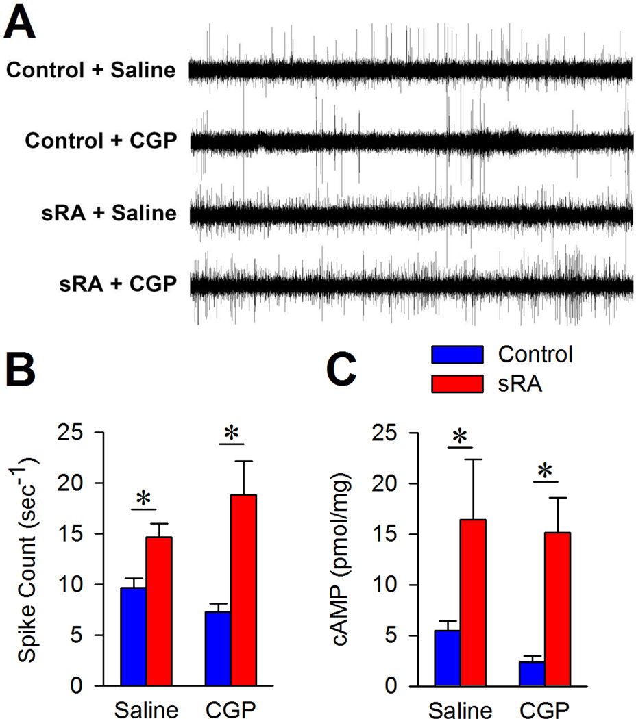
Sympathetic nerve activity (SNA) is known to mediate adaptive thermogenesis and thereby contribute to resting metabolism. Therefore, we hypothesized that AT2 activation may reduce RMR through suppression of SNA. To test this, we recorded inguinal sympathetic nerve activity in littermate control or sRA mice following treatment with saline or the AT2 agonist, CGP (Figure 3A). SNA to the iWAT was increased in sRA mice compared to littermate controls (Figures 3B and S3A). This is consistent with generalized increase in sympathetic activity as we previously documented where renal and BAT SNA were elevated in sRA mice (Grobe et al., 2010). Somewhat unexpectedly given its known suppressive functions within the central nervous system (Gao and Zucker, 2011), AT2 activation via CGP infusion failed to reduce SNA in either genotype (Figure 3B and S3A). Additionally, we determined that while sRA mice exhibit various changes in blood pressure, heart rate and renal renin expression, these endpoints were not significantly affected by CGP treatment in both control and sRA mice, possibly due to the low dose of CGP that was administered (Figure S3B–C).
Figure 3. AT2 activation does not reduce inguinal SNA.
(A) Representative electrograms from inguinal sympathetic nerves. (B) Quantification of thresholded spike count. Con+Sal N=10 (3 males, 7 females); sRA+Sal N=10 (3 males, 7 females); Con+CGP N=8 each (2 males, 6 females); sRA+CGP N=8 each (2 males, 6 females). (C) cAMP levels from iWAT isolated from control and sRA mice treated with saline or CGP (N=3 per group). * p<0.05 was considered significant. All data presented as mean ± sem. See also Figure S3.
Although the sympathetic drive to the iWAT was not suppressed with AT2 activation, we next examined whether β-adrenergic signaling was altered within the iWAT. Therefore, we measured cAMP, which is produced upon β-adrenergic stimulation, within the iWAT. sRA mice exhibited elevated cAMP in the inguinal fat pad, and cAMP levels remain high after AT2 activation (Figure 3C). Hence, AT2 activation does not reduce SNA or the immediate downstream signal of β-adrenergic stimulation within inguinal adipose. Rather, AT2 likely may modulate other intracellular signal downstream of cAMP to suppress UCP1 transcription, glucose flux, and RMR within the iWAT.
AT2 receptor activation blunts induction of UCP1 mRNA in white adipocytes
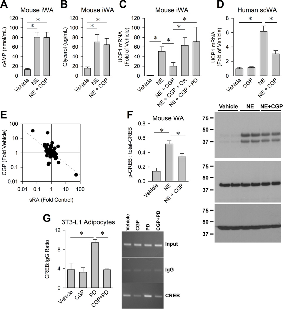
To further investigate the mechanism of RMR modulation by AT2 and to determine the effects of direct adipocyte-specific activation of AT2, we examined second-messenger pathway functions in cultured primary adipocytes. Adipocytes from the inguinal fat pad (iWA) or the interscapular fat pad (BA) were isolated from neonatal wild-type C57BL/6J mice and cultured as reported previously (Markan et al., 2014). To examine the effect of activating AT2 upon β-adrenergic signaling, we first assessed cAMP production in response to norepinephrine (NE; 10 µM) with or without CGP (10 nM) co-treatment. NE increased cAMP as expected in iWA, and CGP did not alter this effect (Figure 4A), which was consistent with our in vivo findings (Figure 3C). NE is also known to induce lipolysis, and liberated fatty acids are required to functionally activate UCP1 protein and to stimulate heat production (Atgié et al., 1997). Therefore, we considered the possibility that AT2 may suppress lipolysis, ultimately suppressing heat production. Glycerol levels in the media of the cells were increased with NE treatment, but not altered with CGP in mouse iWA (Figure 4B) or BA (Figure S4A). We conclude that AT2 does not modulate adipocyte cAMP production or lipolysis in response to NE.
Figure 4. AT2 activation suppresses norepinephrine induced UCP1 in white adipocytes (iWA).
(A) cAMP levels from mouse white adipocytes from iWAT (iWA) following treatment for 6 hours with vehicle, NE (10 µM), or NE and CGP (10 nM) (N=6). (B) Glycerol in the media from mouse iWA following treatment for 6 hours with vehicle, NE, or NE and CGP (N=6). (C) UCP1 mRNA in mouse iWA treatment for 6 hours (N=7–8). (D) UCP1 mRNA in human immortalized adipocytes following treatment for 6 hours with vehicle (N=16), CGP (N=11), NE (N=9), or NE and CGP (N=11). (E) Expression of CREB target genes in iWAT transcriptome dataset. (F) Representative Western blot and quantification of Western blot analysis of total and phosphorylated CREB in mouse iWA treated with vehicle, NE (1 µM), NE + CGP (10 nM; N=5 per group) for 5 min. β-actin served as a loading control. (G) Chromatin immunoprecipitation assay for binding of CREB to the UCP1 promoter with CGP (10 nM) ± PD (10 µM); N=3. * p<0.05 was considered significant. All data presented as mean ± sem. See also Figure S4 and Table S3.
Although lipolysis (and by extension UCP1 activity) is not modulated by AT2 activation, our data above support the modulation of UCP1 mRNA expression by AT2 receptors. Activation of AT2 did not alter NE-induced UCP1 mRNA in cultured mouse BA (Figure S4B). In contrast, the induction of UCP1 mRNA by NE was significantly blunted with CGP treatment in mouse iWA (Figure 4C). In a third adipose cell culture model consisting of immortalized human subcutaneous preadipocytes (Gadupudi et al., 2015; Vu et al., 2013; Vu et al., 2015), we confirmed a suppressive effect of AT2 activation upon UCP1 mRNA (Figure 4D). These data support an action of AT2 receptors to modulate β-adrenergic signaling downstream of cAMP production and the bifurcation of second-messenger modulation of UCP1 expression versus lipolysis. AT2 is known to activate the serine/threonine phosphatase type 2A (PP2A) in other cell types (Shenoy et al., 1999), which led us to hypothesize that the modulatory effect of AT2 activation in adipocytes may be mediated through phosphatase activity. Consistent with this possibility, we found that phosphatase inhibition with okadaic acid (OA) reversed the blunting effect of CGP on NE-induced UCP1 expression (Figure 4C). OA inhibits several phosphatases including PP1, PP2A, PP4, PP5, and PP6 (Swingle et al., 2007), and all of these were expressed at detectable levels in our RNAseq analysis of inguinal fat pads (Table S5). Similarly, the AT2 antagonist, PD-123,319 (PD), also reversed the effects of AT2 activation (Figure 4C). Together these data confirm the AT2-dependent action of CGP in adipocytes to specifically modulate UCP1 expression (not activation) and implicate the activation of an okadaic-acid dependent phosphatase in this mechanism.
cAMP-response element binding protein (CREB) induces UCP1 transcription in response to elevated cAMP signaling (Rim and Kozak, 2002). Examination of known CREB target genes in the iWAT transcriptome via RNA sequencing (RNAseq) uncovered that 15 CREB targets had altered expression levels in control versus sRA mice, and that the expression patterns of 7 of these genes were then significantly reversed with CGP treatment (Figure 4E and Table S6). CREB phosphorylation at Ser133 was increased after NE treatment and significantly attenuated with CGP co-treatment in mouse iWA (Figure 4F). Finally, antagonism of AT2 activation by PD-123,319 disinhibited CREB binding to the UCP1 promoter in differentiated 3T3-L1 adipocytes in a chromatin immunoprecipitation assay (Figure 4G). Therefore, AT2 likely blocks transcription of UCP1 through the phosphatase-mediated dephosphorylation and subsequent inactivation of the CREB transcription factor.
AT2 activation does not decrease canonical natriuretic peptide signaling
Positive implication of an AT2/phosphatase/CREB mechanism does not rule out other potential contributors to elevated RMR in sRA mice or other potential targets of AT2 signaling in iWA. To probe these other potential mechanisms we considered natriuretic peptide signaling which has been reported to increase RMR and induce UCP1 in mice (Bordicchia et al., 2012). Upon atrial natriuretic peptide (ANP) or brain natriuretic peptide (BNP) binding to the signaling receptor, NPR-A, intracellular cGMP is produced. cGMP levels have been shown to be altered by AT2 in other cell types (Bottari et al., 1992) raising the possibility that adipose natriuretic peptide signaling may be modulated by AT2 signaling. Furthermore, chronic hypertension (as previously documented in sRA mice (Grobe et al., 2010; Littlejohn et al., 2013; Sakai et al., 2007)) has been correlated with increased levels of ANP (Sagnella et al., 1986) and BNP (Kohno et al., 1992). Thus, we assessed whether natriuretic peptide signaling is increased in sRA mice, and this may contribute to elevated RMR.
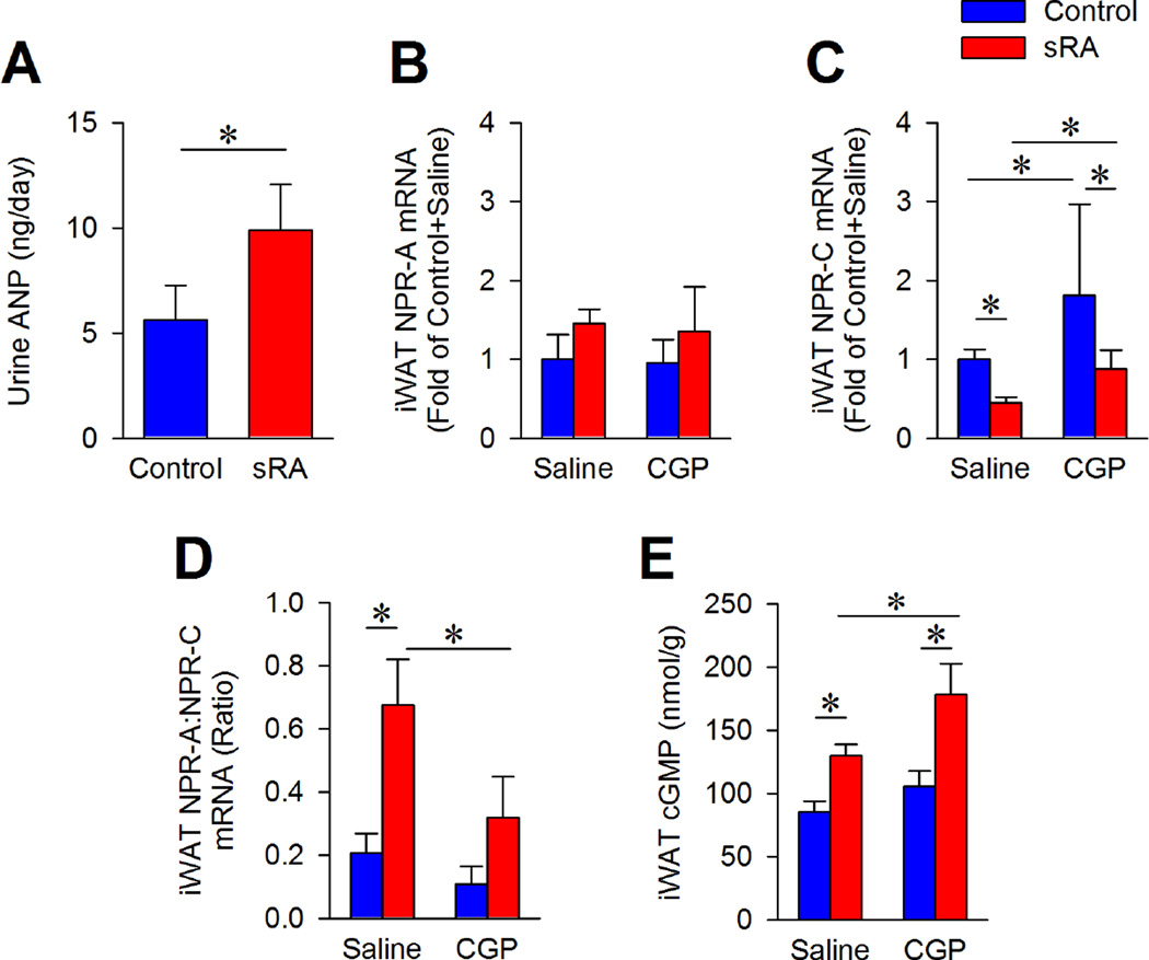
Total daily loss of ANP to urine (Figure 5A) was elevated in sRA mice even though no change in plasma ANP was detected (Figure S5A), supportive of an increased total daily ANP release. To determine if sensitivity to natriuretic peptide in iWAT was altered, we measured the expression of the canonical signaling receptor, NPR-A, and the clearance receptor, NPR-C. While NPR-A expression (Figure 5B) was not different, saline-treated sRA mice have decreased NPR-C mRNA (Figure 5C) compared to control mice. An enhanced signaling-to-clearance receptor mRNA ratio was proposed to indicate elevated sensitivity to natriuretic peptide signaling (Bordicchia et al., 2012). Therefore, the increased ratio of these receptor transcripts in iWAT of sRA mice (Figure 5D) may indicate greater sensitivity to natriuretic peptide signaling in sRA mice. CGP increased the expression of NPR-C and normalized the receptor ratio in sRA iWAT. As expected, iWAT cGMP levels were elevated in sRA mice, but surprisingly CGP did not reduce cGMP (Figure 5E). Thus even though natriuretic peptide signaling appears to be enhanced in sRA iWAT and AT2 activation increased the expression of the NPR-C clearance receptor, AT2 activation does not suppress immediate second-messenger activation due to natriuretic peptide signaling. Therefore, the NPR-A/cGMP pathway is unlikely to mediate the observed effects of AT2 activation upon UCP1 and RMR.
Figure 5. AT2 activation does not suppress canonical natriuretic peptide signaling.
(A) Urine ANP levels (N=6; 3 males, 3 females). (B) NPR-A mRNA in iWAT N=4 per group (2 males, 2 females). (C) NPR-C mRNA in iWAT N=4 per group (2 males, 2 females). (D) NPR-A to NPR-C mRNA ratio in iWAT N=4 per group (2 males, 2 females). (E) cGMP levels in iWAT (N=4; 2 males, 2 females). * p<0.05 was considered significant. All data presented as mean ± sem. See also Figure S5.
AT2 activation reduces RMR via suppression of growth factor signaling
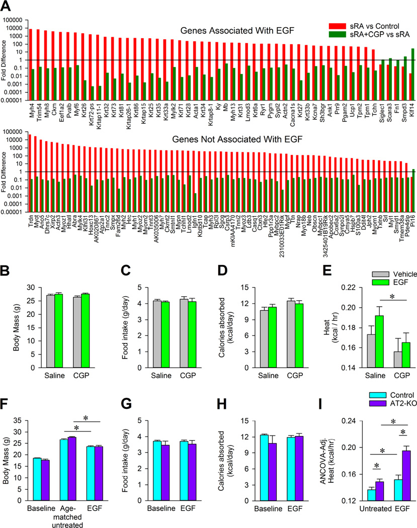
Next, we used an unbiased approach to identify additional signaling pathways that are altered in the iWAT of sRA mice. Specifically, we analyzed the transcriptome of iWAT from control and sRA mice treated with either saline or CGP, using RNA sequencing. From this examination, 123 genes were identified as having significantly altered expression (p<0.001) between control and sRA mice. Intriguingly, the alterations in expression of these same 123 genes were largely reversed with CGP treatment (Figure 6A). Gene Set Enrichment Analysis (GSEA) (Subramanian et al., 2005) of the dataset uncovered that genes in the epidermal growth factor (EGF) signaling pathway were, as a group, upregulated (p-value=0.02) in sRA mice compared to littermate controls, and suppressed (p-value=0.02) by CGP treatment. These data precipitated the hypothesis that AT2 may additionally modulate RMR in sRA mice via interference with EGF signaling within the inguinal fat pad.
Figure 6. AT2 activation suppresses EGF signaling in vivo.
(A) Genes identified through RNA sequencing analysis from the iWAT isolated from control or sRA mice treated with saline or CGP. Top panel includes genes known to be associated with EGF signaling; lower panel genes not associated with EGF. (B–E) Body mass (B), daily food intake (C), calories absorbed via the gastrointestinal tract (D), and heat as measured by respirometry (E) in wild-type C57BL/6J male mice treated for 2 weeks with saline (N=20), EGF (N=20), CGP (N=9), or EGF and CGP (N=17). (F–I) Body mass (F), food intake (G), calories absorbed via the gastrointestinal track (H), or ANCOVA-adjusted heat production (I) in control and AT2-KO mice (all males) at baseline, age-matched untreated control mice (N=11), untreated AT2-KO mice (N=12), or after 2 week EGF treatment in control mice (N=4) and AT2-KO mice (N=4). * p<0.05 was considered significant. All data presented as mean ± sem. See also Figure S6, Table S4, and S5.
To test if AT2 targets EGF signaling to modulate RMR in vivo, we chronically infused wildtype C57BL/6J male mice for 2 weeks with saline, EGF (0.833 µg/hr, s.c.), CGP (50 ng/kg/min, s.c.), or both EGF and CGP. There was no significant change in body mass (Figure 6B), food intake (Figure 6C), or gastrointestinal caloric absorption (Figure 6D) in any treatment group during this relatively short-term treatment. In contrast, fat mass was reduced with EGF treatment, and this corresponded with an increase in lean body mass (Table S7). Critically, CGP co-treatment with EGF significantly reduced heat production compared to EGF alone (Figure 6E), though this co-treatment did not significantly alter the effect of 2-week EGF treatment upon adipose mass (Table S7). Therefore, AT2 activation in wildtype mice is sufficient to reduce EGF-induced heat production.
To further examine the effects of AT2 modulation upon RMR and the interaction between AT2 and EGF in the control of RMR, we examined RMR control in mice genetically deficient for the AT2 receptor (AT2-KO), originally developed by Drs. Victor J. Dzau and Richard E. Pratt (Hein et al., 1995). Mice deficient for the AT2 receptor, originally on the FVB/NCrl genetic background, were backcrossed onto the C57BL/6J background for >7 generations before testing. RMR in a cohort of five male AT2-KO (29.32±1.62g) and five weight-matched male littermate control mice (28.75±1.52g) was examined by respirometry. Although no change was observed in respiratory exchange ratio (RER; control 0.832±0.010, AT2-KO 0.833±0.012), AT2-KO mice exhibited a robust increase in RMR (control 0.217±0.006 vs AT2-KO 0.272±0.019 kcal/hr, p=0.02) (Figure S6).
We next tested the RMR responses of AT2-KO mice and littermate controls to two-week subcutaneous infusion of EGF. Body mass increased after EGF infusion in both littermate control (4.98±0.36 g) and AT2-KO (5.63±0.35 g) mice compared to baseline, but this weight gain was reduced compared to untreated age-matched control and AT2-KO mice (Figure 6F). Food intake (Figure 6G) and total caloric absorption (Figure 6H) were unchanged by treatment or genotype. EGF reduced iWAT and eWAT mass (Table S8), which is consistent with decreased adipose mass in EGF-treated rats (Pedersen et al., 2000; Serrero and Mills, 1991). Because of the wide differences in body weights between groups, we normalized heat production using ANCOVA adjustment. As above, untreated AT2-KO mice exhibited increased heat production compared to littermate controls under baseline conditions, consistent with a tonic inhibitory role for AT2 in RMR control (Figure 6I). In addition, EGF increased heat production in both genotypes, but increased RMR in AT2-KO mice to a much higher level than in littermate controls (Figure 6I). Therefore, we conclude that AT2 receptors act as tonic suppressors of RMR, and this is related to alterations in EGF signaling.
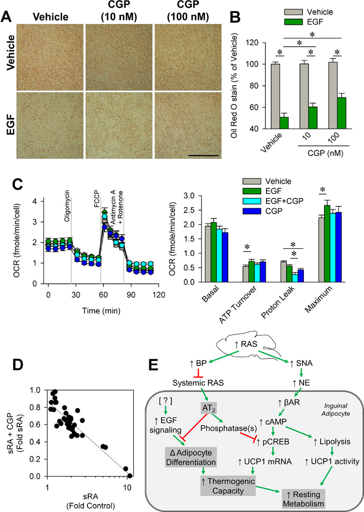
Surprisingly, the mechanism of interaction between AT2 and EGF in the adipose appears to be independent of modulation of UCP1 mRNA. UCP1 mRNA in the iWAT pad was unchanged (Figure S7A) or reduced (Figure S7B) by EGF infusion. In addition, we determined that UCP1 mRNA was unchanged by EGF treatment with or without co-treatment with the AT2 antagonist, PD-123,319, in cultured adipocytes (Figure S7C), similar to our in vivo observations. In contrast, the effects of EGF upon adipocytes have largely been attributed to the modulation of differentiation (Hauner et al., 1995; Lee et al., 2008; Serrero, 1987). Thus, we examined the modulatory effect of CGP upon adipocyte differentiation. EGF treatment during differentiation resulted in a major suppression of adipocyte Oil-Red-O staining (Figure 7A). Consistent with the results above, CGP dose-dependently blunted the inhibitory effect of EGF on adipocyte differentiation (Figure 7B). To determine the functional significance of EGF-altered adipocyte differentiation, we measured oxygen consumption rate (OCR; normalized to cell number) in cells supplemented with EGF and/or CGP during differentiation (Figure 7C). Though EGF treatment had no significant effects upon basal OCR, CGP treatment significantly (main effect p=0.04) suppressed basal OCR. Oligomycin treatment revealed a significant effect of CGP (main effect p<0.01) to reduce proton leak consistent with a suppressive effect upon UCP1 expression in these cells. This major suppression of uncoupled proton leak by CGP (CGP alone was 62% of vehicle, and EGF+CGP was 46% of EGF alone) is consistent with the suppressive effect of CGP upon UCP1 and RMR in vivo (Figures 2F, 2G, 6E, 6I). In addition, EGF increased ATP turnover by 32% in the absence of CGP (EGF x CGP interaction p=0.03), consistent with a cross-talk between EGF and AT2 in the control of electron transport chain activity. GSEA of the iWAT transcriptome from control and sRA mice treated with and without CGP (as presented in Figure 6A) uncovered a significant change in the expression of electron transport chain components (Figure 7D and Table S6). In the absence of CGP, EGF significantly increased maximal OCR responses to carbonyl cyanide-4-phenylhydrazone (FCCP) by 19% compared to vehicle, also supporting a major modulatory effect of EGF upon adipocyte thermogenic capacity. These data confirm physiologically-significant and direct actions of both EGF and AT2 within the inguinal adipocyte to modulate energy turnover, and highlight complex interactions which may represent novel therapeutic targets for controlling adipocyte identity and function.
Figure 7. AT2 activation reverses the suppressed adipogenesis by EGF.
(A) Representative image of Oil Red O staining on day 4 of differentiation in mouse iWA treated with or without EGF (1 ng/mL) and/or CGP (10 or 100 nM). Bar = 1 mm. (B) Quantification of Oil Red O staining (N=5 per group). (C) Oxygen consumption rate (OCR) in primary mouse WA treated with vehicle (N=20), EGF (N=10), or EGF and CGP (N=20) during differentiation. Cells were subjected to a mitochondria stress test by acute stimulation with oligomycin, FCCP, and Antimycin A/Rotenone. (D) Expression of electron transport genes identified as an enrichment set by GSEA in iWAT transcriptome-dataset. (E) Model of elevated brain RAS and subsequent suppressed circulating RAS induction of resting metabolism. * p<0.05 was considered significant. All data presented as mean ± sem. See also Table S6.
DISCUSSION
The implication of the RAS in the control of energy homeostasis in addition to its well-recognized role in cardiovascular functions may help clarify the comorbidity of metabolic and cardiovascular diseases, such as obesity and hypertension. The present study helps to illuminate the mechanisms involved in metabolic control by the RAS. Specifically, we have documented actions of AT2 receptor within inguinal adipocytes to control the molecular identity and thermogenic capacity of these cells (Figure 7E), ultimately to regulate whole-body energy balance. These insights help to clarify the nuanced and multifaceted effects of ANG in the periphery in the control of energy expenditure, and the mechanisms of cross-talk between the brain and peripheral RAS. Future obesity therapeutics targeting the RAS may take advantage of the selective actions of ANG through the AT2 receptor or its downstream targets specifically within subcutaneous adipose to modulate RMR and thereby energy homeostasis.
The adipose RAS contributes to obesity-hypertension, and adipose expansion is responsive to both the local adipose RAS and the circulating RAS (Goossens et al., 2003; Yvan-Charvet and Quignard-Boulange, 2011). For example, transgenic overexpression of angiotensinogen specifically in adipose tissue causes mice to become hypertensive and obese (Yvan-Charvet et al., 2009). Yiannikouris et al. found that adipose-specific disruption of angiotensinogen prevents the induction of hypertension but not weight gain with diet-induced obesity (Yiannikouris et al., 2012a; Yiannikouris et al., 2012b). Consistent with these findings and the current study, Yvan-Charvet et al. previously demonstrated that genetic disruption of the AT2 receptor prevented obesity but had no antihypertensive effect in mice overexpressing angiotensinogen in adipose tissue (Yvan-Charvet et al., 2005; Yvan-Charvet et al., 2009). Intriguingly, the actions of AT2 to modulate UCP1 expression appear to be limited to the inguinal (subcutaneous) adipose pad. This discovery is interesting both mechanistically, and for its clinical implications. As subcutaneous fat is largely considered “beneficial” opposed to inflammatory abdominal/visceral fat (Abate et al., 1995; McLaughlin et al., 2011; Miyazaki and DeFronzo, 2009), and as obesity is associated with increased circulating RAS activity (Engeli et al., 2005; Yasue et al., 2010), these findings implicate the adipose RAS in the modulation of cardio/metabolic risks during obesity.
The discovery of an interaction between AT2 receptor signaling and EGF receptor signaling within adipocytes was not entirely unexpected. Others have previously documented a role for EGF in adipose development, and interactions between EGF and AT2 have been reported in other tissues (Meffert et al., 1996; Nouet et al., 2004; Plouffe et al., 2006). Our data indicate that AT2 stimulation impairs oxidative phosphorylation and highlight an important interaction between EGF and the AT2 within inguinal adipocytes, but the molecular mechanism of this interaction remains elusive. Increased body mass index is correlated with a reduction in electron transport chain components resulting in decreased OCR in human adipocytes (Fischer et al., 2015). Thus, one possible mechanism for the elevated maximal oxidative phosphorylation observed with EGF treatment is an alteration in levels of electron transport chain components. Although further analysis of these mechanisms is warranted to determine how EGF and AT2 modulate oxidative phosphorylation capacity, our data clearly support an interaction between EGF and the AT2 receptor on adipocytes. Given our previous demonstration that the AT2 receptor modulates anaerobic metabolism in vivo (Burnett and Grobe, 2013) and the previously-established role for EGF in the modulation of anaerobic metabolism in cancer cells (Lee et al., 2015; Velpula et al., 2013), we posit that the molecular interaction between AT2 and EGF within adipocytes may prove to be similar to that which occurs in cells exhibiting the Warburg effect. Another possible contributing mechanism may involve futile cycling of substrates such as creatinine, as this pathway has recently been documented as another UCP1-independent pathway of energy expenditure by adipocytes (Kazak et al., 2015).
Although no sex differences were noted in the current study, some data support a complex, sex-dependent contribution of AT2 to the regulation of weight gain. Samuel et al. previously demonstrated that weight gain of AT2-deficient mice on a high fat diet is normal in males but accelerated in females (Samuel et al., 2013). Nag et al. also previously demonstrated that pharmacological activation of the AT2 receptor with C21 attenuated high fat diet-induced weight gain in female mice (Nag et al., 2015). How AT2 may contribute to sex differences in susceptibility to weight gain, in terms of physiological mechanism (intake behavior, digestive efficiency, RMR, etc.) or molecular mechanism (activation of phosphatases, interference with EGFR signaling, etc.) remains unclear. Nonetheless, given the inguinal fat-specific role for AT2 action noted herein, a potential role for AT2 in observed differences in adiposity and patterns of adipose distribution and adipose function (Blaak, 2001; Krotkiewski et al., 1983) between males and females could be speculated.
Ultimately these studies support the general concept that tissue- and receptor-specific actions of the RAS contribute to energy homeostasis (Littlejohn and Grobe, 2015). This overall concept likely helps to explain the lack of efficacy of ‘global’ pharmacological RAS inhibition as a therapeutic approach to human obesity. Human obesity is correlated with increased circulating RAS activity (Engeli et al., 2005; Yasue et al., 2010), which would be expected to increase SNA (de Kloet et al., 2010). However, increased circulating RAS activity would presumably stimulate AT2 signaling in thermogenic (or potentially thermogenic) adipose, resulting in reduced thermogenic capacity and thermogenesis in response to SNA stimulation. While pharmacological inhibition of the peripheral RAS could result in reduced adipose AT2 activation, the loss of compartmentalization during obesity due to increased permeability of the blood-brain barrier (Gustafson et al., 2007) would simultaneously result in reduced brain RAS activation. The net effect on body mass would therefore be expected to be negligible. Future work to develop selective AT2 antagonists which are incapable of crossing even a highly-permeable blood-brain barrier may therefore prove useful for obesity therapeutics.
EXPERIMENTAL PROCEDURES
Experimental procedures are outlined in detail in the associated supplemental text file.
Animals
All procedures performed were approved by the University of Iowa’s Institutional Animal Care and Use Committee, and conform to the guidelines set forth by the National Research Council (National Research Council, 2011). The sRA (sRAflox line 11110/2 × 4284/1) transgenic colony was generated as previously described (Grobe et al., 2010; Sakai et al., 2007). Mice were treated with subcutaneous infusion of saline, CGP-42112a (100 ng/kg/min, s.c.), or epidermal growth factor (EGF; AbD Serotec; 0.833 µg/hr, s.c.) using Alzet osmotic minipumps. AT2 -KO mice, originally developed by Victor J. Dzau and Richard E. Pratt (Hein et al., 1995) were obtained from Charles River Laboratories on the FVB/NCrl background and were backcrossed onto the C57BL/6J background for at least 7 generations. All animals had ad libitum access to standard chow (Harlan Teklad 7013) and water, and were maintained on a standard 12:12 hr lighting cycle.
In Vivo Glucose Uptake Assay
Tail blood was collected from all mice (time=0) followed by an injection (i.p.) of 8–10µCi of [3H]-2-deoxyglucose in a 20% glucose solution. Plasma radioactivity and [3H]-2-deoxyglucose uptake in tissues was analyzed as previously described (Markan et al., 2014).
Sympathetic Nerve Activity
Sympathetic nerve activity to the inguinal fat pad was assessed as previously described (Grobe et al., 2010; Rahmouni et al., 2008).
Primary adipocyte culture
Inguinal white adipose tissue or interscapular brown adipose tissue was collected from 4 day-old pups as previously described (Markan et al., 2014).
Immortalized human preadipocyte/adipocyte culture
Immortalized normal human preadipocytes (NPADs) have been described previously (Vu et al., 2013).
Cellular respiration
Mouse primary inguinal white adipocytes were seeded and grown in XF96 V3 PET cell culture microplates (Seahorse Biosciences, Billerica, MA). Oxygen consumption rate (OCR) values were normalized to cell numbers as previously described (Wagner et al., 2011). The final mitochondrial inhibitor concentrations used were: 2.5 µM Oligomycin, 0.75 µM FCCP, 10 µM Rotenone, 10 µM Antimycin A.
RNA sequencing analysis
Total RNA was isolated from the inguinal fat pad and was used to generate cDNA libraries with each library tagged with a unique sequence barcode. The libraries were sequenced (50 bp paired end reads) with the Illumina HiSeq 2000 at the DNA Facility at Iowa State University. The quality of the sequence reads was verified using the FastQC program. Bowtie (Langmead et al., 2009) was used to align sequences to the mouse genome (mm9, NCBI37) with alignment results saved in BAM/SAM format. The SortSam function of Picard Tools was used to sort SAM files and paired reads not mapping to the same genomic location were removed. The number of sequence reads per gene was calculated using the count function of gfold (Feng et al., 2012). The total counts per gene was normalized using the edgeR (Robinson et al., 2010) package in R/Bioconductor to account for the variance in the number of total sequence reads between samples. Statistical analysis of differential expression was done using the edgeR (Robinson et al., 2010) package and p-value correction for multiple testing was done using the qvalue (Storey and Tibshirani, 2003) package in R/Bioconductor. An adjusted p-value less than 0.001 was considered statistically significant. All RNA sequencing data has been submitted to the public repository at NCBI-GEO (Accession Number GSE77214).
Statistical Analyses
ANOVA (with or without repeated measures as appropriate) followed by Tukey multiple comparisons procedures were used throughout, with p<0.05 considered statistically significant. ANCOVA was used to correct data for body mass, as indicated. Gene expression data were analyzed using the Livak method (Livak and Schmittgen, 2001). Mann-Whitney U, Kruskal-Wallis, or Friedman’s ANOVA were used when data failed normality or equal variance tests, and their applications are noted in figure legends if used. Data are presented as mean±sem throughout.
Supplementary Material
Acknowledgments
The authors gratefully acknowledge technical assistance by the University of Iowa Genome Editing Core Facility, the Office of Animal Resources of the University of Iowa, Brett Wagner for technical assistance, and the intellectual support of Allyn L. Mark, MD. NKL was supported by a predoctoral fellowship from the American Heart Association (14PRE18330015). BJW was supported by undergraduate fellowships from the American Heart Association, the American Physiological Society, and the University of Iowa Center for Research by Undergraduates (ICRU). KEC was supported by a predoctoral fellowship from the American Heart Association (14PRE20380401). KRM was supported by a postdoctoral fellowship from the NIH (F32DK102347). This work was supported by grants from the National Institutes of Health (DK106104 to MJP, HL084207 to KR, CDS and JLG, HL048058 to CDS, HL098276 to JLG), the American Diabetes Association (7-13-JF-49 to MJP, 1-14-BS-079 to JLG), the American Heart Association (14EIA18860041 to KR, 14IRG18710013, 15SFRN23480000 to CDS, and 15SFRN23730000 to JLG), the University of Iowa’s Vice President for Research and Economic Development (JLG), Roy J. Carver Trust (CDS) and Fraternal Order of Eagles’ Diabetes Research Center (AJK, MJP, and JLG).
Footnotes
Publisher's Disclaimer: This is a PDF file of an unedited manuscript that has been accepted for publication. As a service to our customers we are providing this early version of the manuscript. The manuscript will undergo copyediting, typesetting, and review of the resulting proof before it is published in its final citable form. Please note that during the production process errors may be discovered which could affect the content, and all legal disclaimers that apply to the journal pertain.
SUPPLEMENTAL INFORMATION
Supplemental Information includes six figures and six tables and can be found with this article.
AUTHOR CONTRIBUTIONS
N.K.L., H.L.K., B.J.W., K.E.C., K.V.T., K.R.M., M.C.N, F.A.G., N.A.P., X.L., D.A.M., A.J.K. performed research and analyzed data. N.K.L. and J.L.G. drafted the manuscript. N.K.L., A.J.K., M.J.P., K.R., C.D.S., J.L.G. revised manuscript. All authors approved final manuscript.
REFERENCES
- Adult Obesity: Obesity rises among adults. Centers for Disease Control and Prevention [Google Scholar]
- Abate N, Garg A, Peshock RM, Stray-Gundersen J, Grundy SM. Relationships of generalized and regional adiposity to insulin sensitivity in men. J Clin Invest. 1995;96:88–98. doi: 10.1172/JCI118083. [DOI] [PMC free article] [PubMed] [Google Scholar]
- Atgié C, D’Allaire F, Bukowiecki LJ. Role of β1- and β3-adrenoceptors in the regulation of lipolysis and thermogenesis in rat brown adipocytes. American journal of physiology. Cell physiology. 1997 doi: 10.1152/ajpcell.1997.273.4.C1136. [DOI] [PubMed] [Google Scholar]
- Blaak E. Gender differences in fat metabolism. Current opinion in clinical nutrition and metabolic care. 2001;4:499–502. doi: 10.1097/00075197-200111000-00006. [DOI] [PubMed] [Google Scholar]
- Bordicchia M, Dianxin L, Amri E-Z, Aihaud G, Dessi-Fulgheri P, Zhang C, Takahashi N, Sarzani R, Collins S. Cardia natriuretic peptides act via p38 MAPK to induce the brown fat thermogenice program in mouse and human adipocytes. J Clin Invest. 2012;122:1022–1036. doi: 10.1172/JCI59701. [DOI] [PMC free article] [PubMed] [Google Scholar]
- Bottari SP, King IN, Reichlin S, Dahlstroem I, Lydon N, de Gasparo M. The angiotensin AT2 receptor stimulates protein tyrosine phosphatase activity and mediates inhibition of particulate guanylate cyclase. Biochemical and biophysical research communications. 1992;183:206–211. doi: 10.1016/0006-291x(92)91629-5. [DOI] [PubMed] [Google Scholar]
- Burnett CM, Grobe JL. Direct calorimetry identifies deficiencies in respirometry for the determination of resting metabolic rate in C57Bl/6 and FVB mice. American journal of physiology. Endocrinology and metabolism. 2013;305:E916–F924. doi: 10.1152/ajpendo.00387.2013. [DOI] [PMC free article] [PubMed] [Google Scholar]
- Claflin KE, Grobe JL. Control of energy balance by the brain renin-angiotensin system. Current hypertension reports. 2015;17:38. doi: 10.1007/s11906-015-0549-x. [DOI] [PubMed] [Google Scholar]
- de Kloet AD, Krause EG, Scott KA, Foster MT, Herman JP, Sakai RR, Seeley RJ, Woods SC. Central angiotensin II has catabolic action at white and brown adipose tissue. American journal of physiology. Endocrinology and metabolism. 2011;301:E1081–E1091. doi: 10.1152/ajpendo.00307.2011. [DOI] [PMC free article] [PubMed] [Google Scholar]
- de Kloet AD, Krause EG, Woods SC. The renin angiotensin system and the metabolic syndrome. Physiology & behavior. 2010;100:525–534. doi: 10.1016/j.physbeh.2010.03.018. [DOI] [PMC free article] [PubMed] [Google Scholar]
- de Kloet AD, Pati D, Wang L, Hiller H, Sumners C, Frazier CJ, Seeley RJ, Herman JP, Woods SC, Krause EG. Angiotensin type 1a receptors in the paraventricular nucleus of the hypothalamus protect against diet-induced obesity. The Journal of neuroscience : the official journal of the Society for Neuroscience. 2013;33:4825–4833. doi: 10.1523/JNEUROSCI.3806-12.2013. [DOI] [PMC free article] [PubMed] [Google Scholar]
- Engeli S, Bohnke J, Gorzelniak K, Janke J, Schling P, Bader M, Luft FC, Sharma AM. Weight loss and the renin-angiotensin-aldosterone system. Hypertension. 2005;45:356–362. doi: 10.1161/01.HYP.0000154361.47683.d3. [DOI] [PubMed] [Google Scholar]
- Feng J, Meyer CA, Wang Q, Liu JS, Shirley Liu X, Zhang Y. GFOLD: a generalized fold change for ranking differentially expressed genes from RNA-seq data. Bioinformatics. 2012;28:2782–2788. doi: 10.1093/bioinformatics/bts515. [DOI] [PubMed] [Google Scholar]
- Fischer B, Schottl T, Schempp C, Fromme T, Hauner H, Klingenspor M, Skurk T. Inverse relationship between body mass index and mitochondrial oxidative phosphorylation capacity in human subcutaneous adipocytes. Am J Physiol Endocrinol Metab. 2015;309:E380–E387. doi: 10.1152/ajpendo.00524.2014. [DOI] [PubMed] [Google Scholar]
- Gadupudi G, Gourronc FA, Ludewig G, Robertson LW, Klingelhutz AJ. PCB126 inhibits adipogenesis of human preadipocytes. Toxicology in vitro : an international journal published in association with BIBRA. 2015;29:132–141. doi: 10.1016/j.tiv.2014.09.015. [DOI] [PMC free article] [PubMed] [Google Scholar]
- Gao L, Zucker IH. AT2 receptor signaling and sympathetic regulation. Current opinion in pharmacology. 2011;11:124–130. doi: 10.1016/j.coph.2010.11.004. [DOI] [PMC free article] [PubMed] [Google Scholar]
- Goossens GH, Blaak EE, van Baak MA. Possible involvement of the adipose tissue renin-angiotensin system in the pathophysiology of obesity and obesity-related disorders. Obesity reviews : an official journal of the International Association for the Study of Obesity. 2003;4:43–55. doi: 10.1046/j.1467-789x.2003.00091.x. [DOI] [PubMed] [Google Scholar]
- Grassi G, Seravalle G, Dell’Oro R, Trevano FQ, Bombelli M, Scopelliti F, Facchini A, Mancia G, Study C. Comparative effects of candesartan and hydrochlorothiazide on blood pressure, insulin sensitivity, and sympathetic drive in obese hypertensive individuals: results of the CROSS study. Journal of hypertension. 2003;21:1761–1769. doi: 10.1097/00004872-200309000-00027. [DOI] [PubMed] [Google Scholar]
- Grobe JL, Buehrer BA, Hilzendeger AM, Liu X, Davis DR, Xu D, Sigmund CD. Angiotensinergic signaling in the brain mediates metabolic effects of deoxycorticosterone (DOCA)-salt in C57 mice. Hypertension. 2011;57:600–607. doi: 10.1161/HYPERTENSIONAHA.110.165829. [DOI] [PMC free article] [PubMed] [Google Scholar]
- Grobe JL, Grobe CL, Beltz TG, Westphal SG, Morgan DA, Xu D, de Lange WJ, Li H, Sakai K, Thedens DR, et al. The brain Renin-angiotensin system controls divergent efferent mechanisms to regulate fluid and energy balance. Cell metabolism. 2010;12:431–442. doi: 10.1016/j.cmet.2010.09.011. [DOI] [PMC free article] [PubMed] [Google Scholar]
- Grobe JL, Rahmouni K, Liu X, Sigmund CD. Metabolic rate regulation by the renin-angiotensin system: brain vs. body. Pflugers Archiv : European journal of physiology. 2013;465:167–175. doi: 10.1007/s00424-012-1096-9. [DOI] [PMC free article] [PubMed] [Google Scholar]
- Gross V, Milia AF, Plehm R, Inagami T, Luft FC. Long-term blood pressure telemetry in AT2 receptor-disrupted mice. Journal of hypertension. 2000;18:955–961. doi: 10.1097/00004872-200018070-00018. [DOI] [PubMed] [Google Scholar]
- Gustafson DR, Karlsson C, Skoog I, Rosengren L, Lissner L, Blennow K. Mid-life adiposity factors relate to blood-brain barrier integrity in late life. Journal of internal medicine. 2007;262:643–650. doi: 10.1111/j.1365-2796.2007.01869.x. [DOI] [PubMed] [Google Scholar]
- Hauner H, Rohrig K, Petruschke T. Effects of epidermal growth factor (EGF), platelet-derived growth factor (PDGF) and fibroblast growth factor (FGF) on human adipocyte development and function. Eur J Clin Invest. 1995;25:90–96. doi: 10.1111/j.1365-2362.1995.tb01532.x. [DOI] [PubMed] [Google Scholar]
- Hein L, Barsh GS, Pratt RE, Dzau VJ, Kobilka BK. Behavioural and cardiovascular effects of disrupting the angiotensin II type-2 receptor in mice. Nature. 1995;377:744–747. doi: 10.1038/377744a0. [DOI] [PubMed] [Google Scholar]
- Hilzendeger AM, Cassell MD, Davis DR, Stauss HM, Mark AL, Grobe JL, Sigmund CD. Angiotensin type 1a receptors in the subfornical organ are required for deoxycorticosterone acetate-salt hypertension. Hypertension. 2013;61:716–722. doi: 10.1161/HYPERTENSIONAHA.111.00356. [DOI] [PMC free article] [PubMed] [Google Scholar]
- Hines J, Heerding JN, Fluharty SJ, Yee DK. Identification of angiotensin II type 2 (AT2) receptor domains mediating high-affinity CGP 42112A binding and receptor activation. The Journal of pharmacology and experimental therapeutics. 2001;298:665–673. [PubMed] [Google Scholar]
- Hsueh W, Davidai G, Henry R, Mudaliar S. Telmisartan effects on insulin resistance in obese or overweight adults without diabetes or hypertension. Journal of clinical hypertension. 2010;12:746–752. doi: 10.1111/j.1751-7176.2010.00335.x. [DOI] [PMC free article] [PubMed] [Google Scholar]
- Kazak L, Chouchani ET, Jedrychowski MP, Erickson BK, Shinoda K, Cohen P, Vetrivelan R, Lu GZ, Laznik-Bogoslavski D, Hasenfuss SC, et al. A creatine-driven substrate cycle enhances energy expenditure and thermogenesis in beige fat. Cell. 2015;163:643–655. doi: 10.1016/j.cell.2015.09.035. [DOI] [PMC free article] [PubMed] [Google Scholar]
- Kohno M, Horio T, Yokokawa K, Murakawa K, Yasunari K, Akioka K, Tahara A, Toda I, Takeuchi K, Kurihara N, et al. Brain natriuretic peptides as a cardiac hormone in essential hypertension. Am J Med. 1992;92:29–34. doi: 10.1016/0002-9343(92)90011-y. [DOI] [PubMed] [Google Scholar]
- Krotkiewski M, Bjorntorp P, Sjostrom L, Smith U. Impact of obesity on metabolism in men and women. Importance of regional adipose tissue distribution. The Journal of clinical investigation. 1983;72:1150–1162. doi: 10.1172/JCI111040. [DOI] [PMC free article] [PubMed] [Google Scholar]
- Langmead B, Trapnell C, Pop M, Salzberg SL. Ultrafast and memory-efficient alignment of short DNA sequences to the human genome. Genome biology. 2009;10:R25. doi: 10.1186/gb-2009-10-3-r25. [DOI] [PMC free article] [PubMed] [Google Scholar]
- Lee JS, Suh JM, Park HG, Bak EJ, Yoo YJ, Cha JH. Heparin-binding epidermal growth factor-like growth factor inhibits adipocyte differentiation at commitment and early induction stages. Differentiation; research in biological diversity. 2008;76:478–487. doi: 10.1111/j.1432-0436.2007.00250.x. [DOI] [PubMed] [Google Scholar]
- Lee KM, Nam K, Oh S, Lim J, Lee T, Shin I. ECM1 promotes the Warburg effect through EGF-mediated activation of PKM2. Cellular signalling. 2015;27:228–235. doi: 10.1016/j.cellsig.2014.11.004. [DOI] [PubMed] [Google Scholar]
- Lindholm LH, Persson M, Alaupovic P, Carlberg B, Svensson A, Samuelsson O. Metabolic outcome during 1 year in newly detected hypertensives: results of the Antihypertensive Treatment and Lipid Profile in a North of Sweden Efficacy Evaluation (ALPINE study) Journal of hypertension. 2003;21:1563–1574. doi: 10.1097/01.hjh.0000084723.53355.76. [DOI] [PubMed] [Google Scholar]
- Littlejohn NK, Grobe JL. Opposing tissue-specific roles of angiotensin in the pathogenesis of obesity, and implications for obesity-related hypertension. Am J Physiol Regul Integr Comp Physiol. 2015;309:R1463–R1473. doi: 10.1152/ajpregu.00224.2015. [DOI] [PMC free article] [PubMed] [Google Scholar]
- Littlejohn NK, Siel RB, Jr, Ketsawatsomkron P, Pelham CJ, Pearson NA, Hilzendeger AM, Buehrer BA, Weidemann BJ, Li H, Davis DR, et al. Hypertension in mice with transgenic activation of the brain renin-angiotensin system is vasopressin dependent. American journal of physiology. Regulatory, integrative and comparative physiology. 2013;304:R818–R828. doi: 10.1152/ajpregu.00082.2013. [DOI] [PMC free article] [PubMed] [Google Scholar]
- Livak KJ, Schmittgen TD. Analysis of relative gene expression data using real-time quantitative PCR and the 2(-Delta Delta C(T)) Method. Methods (San Diego, Calif.) 2001;25:402–408. doi: 10.1006/meth.2001.1262. [DOI] [PubMed] [Google Scholar]
- Markan KR, Naber MC, Ameka MK, Anderegg MD, Mangelsdorf DJ, Kliewer SA, Mohammadi M, Potthoff MJ. Circulating FGF21 is liver derived and enhances glucose uptake during refeeding and overfeeding. Diabetes. 2014;63:4057–4063. doi: 10.2337/db14-0595. [DOI] [PMC free article] [PubMed] [Google Scholar]
- McLaughlin T, Lamendola C, Liu A, Abbasi F. Preferential fat deposition in subcutaneous versus visceral depots is associated with insulin sensitivity. The Journal of clinical endocrinology and metabolism. 2011;96:E1756–E1760. doi: 10.1210/jc.2011-0615. [DOI] [PMC free article] [PubMed] [Google Scholar]
- Meffert S, Stoll M, Steckelings UM, Bottari SP, Unger T. The angiotensin II AT2 receptor inhibits proliferation and promotes differentiation in PC12W cells. Molecular and cellular endocrinology. 1996;122:59–67. doi: 10.1016/0303-7207(96)03873-7. [DOI] [PubMed] [Google Scholar]
- Miyazaki Y, DeFronzo RA. Visceral fat dominant distribution in male type 2 diabetic patients is closely related to hepatic insulin resistance, irrespective of body type. Cardiovascular diabetology. 2009;8:44. doi: 10.1186/1475-2840-8-44. [DOI] [PMC free article] [PubMed] [Google Scholar]
- Nag S, Khan MA, Samuel P, Ali Q, Hussain T. Chronic angiotensin AT2R activation prevents high-fat diet-induced adiposity and obesity in female mice independent of estrogen. Metabolism: clinical and experimental. 2015;64:814–825. doi: 10.1016/j.metabol.2015.01.019. [DOI] [PMC free article] [PubMed] [Google Scholar]
- National Research Council. Guide for the Care and Use of Laboratory Animals. Washington, D.C: National Acadamies Press; 2011. [Google Scholar]
- Nedergaard J, Cannon B. UCP1 mRNA does not produce heat. Biochimica et biophysica acta. 2013;1831:943–949. doi: 10.1016/j.bbalip.2013.01.009. [DOI] [PubMed] [Google Scholar]
- Nouet S, Amzallag N, Li JM, Louis S, Seitz I, Cui TX, Alleaume AM, Di Benedetto M, Boden C, Masson M, et al. Trans-inactivation of receptor tyrosine kinases by novel angiotensin II AT2 receptor-interacting protein, ATIP. The Journal of biological chemistry. 2004;279:28989–28997. doi: 10.1074/jbc.M403880200. [DOI] [PubMed] [Google Scholar]
- Pedersen SB, Kristensen K, Bruun JM, Flyvbjerg A, Vinter-Jensen L, Richelsen B. Systemic administration of epidermal growth factor increases UCP3 mRNA levels in skeletal muscle and adipose tissue in rats. Biochemical and biophysical research communications. 2000;279:914–919. doi: 10.1006/bbrc.2000.4022. [DOI] [PubMed] [Google Scholar]
- Plouffe B, Guimond MO, Beaudry H, Gallo-Payet N. Role of tyrosine kinase receptors in angiotensin II AT2 receptor signaling: involvement in neurite outgrowth and in p42/p44mapk activation in NG108-15 cells. Endocrinology. 2006;147:4646–4654. doi: 10.1210/en.2005-1315. [DOI] [PubMed] [Google Scholar]
- Porter JP, Anderson JM, Robison RJ, Phillips AC. Effect of central angiotensin II on body weight gain in young rats. Brain research. 2003;959:20–28. doi: 10.1016/s0006-8993(02)03676-4. [DOI] [PubMed] [Google Scholar]
- Porter JP, Potratz KR. Effect of intracerebroventricular angiotensin II on body weight and food intake in adult rats. American journal of physiology. Regulatory, integrative and comparative physiology. 2004;287:R422–R428. doi: 10.1152/ajpregu.00537.2003. [DOI] [PubMed] [Google Scholar]
- Rahmouni K, Fath MA, Seo S, Thedens DR, Berry CJ, Weiss R, Nishimura DY, Sheffield VC. Leptin resistance contributes to obesity and hypertension in mouse models of Bardet-Biedl syndrome. Journal of Clinical Investigation. 2008;118:1458–1467. doi: 10.1172/JCI32357. [DOI] [PMC free article] [PubMed] [Google Scholar]
- Rim JS, Kozak LP. Regulatory motifs for CREB-binding protein and Nfe2l2 transcription factors in the upstream enhancer of the mitochondrial uncoupling protein 1 gene. The Journal of biological chemistry. 2002;277:34589–34600. doi: 10.1074/jbc.M108866200. [DOI] [PubMed] [Google Scholar]
- Robinson MD, McCarthy DJ, Smyth GK. edgeR: a Bioconductor package for differential expression analysis of digital gene expression data. Bioinformatics. 2010;26:139–140. doi: 10.1093/bioinformatics/btp616. [DOI] [PMC free article] [PubMed] [Google Scholar]
- Sagnella GA, Markandu ND, Shore AC, MacGregor GA. Raised circulating levels of atrial natriuretic peptides in essential hypertension. Lancet. 1986:179–181. doi: 10.1016/s0140-6736(86)90653-7. [DOI] [PubMed] [Google Scholar]
- Sakai K, Agassandian K, Morimoto S, Sinnayah P, Cassell MD, Davisson RL, Sigmund CD. Local production of angiotensin II in the subfornical organ causes elevated drinking. The Journal of Clinical Investigation. 2007;117:1088–1095. doi: 10.1172/JCI31242. [DOI] [PMC free article] [PubMed] [Google Scholar]
- Samuel P, Khan MA, Nag S, Inagami T, Hussain T. Angiotensin AT2 Receptor Contributes towards Gender Bias in Weight Gain. PLoS ONE. 2013;8:e48425. doi: 10.1371/journal.pone.0048425. [DOI] [PMC free article] [PubMed] [Google Scholar]
- Serrero G. EGF inhibits the differentiation of adipocyte precursors in primary cultures. Biochemical and biophysical research communications. 1987;146:194–202. doi: 10.1016/0006-291x(87)90710-8. [DOI] [PubMed] [Google Scholar]
- Serrero G, Mills D. Physiological role of epidermal growth factor on adipose tissue development in vivo . Proceedings of the National Academy of Sciences of the United States of America. 1991;88:3912–3916. doi: 10.1073/pnas.88.9.3912. [DOI] [PMC free article] [PubMed] [Google Scholar]
- Shabalina IG, Petrovic N, de Jong JM, Kalinovich AV, Cannon B, Nedergaard J. UCP1 in brite/beige adipose tissue mitochondria is functionally thermogenic. Cell reports. 2013;5:1196–1203. doi: 10.1016/j.celrep.2013.10.044. [DOI] [PubMed] [Google Scholar]
- Shenoy UV, Richards EM, Huang X, Sumners C. Angiotensin II Type 2 Receptor-mediated apoptosis of cultured neurons from newborn rat brain. Endocrinology. 1999;140:500–509. doi: 10.1210/endo.140.1.6396. [DOI] [PubMed] [Google Scholar]
- Shimabukuro M, Tanaka H, Shimabukuro T. Effects of telmisartan on fat distribution in individuals with the metabolic syndrome. Journal of hypertension. 2007;25:841–848. doi: 10.1097/HJH.0b013e3280287a83. [DOI] [PubMed] [Google Scholar]
- Storey JD, Tibshirani R. Statistical significance for genomewide studies. Proceedings of the National Academy of Sciences of the United States of America. 2003;100:9440–9445. doi: 10.1073/pnas.1530509100. [DOI] [PMC free article] [PubMed] [Google Scholar]
- Subramanian A, Tamayo P, Mootha VK, Mukherjee S, Ebert BL, Gillette MA, Paulovich A, Pomeroy SL, Golub TR, Lander ES, et al. Gene set enrichment analysis: a knowledge-based approach for interpreting genome-wide expression profiles. Proceedings of the National Academy of Sciences of the United States of America. 2005;102:15545–15550. doi: 10.1073/pnas.0506580102. Epub 12005 Sep 15530. [DOI] [PMC free article] [PubMed] [Google Scholar]
- Swingle M, Ni L, Honkanen RE. Small-molecule inhibitors of ser/thr protein phosphatases: specificity, use and common forms of abuse. Methods in molecular biology (Clifton, N.J.) 2007;365:23–38. doi: 10.1385/1-59745-267-X:23. [DOI] [PMC free article] [PubMed] [Google Scholar]
- Velpula KK, Bhasin A, Asuthkar S, Tsung AJ. Combined targeting of PDK1 and EGFR triggers regression of glioblastoma by reversing the Warburg effect. Cancer research. 2013;73:7277–7289. doi: 10.1158/0008-5472.CAN-13-1868. [DOI] [PubMed] [Google Scholar]
- Vidal-Puig A, Solanes G, Grujic D, Flier FS, Lowell BB. UCP3: An uncoupling protein homologue expressed preferentially and aundantly in skeletal muscle and brown adipose tissue. Biochemical and biophysical research communications. 1997;235:79–82. doi: 10.1006/bbrc.1997.6740. [DOI] [PubMed] [Google Scholar]
- Vu BG, Gourronc FA, Bernlohr DA, Schlievert PM, Klingelhutz AJ. Staphylococcal superantigens stimulate immortalized human adipocytes to produce chemokines. PLoS One. 2013;8:e77988. doi: 10.1371/journal.pone.0077988. [DOI] [PMC free article] [PubMed] [Google Scholar]
- Vu BG, Stach CS, Kulhankova K, Salgado-Pabon W, Klingelhutz AJ, Schlievert PM. Chronic superantigen exposure induces systemic inflammation, elevated bloodstream endotoxin, and abnormal glucose tolerance in rabbits: possible role in diabetes. mBio. 2015;6:e02554. doi: 10.1128/mBio.02554-14. [DOI] [PMC free article] [PubMed] [Google Scholar]
- Wagner BA, Venkataraman S, Buettner GR. The rate of oxygen utilization by cells. Free radical biology & medicine. 2011;51:700–712. doi: 10.1016/j.freeradbiomed.2011.05.024. [DOI] [PMC free article] [PubMed] [Google Scholar]
- Watanabe T, Hashimoto M, Okuyama S, Inagami T, Nakamura S. Effects of targeted disruption of the mouse angiotensin II type 2 receptor gene on stress-induced hyperthermia. The Journal of physiology. 1999;515(Pt 3):881–885. doi: 10.1111/j.1469-7793.1999.881ab.x. [DOI] [PMC free article] [PubMed] [Google Scholar]
- Wu J, Bostrom P, Sparks LM, Ye L, Choi JH, Giang AH, Khandekar M, Virtanen KA, Nuutila P, Schaart G, et al. Beige adipocytes are a distinct type of thermogenic fat cell in mouse and human. Cell. 2012;150:366–376. doi: 10.1016/j.cell.2012.05.016. [DOI] [PMC free article] [PubMed] [Google Scholar]
- Yasue S, Masuzaki H, Okada S, Ishii T, Kozuka C, Tanaka T, Fujikura J, Ebihara K, Hosoda K, Katsurada A, et al. Adipose tissue-specific regulation of angiotensinogen in obese humans and mice: impact of nutritional status and adipocyte hypertrophy. American journal of hypertension. 2010;23:425–431. doi: 10.1038/ajh.2009.263. [DOI] [PMC free article] [PubMed] [Google Scholar]
- Yiannikouris F, Gupte M, Putnam K, Thatcher S, Charnigo R, Rateri DL, Daugherty A, Cassis LA. Adipocyte deficiency of angiotensinogen prevents obesity-induced hypertension in male mice. Hypertension. 2012a;60:1524–1530. doi: 10.1161/HYPERTENSIONAHA.112.192690. [DOI] [PMC free article] [PubMed] [Google Scholar]
- Yiannikouris F, Karounos M, Charnigo R, English VL, Rateri DL, Daugherty A, Cassis LA. Adipocyte-specific deficiency of angiotensinogen decreases plasma angiotensinogen concentration and systolic blood pressure in mice. American journal of physiology. Regulatory, integrative and comparative physiology. 2012b;302:R244–R251. doi: 10.1152/ajpregu.00323.2011. [DOI] [PMC free article] [PubMed] [Google Scholar]
- Yvan-Charvet L, Even P, Bloch-Faure M, Guerre-Millo M, Moustaid-Moussa N, Ferre P, Quignard-Boulange A. Deletion of the angiotensin type 2 receptor (AT2R) reduces adipose cell size and protects from diet-induced obesity and insulin resistance. Diabetes. 2005;54:991–999. doi: 10.2337/diabetes.54.4.991. [DOI] [PubMed] [Google Scholar]
- Yvan-Charvet L, Massiera F, Lamande N, Ailhaud G, Teboul M, Moustaid-Moussa N, Gasc JM, Quignard-Boulange A. Deficiency of angiotensin type 2 receptor rescues obesity but not hypertension induced by overexpression of angiotensinogen in adipose tissue. Endocrinology. 2009;150:1421–1428. doi: 10.1210/en.2008-1120. [DOI] [PubMed] [Google Scholar]
- Yvan-Charvet L, Quignard-Boulange A. Role of adipose tissue renin-angiotensin system in metabolic and inflammatory diseases associated with obesity. Kidney international. 2011;79:162–168. doi: 10.1038/ki.2010.391. [DOI] [PubMed] [Google Scholar]
Associated Data
This section collects any data citations, data availability statements, or supplementary materials included in this article.


