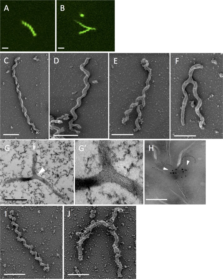
FIG 1 .
Presence of Y-shaped S. poulsonii in Drosophila hemolymph samples. (A and B) Fluorescence microscopy images showing SYTO9-stained S. poulsonii from freshly extracted hemolymph from 1-week-old Drosophila melanogaster flies. Bars, 1 µm. (C to F) SEM of S. poulsonii extracted from 1-week-old Spiroplasma-infected female flies. S. poulsonii can be found as one elongated body (C) or with a Y-shape conformation (D to F) with variation in the length of the arms. Bars, 1 µm. (G and zoom in G′) TEM of S. poulsonii from freshly extracted Drosophila hemolymph. The white arrowhead shows the branching. Bar, 1 µm. (H) Immunogold labeling pattern of the FtsZ protein with an anti-FtsZ antibody. Y-shaped bacteria have two FtsZ protein aggregates. Bar, 200 nm. (I and J) SEM of in vitro-cultured S. citri. S. citri can be found as one elongated body (I) or with a Y-shaped conformation (J). Bars, 1 µm. The presence of aggregates might be due to protein enrichment in the medium used to cultivate S. citri.

