Abstract
Background:
Intracranial aneurysms most commonly present following rupture causing subarachnoid hemorrhage. Mental disorders are common among patients with unruptured intracranial aneurysms and in aneurysmal subarachnoid hemorrhage survivors. However, to the best of our knowledge, there is no published report of unruptured intracranial aneurysm presenting as a mental disorder.
Case Description:
A 69-year-old male without a past history of mental disorders and neurological symptoms presented with a 2-month history of anxiety, sadness, lack of pleasure in usual activities, fatigue, difficulties falling asleep and waking up early in the morning, reduced appetite, and weight loss. The patient was diagnosed with major depressive disorder and antidepressant treatment was initiated. Subsequent non-contrast computed tomography (CT) of the head demonstrated hypointense oval-shaped lesion within the projection of the anterior communicating artery. CT angiography confirmed the diagnosis of a 0.8 × 0.6 cm saccular aneurysm originating from the anterior communicating artery and anterior cerebral artery. The patient underwent microsurgical clipping of the aneurysm. On psychiatric assessment 1 month after the surgery, there were no signs of depressive disorder and antidepressive treatment was discontinued. On follow-up visit 1 year after the surgery, the patient did not have any mood symptoms.
Conclusions:
The case indicates that organic brain lesions, including intracranial aneurysms, should be suspected in elderly patients presenting with their first episode of mental disorder.
Keywords: Intracranial aneurysm, major depressive episode, unruptured
INTRODUCTION
Aneurysm of intracranial arteries is rare but potentially fatal disorder affecting up to 5% of the general population.[2,12] Subarachnoid hemorrhage (SAH) due to aneurysm rupture is the most common initial presentation and is associated with significant morbidity and mortality. Estimated 30-day mortality rate following intracranial aneurysm rupture reaches 45%, and one-third of the surviving patients suffer from moderate to severe disability.[2,6]
Rarely, intracranial aneurysms are diagnosed prior to rupture. The most common presenting signs and symptoms of unruptured intracranial aneurysms include headache, reduced visual acuity, double vision, and other cranial nerve neuropathies that are usually attributed to mass effect imposed by the aneurysm to nearby neural structures.[5] Brain ischemic events caused by microemboli originating from unruptured intracranial aneurysms have also been reported.[5] It has been documented that patients with unruptured intracranial aneurysm suffer from poor mental health.[7] However, to the best of our knowledge, there are no published reports demonstrating intracranial aneurysms presenting as a psychiatric disorder. We present a patient who presented with symptoms consistent with major depressive episode and was subsequently diagnosed with unruptured anterior communicating artery aneurysm.
CASE REPORT
Mr. G is a 69-year-old male who presented to his general practitioner with a 2-month history of anxiety, sadness, lack of pleasure in usual activities, fatigue, difficulties falling asleep and waking up early in the morning, reduced appetite, and significant weight loss. He has been working as a transport manager but had retired 1 month prior to presentation due to his current health problems. He was living alone in the countryside. The patient denied a past history of psychiatric or somatic disorders and was not taking any medication at the time of presentation. Physical examination revealed no abnormalities. Routine blood work that included clinical blood count and blood chemistry was evident for dyslipidemia.
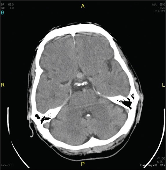
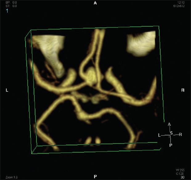
The patient was referred for neurology and psychiatry consultations. On psychiatric interview, the patient was alert, oriented, and was noted to have symptoms of psychomotor retardation, tearfulness, depressed mood, feelings of worthlessness, and pessimistic thoughts about the future. The patient denied suicidal thoughts. Neuropsychological testing demonstrated mild cognitive impairment due to concentration difficulties, and moderately severe depressive symptoms as well as mild anxiety symptoms. The patient's symptoms were mostly consistent with major depressive episode, and antidepressant treatment with mirtazapine 15 mg once daily was initiated. Neurological examination did not reveal any cranial nerve deficits. Non-contrast computed tomography (CT) of the head was ordered to rule out organic brain lesions. Rather unexpectedly, non-contrast head CT demonstrated hypointense oval-shaped lesion of 0.9 cm in diameter within the projection of the anterior communicating artery that was mostly consistent with a saccular aneurysm [Figure 1]. On head CT scan, there were also multiple lacunar ischemic lesions and signs of chronic ischemic encephalopathy. There were no signs of subarachnoid bleeding. Subsequent CT angiography confirmed the diagnosis of 0.8 × 0.6 cm saccular aneurysm originating from the anterior communicating artery and from the A2 segment of the left anterior cerebral artery [Figure 2]. The patient underwent microsurgical clipping of the aneurysm. Postoperative period was uneventful without postoperative complications. On psychiatric assessment 10 days after the surgery, the patient reported improved sleep quality and reduced anxiety and fatigue symptom severity. Depressive symptoms were not identified, and only mildly increased emotional lability and mild distraction were noted. Mirtazapine treatment was discontinued. On neurological assessment, the patient was alert and oriented, without any neurological signs or symptoms. During the psychiatrist consultation 1 month after the surgery, the patient reported improved mood symptoms. He enjoyed his everyday activities again. On neuropsychological assessment, cognitive functioning was within age limits, and he did not express mood and anxiety symptoms. There were no neurological signs and symptoms. On follow-up 1 year after the surgery, the patient did not report mood and anxiety symptoms.
Figure 1.

Axial non-contrast computed tomography of the head demonstrating oval shaped hyperintense lesion within the projection of the anterior communicating artery
Figure 2.

Computed tomography angiography demonstrating a 0.8 × 0.6 cm saccular aneurysm originating from the anterior communicating artery and A2 segment of the left anterior cerebral artery
DISCUSSION
The current case report presents an uncommon presentation of an unruptured intracranial aneurysm as major depressive episode in an otherwise asymptomatic elderly patient without a past history of psychiatric disorders.
Possible biological mechanisms underlying the association between depressive symptoms and anterior communicating artery aneurysm cannot be discerned from the present case. Anterior communicating artery lies beneath the frontal lobes in the anterior aspect of the limbic system and connects two anterior cerebral arteries that provide major oxygenated blood supply to medial the portions of the frontal and parietal lobes and to the limbic system. It is well-described that structural abnormalities and organic lesions within the frontal lobe and limbic system can produce depressive symptoms.[3,10] However, it should be noted that, in the present case, there were no signs of the frontal lobe compression by the aneurysm on non-contrast brain CT scan; therefore, brain compression is not a likely culprit of psychiatric symptoms observed in our patient. Anterior cerebral artery aneurysms can also be associated with impaired blood supply to the frontal lobes and limbic system. Thrombosis and microemboli originating from intracranial artery aneurysms causing brain ischemia and stroke has been described.[5,8] Therefore, there is a possibility that our patient experienced microembolism originating from the aneurysm causing silent minor ischemic lesions in the frontal lobes and the limbic system; however, the patient was not specifically evaluated for aneurysmal thrombosis and ischemic brain lesions.
On the other hand, it is also possible that the patient had two unrelated conditions. However, it should be noted that the patient's presentation was not typical because depression usually manifests at a younger age and the patient denied a past history of psychiatric disorders. From a clinical perspective, the present case illustrates that careful neurologic and radiologic evaluation should be performed in middle-aged and elderly patients presenting with their first episode of psychiatric disorder. Detailed radiological investigation should be considered to rule out organic brain lesions. In the present case, an aneurysm was detected on non-contrast head CT scan. Low cost and short testing timing are the most important advantages of non-contrast head CT scan making it the most commonly employed imaging modality in routine clinical settings for initial screening purposes of patients with suspected organic brain lesions. Non-contrast CT scan can reliably detect certain brain tumors, hydrocephalus, and intracranial bleeding, such as SAH; however, it should not be used for intracranial aneurysm screening purposes. Magnetic resonance angiography (MRA) and CTA are recommended as reasonable diagnostic imaging modalities for intracranial aneurysm screening in asymptomatic patients.[2] To this end, patients with suspected organic brain lesions and non-contrast CT scan results without abnormalities should be considered for more detailed radiological evaluation, including MRI and MRA.
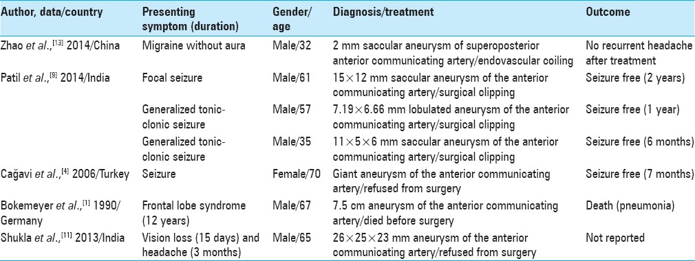
Aneurysm rupture causing SAH-associated symptoms remains the most common presentation of intracranial aneurysms. Unruptured intracranial aneurysms usually present with headache, reduced visual acuity, double vision, and other cranial nerve neuropathies that are usually caused by mass effect imposed by an aneurysm to nearby neural structures. However, it should be remembered that rarely unruptured intracranial aneurysms can cause other neurologic and neuropsychiatric symptoms and/or disorders. Other previously described unusual presentations of unruptured intracranial aneurysms include migraine without aura,[13] focal and generalized seizures,[4,9] frontal lobe syndrome,[1] and sudden vision loss[11] [Table 1]. Treatment of an underlying aneurysm can alleviate these symptoms and improve patient's health status.
Table 1.
Case reports describing atypical presentation of unruptured anterior communicating artery aneurysms

CONCLUSION
We describe an uncommon association between depressive disorder and unruptured intracranial artery aneurysm in an elderly patient without a past history of psychiatric disorder. The case illustrates that elderly patients presenting with the first episode of psychiatric disorder should be carefully evaluated for underlying intracranial pathology, including unruptured aneurysm of intracranial arteries.
Financial support and sponsorship
Nil.
Conflicts of interest
There are no conflicts of interest.
Footnotes
Contributor Information
Adomas Bunevicius, Email: a.bunevicius@yahoo.com.
Paulius Cikotas, Email: paulius.cikotas@kaunoklinikos.lt.
Vesta Steibliene, Email: Vesta.Steibliene@kaunoklinikos.lt.
Vytenis P. Deltuva, Email: vytrnis.deltuva@kaunoklinikos.lt.
Arimantas Tamsauskas, Email: arimantas.tamasauskas@kaunoklinikos.lt.
REFERENCES
- 1.Bokemeyer C, Frank B, Brandis A, Weinrich W. Giant aneurysm causing frontal lobe syndrome. J Neurol. 1990;237:47–50. doi: 10.1007/BF00319669. [DOI] [PubMed] [Google Scholar]
- 2.Brisman JL, Song JK, Newell DW. Cerebral aneurysms. N Eng J Med. 2006;355:928–39. doi: 10.1056/NEJMra052760. [DOI] [PubMed] [Google Scholar]
- 3.Bunevicius A, Deltuva VP, Deltuviene D, Tamasauskas A, Bunevicius R. Brain lesions manifesting as psychiatric disorders: Eight cases. CNS spectr. 2008;13:950–8. doi: 10.1017/s1092852900014000. [DOI] [PubMed] [Google Scholar]
- 4.Cağavi F, Kalayci M, Unal A, Atasoy HT, Cagavi Z, Acikgoz B. Giant unruptured anterior communicating artery aneurysm presenting with seizure. J Clin Neurosci. 2006;13:390–4. doi: 10.1016/j.jocn.2005.04.024. [DOI] [PubMed] [Google Scholar]
- 5.Friedman JA, Piepgras DG, Pichelmann MA, Hansen KK, Brown RD, Jr, Wiebers DO. Small cerebral aneurysms presenting with symptoms other than rupture. Neurology. 2001;57:1212–6. doi: 10.1212/wnl.57.7.1212. [DOI] [PubMed] [Google Scholar]
- 6.Johnston SC, Selvin S, Gress DR. The burden, trends, and demographics of mortality from subarachnoid hemorrhage. Neurology. 1998;50:1413–8. doi: 10.1212/wnl.50.5.1413. [DOI] [PubMed] [Google Scholar]
- 7.King JT, Jr, Kassam AB, Yonas H, Horowitz MB, Roberts MS. Mental health, anxiety, and depression in patients with cerebral aneurysms. J Neurosurg. 2005;103:636–41. doi: 10.3171/jns.2005.103.4.0636. [DOI] [PubMed] [Google Scholar]
- 8.Kobayashi H, Hayashi M, Kawano H, Handa Y, Kabuto M, Ishii Y. Magnetic resonance imaging of embolism from intracranial aneurysms. Surg Neurol. 1989;32:225–30. doi: 10.1016/0090-3019(89)90183-3. [DOI] [PubMed] [Google Scholar]
- 9.Patil A, Menon GR, Nair S. Unruptured anterior communicating artery aneurysms presenting with seizure: Report of three cases and review of literature. Asian J Neurosurg. 2013;8:164. doi: 10.4103/1793-5482.121693. [DOI] [PMC free article] [PubMed] [Google Scholar]
- 10.Sexton CE, Mackay CE, Ebmeier KP. A systematic review and meta-analysis of magnetic resonance imaging studies in late-life depression. Am J Geriatr Psychiatry. 2013;21:184–95. doi: 10.1016/j.jagp.2012.10.019. [DOI] [PubMed] [Google Scholar]
- 11.Shukla DP, Bhat DI, Devi BI. Anterior communicating artery aneurysm presenting with vision loss. J Neurosci Rural Pract. 2013;4:305–7. doi: 10.4103/0976-3147.118765. [DOI] [PMC free article] [PubMed] [Google Scholar]
- 12.Wiebers DO, Whisnant JP, Huston J, 3rd, Meissner I, Brown RD, Jr, Piepgras DG, et al. Unruptured intracranial aneurysms: Natural history, clinical outcome, and risks of surgical and endovascular treatment. Lancet. 2003;362:103–10. doi: 10.1016/s0140-6736(03)13860-3. [DOI] [PubMed] [Google Scholar]
- 13.Zhao M, Liu CS, Xu XY, Xiao YP, Fang C. Unruptured saccular aneurysm presenting migraine. Genet Mol Res. 2014;13:4046–9. doi: 10.4238/2014.January.24.19. [DOI] [PubMed] [Google Scholar]


