Pododermatitis is defined as inflammation of the skin of the paw. Affected tissues may include interdigital spaces, footpads, nail folds (paronychia), and nails. Cases of canine pododermatitis are common in general practice. One or more feet may be affected. Lesions can spontaneously resolve, wax and wane, or may persist indefinitely (1). Also called pedal folliculitis and furunculosis, the condition is complex, multifactorial, and may be frustrating to diagnose and treat (2).
Pododermatitis is not a diagnosis, rather a clinical presentation, which may or may not be accompanied by other dermatologic or systemic clinical symptoms. Due to the numerous potential underlying causes and progression of pododermatitis due to external trauma or secondary infections, it is important to thoroughly evaluate each patient presenting with pododermatitis. With few variations, the clinical findings related exclusively to the paws can appear to be the same despite the various underlying conditions. The age, breed (2,3), body and paw conformation (3,4), presence of additional clinical symptoms, chronicity (3,5), and the number of paws affected (6) are factors that should be evaluated in order to establish a diagnostic and treatment approach.
Trauma or underlying pruritic disease can lead to intense licking of the paws, which heightens the paw irritation. The feet are subject to a great variety and intensity of trauma, the front feet more so than the rear (2–5). One report (7) suggested that the flat foot and the scoop-shaped web of breeds such as the Pekingese and some terriers predispose the area to folliculitis and pedal dermatitis. Another study (3) suggested that Labradors had wide-based paws with greater distance between pads, compared with Greyhound dogs, predisposing them to paw disease (8). This characteristic suggests the possibility that weight bearing, especially in a heavy dog, might be more likely to be distributed to haired interdigital skin adjacent to digital pads, leading to plantar paw trauma and irritation.
Clinical presentation
Affected paw tissue may present with pruritis, erythema, edema with or without nodules, paronychia, alopecia, ulceration, paw pad involvement, comedones, and draining serosanguineous or seropurulent exudates. The feet may be grossly swollen (2). The skin may be moist from constant licking and/or infection, and varying degrees of pain, pruritus, and lameness may be evident (1,2). In some cases, the interdigital nodules are not tender and are unresponsive to treatment and may be scars from previous lesions (2). A single lesion on one paw may be the only presenting complaint in some patients, whereas other patients may be affected with a variety of lesions affecting multiple paws (6). Many of the differential diagnoses for canine pododermatitis (Table 1) may be presented to the clinician with paw lesions being part of a generalized dermatologic disease. The same diseases may also present as pedal disease only.
Table 1.
Differential diagnoses to be considered when evaluating a canine patient presented with pododermatitisa
| Infection | Superficial bacterial pyoderma Deep pyoderma and furunculosis (actinomycosis, actinobacillosis, nocardiosis, mycobacteriosis) Superficial fungal (dermatophytosis, Malassezia, candidiasis) Deep fungal (phaeohyphomycosis, sporotrichosis, blastomycosis, cryptococcosis) Parasitic (demodicosis, trobiculiasis, hookworm dermatitis, Pelodera dermatitis, tick infestation) Viral (canine distemper) |
| Allergic | Atopic dermatitis, cutaneous adverse food reaction, contact dermatitis, flea allergy dermatitis |
| Immune-mediated | Pemphigus foliaceus, systemic lupus erythematosus, vasculitis, adverse cutaneous drug reaction, lymphocytic plasmacytic pododermatitis |
| Endocrine | Hypothyroidism, hyperadrenocorticism |
| Acquired/Traumatic | Sterile interdigital pyogranulomatous pododermatitis Foreign body pododermatitis (plants, wood splinters, nail, thorn, foxtails, wood slivers) |
| Genetic/Inherited | Familial paw pad hyperkeratosis, lethal acrodermatitis of bull terriers |
| Metabolic | Superficial necrolytic dermatitis |
| Neoplastic | Nail bed squamous cell carcinoma, epitheliotrophic lymphoma |
Common forms of canine pododermatitis
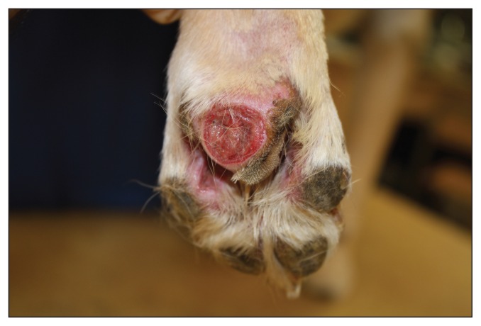
Allergic pododermatitis and secondary infection is most commonly observed in both primary care and referral dermatology practice. Alongside atopic dermatitis, cutaneous adverse food reaction and contact dermatitis can cause pododermatitis as well as secondary bacterial (Figure 1) and superficial fungal infections. It is important to address both the primary underlying disease and the secondary infections simultaneously in order to resolve the pododermatitis. Pedal disease may often become chronic and unresponsive to therapy if not adequately treated initially.
Parasitic pododermatitis is particularly common, with demodex mites being the most challenging. Every case of chronic interdigital pyoderma must be evaluated carefully for demodex mites (2). While skin scrapings and hair plucks are reliable diagnostics for demodicosis, skin biopsy may be required to make a diagnosis in chronically inflamed and fibrosed pedal lesions. Demodectic pododermatitis can be present on the feet of dogs without generalized lesions (9). Demodicosis involving the feet in dogs older than 4 years is one of the most commonly misdiagnosed skin diseases (8).
Sterile pyogranulomatous pododermatitis occurs most commonly in smooth, short-coated breeds such as English bulldogs, dachshunds, great Danes, and boxers (2). Also referred to as interdigital follicular cysts in the literature (8), interdigital nodules with or without draining lesions recur repeatedly and are especially non-responsive to therapy.
Figure 1.
Streptococcal pedal pyoderma in a young Labrador retriever dog.
The front paws are most commonly affected, most often at the lateral space between digits 4 and 5. Histopathology is diagnostic and laser surgery or traditional surgery can be curative (2,4).
Diagnostic approach
Alongside careful history taking and physical examination, basic dermatologic tests such as examination of deep skin scrapings or hair plucks (10), skin cytology, and bacterial culture and sensitivity should be pursued. With the recent advent of drug-resistant strains of staphylococci causing secondary pyoderma, empirical use of systemic antibiotic therapy is discouraged in the absence of confirmed presence of bacterial infection. Fungal culture and skin biopsy may follow the initial workup, or may be pursued as part of the initial diagnostic workup based on the initial differential list. Response to cytology and culture-based antimicrobial therapy should be assessed and usually forms part of the diagnostic workup. Delays in appropriate antimicrobial therapy and workup for the primary underlying cause lead to perpetuation of the condition and increase the scarring potential, so the diagnostic and therapeutic effort should be maximal during the early phases of investigation in cases of pododermatitis.
Direct impression cytology provides the clinician with information regarding the presence or absence of neutrophils, other inflammatory cells, and phagocytosed bacteria (2,11). Yeast, pseudomycetoma grains, and fungal hyphae may also be noticed (2). Skin scrapings and hair plucks are important tests used to diagnose parasitic pododermatitis.
Culture and sensitivity from draining lesions, or tissue culture alongside biopsy sampling is helpful in pursuing appropriate systemic and topical antibacterial therapy. In some cases, radiographs may be required to identify opaque foreign bodies or to demonstrate bony changes. Evaluation of thyroid function and the adrenal glands is usually indicated for adult and geriatric patients, especially if systemic signs suggestive of endocrine disease are noticed in the presence of pododermatitis.
In patients with persistent or recurrent pododermatitis of one or multiple paws, histopathology may be required to demonstrate exogenous or endogenous foreign bodies (including free hair shafts or keratin within the tissue), deep bacterial infection, parasites, fungi, and neoplasia, as well as to evaluate the cellular response. Special stains are usually used during histopathologic evaluation. In general, the histologic response is that of perifolliculitis, folliculitis, or furunculosis; nodular to diffuse pyogranulomatous inflammation being the most common (2,11). If biopsy samples are collected, sampling for bacterial and fungal culture should be strongly considered if adequate amount of tissue is available.
Clinical management and treatment
Canine pododermatitis can often be self-perpetuating, multi-factorial, and resistant to empirical therapy. Such factors can make this a frustrating condition to deal with, for both the pet owner and the clinician. Lesions heal with scarring, which makes the foot more susceptible to future infections. Because the condition has potential for chronic changes within the paw (4), substantial effort should be made to diagnose the underlying primary disease or predisposing factor. Patients should be treated aggressively early while pursuing appropriate diagnostics and therapeutic interventions. A systematic approach towards ruling out differential diagnoses and follow-up examinations will generally be rewarded by reaching the correct diagnosis (6). Client education is paramount to ensure that owners comply with recommended diagnostic testing, treatments, and follow-up visits.
Prolonged antibiotic treatment, usually for 8 to 12 weeks, is needed in cases of deep bacterial pododermatitis (2,11). A dramatic improvement in the first 2 to 4 weeks may be noted but it is essential that antibiotic therapy not be discontinued too soon. For chronic or draining lesions, topical washes and foot soaks are indicated. Monitoring for self-trauma (pruritus), visual changes in lesions and palpation of interdigital lesions are essential for monitoring response to therapy and in ensuring complete resolution.
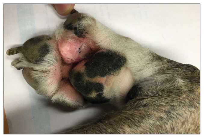
Where plantar comedones or large interdigital lesions are noted (Figure 2), restricting the animal’s activity, keeping the patient on smooth surfaces, and protecting paw trauma by using paw booties are encouraged and help with response to primary therapy. Cases with advanced disease at the onset of treatment have varying degrees of scarring of the digital and interdigital skin and may have sterile dermal granulomas due to endogenous foreign bodies (2). Such lesions may be amenable to surgical removal (1,2,4). Some cases, especially those in which the infection includes secondary Gram-negative organisms, are resistant to medical treatment alone. Traditional surgical debridement of all devitalized tissue or CO2 laser surgery can make medical treatment more effective (1,2,4).
Figure 2.
Interdigital sterile granulomatous pododermatitis with palmar comedone formation.
In severe cases, fusion podoplasty can be beneficial (12). Laser surgery and fusion podoplasty may also be helpful when abnormalities of pedal anatomy lead to weight-bearing on hairy pedal skin, with consequent follicular damage (4,9,12).
Prognosis
The prognosis is good to guarded (1), depending on whether the underlying cause can be identified and corrected. Only skin scrapings should be used for therapeutic monitoring of cases of demodectic pododermatitis (8). Trichograms lose any value they have once treatment is begun. Impression cytology can be helpful in treating and monitoring response to acute or superficial pedal lesions, but is not always helpful for deep, fibrosed lesions. Some dogs may need ongoing, lifelong topical or systemic therapy as well as management of the primary underlying disease in order to prevent further recurrence of pododermatitis episodes.
Footnotes
Use of this article is limited to a single copy for personal study. Anyone interested in obtaining reprints should contact the CVMA office (hbroughton@cvma-acmv.org) for additional copies or permission to use this material elsewhere.
References
- 1.Hnilca KA. Small Animal Dermatology: A color Atlas and Therapeutic Guide. 3rd ed. St. Louis, Missouri: Elsevier Saunders; 2011. pp. 60–62. [Google Scholar]
- 2.Miller WH, Griffin CE, Campbell KL. Muller and Kirk’s Small Animal Dermatology. 7th ed. St. Louis, Missouri: Elsevier; 2013. pp. 201–203. [Google Scholar]
- 3.Besancon MF, Conzemius MG, Evans RB, Ritter MJ. Distribution of vertical forces in the pads of greyhounds and Labrador retrievers during walking. Am J Vet Res. 2004;65:1497–1501. doi: 10.2460/ajvr.2004.65.1497. [DOI] [PubMed] [Google Scholar]
- 4.Duclos DD, Hargis AM, Hanley PW. Pathogenesis of canine interdigital palmar and plantar comedones and follicular cysts, and their response to laser surgery. Vet Dermatol. 2008;19:134–141. doi: 10.1111/j.1365-3164.2008.00662.x. [DOI] [PubMed] [Google Scholar]
- 5.Kovacs MS, McKiernan S, Potter DM, Chilappagari S. An epidemiological study of interdigital cysts in a research beagle colony. Contemp Top Lab Anim Sci. 2005;44:17–21. [PubMed] [Google Scholar]
- 6.Miller WH, Griffin CE, Campbell KL. Muller and Kirk’s Small Animal Dermatology. 7th ed. St. Louis, Missouri: Elsevier; 2013. pp. 106–107. [Google Scholar]
- 7.Whitney JC. Some aspects of interdigital cysts in the dog. J Small Anim Pract. 1970;11:83. doi: 10.1111/j.1748-5827.1970.tb06133.x. [DOI] [PubMed] [Google Scholar]
- 8.Duclos DD. Canine pododermatitis. Vet Clin Small Anim Pract. 2013;43:57–87. doi: 10.1016/j.cvsm.2012.09.012. [DOI] [PubMed] [Google Scholar]
- 9.Miller WH, Griffin CE, Campbell KL. Muller and Kirk’s Small Animal Dermatology. 7th ed. St. Louis, Missouri: Elsevier; 2013. p. 309. [Google Scholar]
- 10.Saridomichelakis MN, Koutinas AF, Farmaki R, Leontides LS, Kasabalis D. Relative sensitivity of hair pluckings and exudate microscopy for the diagnosis of canine demodicosis. Vet Dermatol. 2007;18:138–141. doi: 10.1111/j.1365-3164.2007.00570.x. [DOI] [PubMed] [Google Scholar]
- 11.Nuttal T, Harvey RG, McKeever OJ. A Colour Handbook of Skin Diseases of the Dog and Cat. 2nd ed. London, UK: Manson Publishing; 2009. pp. 168–169. [Google Scholar]
- 12.Swaim SF, Lee AH, MacDonald JM, Angarano DW, Cox NR, Hathcock JT. Fusion podoplasty for the treatment of chronic fibrosing interdigital pyoderma in the dog. J Am Anim Hosp Assoc. 1991;27:264. [Google Scholar]
- 13.Gross TL, Ihrke PJ. Skin Diseases of the Dog and Cat — Clinical and Histopathologial Diagnosis. 2nd ed. Hoboken, New Jersey: Wiley-Blackwell; 2005. pp. 9–10. [Google Scholar]
- 14.Breathnach RM, Fanning S, Mulcahy G, Bassett HF, Jones BR, Daly P. Evaluation of Th1-like, Th2-like and immunomodulatory cytokine mRNA expression in the skin of dogs with immunomodulatory-responsive lymphocytic–plasmacytic pododermatitis. Vet Dermatol. 2006;17:313–321. doi: 10.1111/j.1365-3164.2006.00534.x. [DOI] [PubMed] [Google Scholar]


