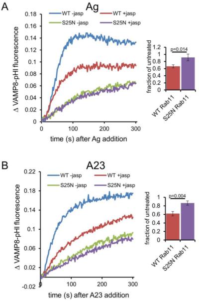
Figure 5. Jasplakinolide (jasp) inhibits stimulated VAMP8-pHl exocytosis.
(A) RBL-2H3 cells were co-transfected with VAMP8-pHluorin and either wild-type (WT) or dominant negative (S25N) mCh-Rab11, and sensitized with anti-DNP IgE (for stimulation with Ag) before harvest. Cells were treated with 3 μM jasp or vehicle (DMSO, -jasp) for 5 minutes before exocytosis was stimulated with 100 ng/ml DNP-BSA (Ag). The data were collected and analyzed as in Figure 4. Bar graph shows integrated fluorescence values expressed as a ratio (jasp treated/untreated) to quantify jasp-mediated inhibition of stimulated exocytosis in the context of WT Rab11 but not S25N Rab11. (B) Cells were prepared and analyzed as in (A) except 1 μM A23187 (A23) was added instead of Ag. On the right side, the average of three experiments is shown for each condition at t=300 s, error bars are standard deviations, and p-values were calculated using the unpaired t-test.

