Abstract
Objective:
To investigate the achievable radiation dose reduction for coronary CT angiography (CCTA) with iterative reconstruction (IR) in adults and the effects on image quality.
Methods:
PubMed and EMBASE were searched, and original articles concerning IR for CCTA in adults using prospective electrocardiogram triggering were included. Primary outcome was the effective dose using filtered back projection (FBP) and IR. Secondary outcome was the effect of IR on objective and subjective image quality.
Results:
The search yielded 1616 unique articles, of which 10 studies (1042 patients) were included. The pooled routine effective dose with FBP was 4.2 mSv [95% confidence interval (CI) 3.5–5.0]. A dose reduction of 48% to a pooled effective dose of 2.2 mSv (95% CI 1.3–3.1) using IR was reported. Noise, contrast-to-noise ratio and subjective image quality were equal or improved in all but one study, whereas signal-to-noise ratio was decreased in two studies with IR at reduced dose.
Conclusion:
IR allows for CCTA acquisition with an effective dose of 2.2 mSv with preserved objective and subjective image quality.
INTRODUCTION
The number of CT examinations has increased rapidly over the past decades leading to increased radiation exposure.1 This is especially a concern for coronary CT angiography (CCTA) since, retrospectively, electrocardiogram (ECG)-gated CCTA used to be associated with relatively high radiation doses of >10 mSv.2 These high CCTA radiation doses have led to the development of new techniques such as prospective ECG-triggering, high-pitch spiral acquisition and, more recently, iterative reconstruction (IR) to reduce radiation dose.3 Despite these advances, radiation dose remains an important issue for CCTA because the number of indications and eligible patients has increased rapidly over the past few years.4–6
IR offers the possibility to reduce radiation dose and was already used on the first CT systems.7,8 However, owing to the limited computational power at that time, it could not be used in clinical practice. With recent improvements in computer processing, IR has become feasible in a clinical setting. Currently, the most commonly used reconstruction technique is filtered back projection (FBP), which is a fast reconstruction technique that suffers from impaired image quality when radiation dose is lowered. IR is a noise-suppressing technique that allows for a decrease in radiation dose compared with FBP while maintaining image quality.9
Recently, new IR algorithms were introduced which led to a surge in the number of publications. Therefore, we present the results of a systematic review and meta-analysis to determine the achievable radiation dose reduction for prospective ECG-gated CCTA acquisitions using IR. Furthermore, the effect of IR on objective and subjective image quality was investigated.
METHODS AND MATERIALS
Search
A systematic search in PubMed and EMBASE was performed on 2 May 2014 for studies investigating IR for CCTA without a publication date limitation. English language restriction was applied. Synonyms for “IR techniques” and “CT” were combined. The search syntax is provided in Appendix A. Duplicates were removed. Hereafter, a manual search of the reference lists of included articles and review articles was performed after which review articles were removed.
Inclusion and exclusion criteria
All articles were screened by two authors (AH and MW). In case of discrepancy, a consensus had to be reached between authors on whether to include the study. Original research articles concerning IR techniques for CCTA using prospective ECG-triggering were included. Studies comparing routine dose acquisitions with FBP to reduced dose acquisitions with IR using the same CT system, contrast medium and dose modulation techniques were included, whereas studies investigating only one dose level without comparison to FBP were excluded. Furthermore, studies only investigating non-enhanced CCTA, ex vivo, in vitro and animal studies as well as studies performed in children were excluded. Case reports and reviews were excluded as well. Case reports were defined as studies including less than five patients.
Data extraction
Data were extracted to a standardized data sheet, which included first author, title, publication date, journal, study design, participant characteristics, reconstruction technique, scan indication, type of scan, type of CT system, and reported dose and image quality measurements.
The primary outcome was the effective dose reduction with IR. The effective dose was calculated as the dose–length product (DLP) times the conversion factor for chest CT (0.014 mSv mGy−1 cm−1).10 This conversion factor was chosen because it was the most commonly used conversion factor in the included articles. In case a different conversion factor was used, the effective dose was recalculated using the DLP. If the effective dose was reported without conversion factor or DLP, the corresponding author was contacted. The corresponding authors were also contacted if both the effective dose and the DLP were not reported.
Secondary outcome was the influence of IR on objective and subjective image quality. Noise, contrast-to-noise ratio (CNR) and signal-to-noise ratio (SNR) were investigated. Image quality was specified as improved, equal or deteriorated compared with FBP. Improved was defined as a statistically significant improvement of image quality with IR compared with FBP. Non-significant differences were classified as the same, and a significant decrease in image quality was classified as deteriorated. If multiple IR levels were studied, the IR level with the most favourable outcome was used for further analysis.
From each article, the mean and standard deviation (SD) of the effective dose were extracted. If only the median and the interquartile range (IQR) were reported, the mean and SD were recalculated. The median was considered to be equal to the mean if the number of patients exceeded 25.11 The IQR was converted to the SD using the formula 1.35 × SD = IQR.
Statistical analysis
Statistical analysis was performed using SPSS® v. 20.0 (IBM Corp., New York, NY; formerly SPSS Inc., Chicago, IL) [for Microsoft® Windows® (Microsoft, Redmond, WA)] and the RStudio statistical environment v. 0.98.1025 (RStudio, Inc., 2009–13) with “meta” package v. 3.7-1.12 For every study, a mean and 95% confidence interval (CI) was calculated. Heterogeneity was assessed by the I2 statistic, and random effect models were used in case of large interstudy variance (I2 ≥ 65%). Results were presented as mean with 95% CI. Both the normal dose data with FBP and the reduced dose data with IR were calculated and pooled. A two-tailed p-value <0.05 was considered statistically significant.
RESULTS
Study selection
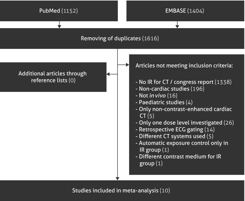
In total, 2556 articles were identified. A flowchart is provided in Figure 1. After removing of duplicates, 1616 articles were screened based on title and abstract. 1606 articles were excluded, because the articles did not investigate IR for CT (n = 1338), were non-cardiac (n = 196), were not in vivo (n = 16), were paediatric studies (n = 4), only concerned non-contrast-enhanced coronary CT (n = 5), only 1 dose level was investigated (n = 26), used retrospective ECG-gating (n = 14), used different CT systems for the FBP and IR groups (n = 5), used automatic exposure control only in the IR group (n = 1) or used a different contrast medium for the IR group (n = 1). Articles investigating one dose level are mentioned in Appendix B. 10 articles remained with a total of 1042 patients. One corresponding author was contacted because the reported information about radiation dose was insufficient.13
Figure 1.
Flowchart of included studies. ECG, electrocardiogram; IR, iterative reconstruction.
Study characteristics
The baseline characteristics are provided in Table 1. Studies were published in 2011 (n = 1), 2012 (n = 3), 2013 (n = 5) and 2014 (n = 1). Different IR techniques were used namely Adaptive Statistical Iterative Reconstruction (ASIR; GE Healthcare, Milwaukee, WI, n = 3), Sinogram-Affirmed Iterative Reconstruction (Siemens Healthcare, Erlangen, Germany, n = 3), iDose (Philips Healthcare, Best, Netherlands, n = 2), Iterative Reconstruction in Image Space (Siemens Healthcare, n = 1) and Adaptive Iterative Dose Reduction (Toshiba Medical Systems Co Ltd., Otowara, Japan, n = 1). One study compared Model-Based Iterative Reconstruction-Veo (GE Healthcare) with ASIR,22 since FBP was replaced by ASIR as the clinically implemented routine reconstruction method. CT systems used were Aquilion One™ (Toshiba Medical Systems Co Ltd., n = 1), Brilliance iCT (Philips Healthcare, n = 2), Discovery™ HD 750 (GE Healthcare, n = 3) and Somatom® Definition Flash (Siemens Medical Systems, Forchheim, Germany, n = 4).
Table 1.
Baseline characteristics of the included studies
| Study characteristics |
Image quality |
Routine dose group |
Reduced dose group |
||||||||||||||
|---|---|---|---|---|---|---|---|---|---|---|---|---|---|---|---|---|---|
| Study | Total number of patients | IR technique | Noise | CNR | SNR | Subjective image quality | BMI (routine dose group, kg m−2) | Number of patients | Tube current | Tube voltage (kV) | Mean effective dose (normal, mSv) | BMI (reduced dose group, kg m−2) | Number of patients | Tube current | Tube voltage (kV) | Mean effective dose (reduced, mSv) | |
| Renker et al14 | 24 | IRIS | Somatom® Definition Flash | + | NR | NR | + | 30.7 | 12 | NR | 120 | 9.6 | 30.2 | 12 | NR | 80/100 | 3.7 |
| Wang et al15 | 78 | SAFIRE | Somatom Definition Flash | ± | ± | ± | ± | 31.7 | 39 | 354–430 mAs | 120 | 8.8 | 32.3 | 39 | 286–370 mAs | 100 | 4.4 |
| Tomizawa et al16 | 100 | AIDR | Aquilion One™ | + | ± | ± | ± | 24.0 | 50 | 483 mA | 120 | 5.5 | 23.9 | 50 | 289 mA | 120 | 4.2 |
| Hou et al13 | 110 | iDose | Brilliance iCT | ± | ± | ± | ± | 25.1 | 21 | 210 mAs | 120 | 3.2 | 25.5 | 16 | 65 mAs | 120 | 0.9 |
| Gebhard et al17 | 70 | ASIR | Discovery™ HD 750 | + | ± | ± | + | 33.8 | 35 | 646 mA | 116 | 2.5 | 32.9 | 35 | 633 mA | 112 | 2.3 |
| Yin et al18 | 60 | SAFIRE | Somatom Definition Flash | ± | ± | ± | ± | NR | 60 | 320–400 mAs | 80–120 | 1.5 | NR | 60 | 160–200 mAs | 80–120 | 0.7 |
| Carrascosa et al19 | 200 | iDose | Brilliance iCT | ± | NR | NR | ± | 27.2 | 100 | 203 mAs | 119 | 4.6 | 26.3 | 100 | 196 mAs | 109 | 2.8 |
| Takx et al20 | 20 | SAFIRE | Somatom Definition Flash | − | − | − | − | 33.1 | 20 | 331 mAs | 100–120 | 5.5 | 33.1 | 20 | 66 mAs | 100–120 | 1.1 |
| Shen et al21 | 338 | ASIR | Discovery HD 750 | ± | NR | − | + | 25.5 | 109 | 600 mA | 120 | 2.9 | 25.2 | 116 | 300 mA | 120 | 1.8 |
| Fuchs et al22 | 42 | ASIR/MBIR | Discovery HD 750 | + | NR | + | ± | 25.2 | 42 | 450–700 mA | 100–120 | 1.2 | 25.2 | 42 | 150–210 mA | 80–100 | 0.2 |
−, decreased with IR; +, improved with IR; ±, no difference; AIDR, Adaptive Iterative Dose Reduction; BMI, body mass index; CNR, contrast-to-noise ratio; IR, iterative reconstruction; IRIS, Iterative Reconstruction in Image Space; MBIR, Model-Based Iterative Reconstruction; NR, not reported; SAFIRE, Sinogram-Affirmed Iterative Reconstruction; SNR, signal-to-noise ratio.
The Somatom Definition Flash was obtained from Siemens Medical Systems, Forchheim, Germany; Aquilion One™ was obtained from Toshiba Medical Systems Co. Ltd, Ottowara, Japan; Brilliance iCT was obtained from Philips Healthcare, Best, Netherlands; Discovery™ HD 750 was obtained from GE Healthcare, Milwaukee, WI.
The median number of patients per study was 74 (range 20–338). In total, data of 1042 patients were included in this study. Most studies (n = 7) used different study populations to compare FBP with IR; however, three studies compared different dose levels in the same patients.18,20,22 This was achieved by using data from only one source of a dual-source CT scanner20 or by making additional scans of the same patient.18,22 Mean body mass index (BMI) varied between studies from 23.9 to 33.8 kg m−2 with heart rates of 57–74 beats per minute. The contrast rate and volume was the same between the FBP and IR groups in all studies. In three studies, tube current modulation was used.18–20
Effective dose
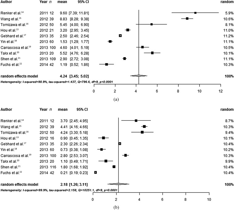
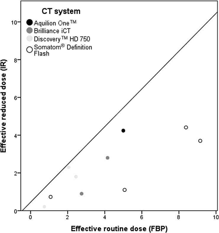
Interstudy variance was high for effective dose pooling (I2 98.9%), therefore random effects models were used. In three studies, the effective dose was (re)calculated using the DLP, because the effective dose was not provided or calculated with a different conversion factor.16,18,20 The pooled routine effective dose using FBP was 4.2 (95% CI 3.5–5.0) mSv. At reduced dose level using IR, the pooled effective dose was 2.2 (95% CI 1.3–3.1) mSv (Figure 2). Standard effective dose varied highly between studies from 1.2 to 9.6 mSv, whereas differences were smaller with IR (0.2–4.4 mSv). The relationship between routine effective dose and reduced effective dose for each CT system is illustrated in Figure 3.
Figure 2.
Forest plot of routine effective dose with filtered back projection (upper panel) and the reduced effective dose using IR (lower panel) with pooled estimate. n = number of patients. CI, confidence interval.
Figure 3.
Relationship between normal effective dose and reduced effective dose in studies comparing multiple dose levels. FBP, filtered back projection; IR, iterative reconstruction. The Somatom Definition Flash was obtained from Siemens Medical Systems, Forcheim, Germany; Aquilion One™, was obtained from Toshiba Medical System Co. Ltd, Ottowara, Japan; Brilliance iCT was obtained from Philips Healthcare, Best, Netherlands; Discovery™ HD 750 was obtained from GE Healthcare, Milwaukee, WI.
Image quality
Objective image quality was scored using image noise (n = 10), CNR (n = 6) and/or SNR (n = 8). Subjective image quality was scored by two observers in all studies, mostly by using a Likert scale. The results are shown in Table 1.
Noise was improved (n = 4) or equal (n = 5) with IR at reduced dose compared with FBP at routine dose in all but one study.20 CNR was equal (n = 5) in all but one study, whereas SNR was improved (n = 1), equal (n = 5) or deteriorated (n = 2) with IR at reduced dose.
Subjective image quality was improved (n = 3) or equal (n = 6) in all but one study.20 Furthermore, Renker et al14 and Takx et al20 reported that IR resulted in a reduction of blooming artefacts. The study of Takx et al20 was the only study reporting a decrease in both objective and subjective image quality which was possibly due to the large dose reduction (80%, compared with a median dose reduction of 50% in the other included studies).
DISCUSSION
This meta-analysis showed that dose reduction is feasible using IR techniques for CCTA with preserved image quality compared with conventional FBP techniques.
Our results, indicating an achievable dose reduction of 48%, are in the range of dose reductions reported in a prior systematic review.23 49 studies were included, and reported achievable dose reduction varied from 23% to 76%. In that review, data were not pooled and only the percentage of achieved dose reduction for each study was reported. In this previous review, CCTA was not specifically studied since the review focused on all body regions. Also, no meta-analysis was performed. Furthermore, a lot of included studies concerned ex vivo data, new IR algorithms have become available and a substantial number of new studies have been published about IR for CT since the publication of the aforementioned review.23
In the present review, the effective dose reported in individual studies was pooled. Furthermore, only effective dose was used with the most commonly used conversion factor to achieve a uniform quantity to report dose. As can be seen in the forest plots (Figure 2), effective doses varied widely between studies. In addition, the 95% CI was high in some studies. This is partly due to the variation in BMI between study samples, but also shows the differences in routine scan protocols between hospitals.
Most studies did not report whether the dose of the localizer and bolus-tracking images were included in the effective dose, which might have influenced results. However, the additional dose of the localizer and bolus-tracking images might be small and therefore less likely to have influenced the dose significantly.
All major vendors have developed and are marketing a variety of slightly different IR algorithms. One study found an ultra-low dose of 0.2 mSv in patients with a mean BMI of 25.2 kg m−2 using Model-Based Iterative Reconstruction, which is a model-based IR algorithm.22 All other studies investigated less advanced hybrid IR algorithms. Therefore with the development of model-based IR algorithms, the radiation dose is expected to decrease even further.
This study has several limitations. First, owing to our strict inclusion and exclusion criteria, we excluded many articles. By only including articles using the same CT system, contrast medium and scan parameters (except for variation in tube voltage and/or current to create dose reduction) for both the routine dose scan and the reduced dose scan, it was possible to investigate the true potential of IR. Second, there are different quantities to report dose but only the effective dose was used in the present study. Volume CT dose index might be more appropriate, because it is independent of anatomical length and dose conversion factors. In this review, the effective dose was used since most studies only reported DLP and/or effective dose. We felt this was appropriate because using a standard conversion factor eliminated the influence of different conversion factors. A conversion factor of 0.014 was used because this was the most commonly used factor in the included articles. However, this factor was designed for chest rather than cardiac CT and a different conversion factor might therefore be more appropriate. Efforts were made to ensure effective dose data concerned only CCTA studies and did not include non-enhanced acquisitions; however, this cannot be guaranteed and could be a potential limitation. Third, we used an English language restriction.
A major limitation is the inability to determine whether the diagnostic accuracy remains acceptable at reduced dose levels. Since this was not reported in most studies, we were not able to investigate this. Therefore, the current meta-analysis only provides an overview of dose reductions reported in the literature and does not focus on diagnostic acceptability. However, it is difficult to investigate the diagnostic accuracy because of IR alone, since this is also influenced by factors as the used CT system and other dose-reduction techniques.
Ideally, different dose levels should be compared within patients. However, performing multiple scans in one patient can lead to difficulties with contrast enhancement. This explains why only two studies performed additional scans for research purposes in the same patients.18,22 Another study tried to simulate a within-patient comparison by using data from one detector of a dual source CT system.20
This meta-analysis provides the possible dose reduction with IR as reported in the literature. However, the lowest possible dose remains unclear. Most studies only investigated one or two different dose levels, and we found that it is feasible to reduce the dose below 3 mSv using prospective ECG-triggering and state-of-the-art CT systems. Other dose-reduction techniques have been developed in the past years such as automatic tube current modulation, which was used in only three of the included studies. It is likely that radiation dose can be reduced even further by combining techniques. Future research investigating dose reduction with IR should focus on radiation doses of 3 mSv and lower. Furthermore, we recommend a uniform way of reporting radiation dose. Both volume CT dose index and DLP should be reported, making it possible to calculate the effective dose with a consistent conversion factor. In addition, authors should be clear about whether scout views and non-enhanced scans were included in the total reported dose.
In conclusion, this meta-analysis provides an overview of currently available dose reduction for CCTA with IR. Pooled data suggested that CCTA acquisition with an effective dose <3 mSv is possible with preserved image quality. Future research should determine if radiation dose can be reduced even further with model-based IR techniques.
Conflicts of interest
The University Medical Center Utrecht–Department of Radiology received research support from Philips Healthcare.
APPENDIX A
SEARCH SYNTAX PUBMED
((((((iterative[Title/Abstract]) AND reconstruction[Title/Abstract])) OR (((iterative[Title/Abstract]) AND dose[Title/Abstract]) AND reduction[Title/Abstract])) OR (((((((((ASIR[Title/Abstract]) OR iDose[Title/Abstract]) OR IRIS[Title/Abstract]) OR AIDR[Title/Abstract]) OR IMR[Title/Abstract]) OR MBIR[Title/Abstract]) OR Veo[Title/Abstract]) OR SAFIRE[Title/Abstract]) OR ADMIRE[Title/Abstract]))) AND (((((CT [Title/Abstract] OR “Tomography, X-Ray Computed” [Mesh] OR “Cone-Beam Computed Tomography” [Mesh] OR “Four-Dimensional Computed Tomography” [Mesh] OR “Spiral Cone-Beam Computed Tomography” [Mesh] OR “Tomography Scanners, X-Ray Computed” [Mesh] OR “Tomography, Spiral Computed” [Mesh])))) OR ((computed[Title/Abstract]) AND tomography[Title/Abstract])) Filters: English
SEARCH SYNTAX EMBASE
(((iterative:ab,ti AND reconstruction:ab, ti) OR (iterative:ab,ti AND dose:ab,ti AND reduction:ab,ti) OR (ASIR:ab,ti OR iDose:ab,ti OR IRIS:ab,ti OR AIDR:ab,ti OR IMR:ab,ti OR MBIR:ab,ti OR Veo:ab,ti OR SAFIRE:ab,ti OR ADMIRE:ab,ti)) AND ((computed:ab,ti AND tomography:ab,ti) OR (CT:ab,ti OR ‘tomography, x-ray computed’:ab,ti OR ‘cone-beam computed tomography’:ab,ti OR ‘four-dimensional computed tomography’:ab,ti OR ‘spiral cone-beam computed tomography’: ab,ti OR ‘tomography scanners, x-ray computed’:ab, ti OR ‘tomography, spiral computed’:ab,ti))) AND [english]/lim
APPENDIX B
LIST OF EXCLUDED STUDIES INVESTIGATING ONE DOSE LEVEL
Leipsic J, LaBounty TM, Heilbron B, Min JK, Mancini GBJ, Lin FY, et al. Adaptive statistical iterative reconstruction: assessment of image noise and image quality in coronary CT angiography. Am J Roentgenol 2010; 195: 649–54.
Renker M, Nance JW Jr, Schoepf UJ, O'Brien TX, Zwerner PL, Meyer M, et al. Evaluation of heavily calcified vessels with coronary CT angiography: comparison of iterative and filtered back projection image reconstruction. Radiology 2011; 260: 390–9.
Bittencourt MS, Schmidt B, Seltmann M, Muschiol G, Ropers D, Daniel WG, et al. Iterative reconstruction in image space (IRIS) in cardiac computed tomography: initial experience. Int J Card Imaging 2011; 27: 1081–7.
Tatsugami F, Matsuki M, Nakai G, Inada Y, Kanazawa S, Takeda Y, et al. The effect of adaptive iterative dose reduction on image quality in 320-detector row CT coronary angiography. Br J Radiol 2012; 85: e378–82.
Pontone G, Andreini D, Bartorelli AL, Bertella E, Mushtaq S, Foti C, et al. Feasibility and diagnostic accuracy of a low radiation exposure protocol for prospective ECG-triggering coronary MDCT angiography. Clin Radiol 2012; 67: 207–15.
Oda S, Utsunomiya D, Funama Y, Yonenaga K, Namimoto T, Nakaura T, et al. A hybrid iterative reconstruction algorithm that improves the image quality of low-tube-voltage coronary CT angiography. AJR Am J Roentgenol 2012; 198: 1126–31.
Hosch W, Stiller W, Mueller D, Gitsioudis G, Welzel J, Dadrich M, et al. Reduction of radiation exposure and improvement of image quality with BMI-adapted prospective cardiac computed tomography and iterative reconstruction. Eur J Radiol 2012; 81: 3568–76.
Maffei E, Martini C, Rossi A, Mollet N, Lario C, Castiglione Morelli M, et al. Diagnostic accuracy of second-generation dual-source computed tomography coronary angiography with iterative reconstructions: a real-world experience. Radiol Med 2012; 117: 725–38.
Utsunomiya D, Weigold WG, Weissman G, Taylor AJ. Effect of hybrid iterative reconstruction technique on quantitative and qualitative image analysis at 256-slice prospective gating cardiac CT. Eur Radiol 2012; 22: 1287–94.
Morsbach F, Desbiolles L, Plass A, Leschka S, Schmidt B, Falk V, et al. Stenosis quantification in coronary CT angiography: impact of an integrated circuit detector with iterative reconstruction. Invest Radiol 2013; 48: 32–40.
Gebhard C, Fiechter M, Fuchs TA, Stehli J, Muller E, Stahli BE, et al. Coronary artery stents: influence of adaptive statistical iterative reconstruction on image quality using 64-HDCT. Eur Heart J Cardiovasc Imaging 2013; 14: 969–77.
Hou Y, Zheng J, Wang Y, Yu M, Vembar M, Guo Q. Optimizing radiation dose levels in prospectively electrocardiogram-triggered coronary CT angiography using iterative reconstruction techniques: a phantom and patient study. PLoS One 2013; 8: e56295.
Eisentopf J, Achenbach S, Ulzheimer S, Layritz C, Wuest W, May M, et al. Low-dose dual-source CT angiography with iterative reconstruction for coronary artery stent evaluation. JACC Cardiovasc Imaging 2013; 6: 458–65.
Yin WH, Lu B, Hou ZH, Li N, Han L, Wu YJ, et al. Detection of coronary artery stenosis with sub-milliSievert radiation dose by prospectively ECG-triggered high-pitch spiral CT angiography and iterative reconstruction. Eur Radiol 2013; 23: 2927–33.
Hassan TA, Abdalaal M. Coronary CT angiography with iterative reconstruction in early triage of patients with acute chest pain. Egypt J Radiol Nucl Med 2013; 44: 755–63.
Yoo RE, Park EA, Lee W, Shim H, Kim YK, Chung JW, et al. Image quality of adaptive iterative dose reduction 3D of coronary CT angiography of 640-slice CT: comparison with filtered back-projection. Int J Cardiovasc Imaging 2013; 29: 669–76.
Schuhbaeck A, Achenbach S, Layritz C, Eisentopf J, Hecker F, Pflederer T, et al. Image quality of ultra-low radiation exposure coronary CT angiography with an effective dose <0.1 mSv using high-pitch spiral acquisition and raw data-based iterative reconstruction. Eur Radiol 2013; 23: 597–606.
Wuest W, May MS, Scharf M, Layritz C, Eisentopf J, Ropers D, et al. Stent evaluation in low-dose coronary CT angiography: effect of different iterative reconstruction settings. J Cardiovasc Comput Tomogr 2013; 7: 319–25.
Fuchs TA, Fiechter M, Gebhard C, Stehli J, Ghadri JR, Kazakauskaite E, et al. CT coronary angiography: impact of adapted statistical iterative reconstruction (ASIR) on coronary stenosis and plaque composition analysis. Int J Cardiovasc Imaging 2013; 29: 719–24.
Nakaura T, Kidoh M, Sakaino N, Utsunomiya D, Oda S, Kawahara T, et al. Low contrast- and low radiation dose protocol for cardiac CT of thin adults at 256-row CT: usefulness of low tube voltage scans and the hybrid iterative reconstruction algorithm. Int J Cardiovasc Imaging 2013; 29: 913–23.
Kropil P, Bigdeli AH, Nagel HD, Antoch G, Cohnen M. Impact of increasing levels of advanced iterative reconstruction on image quality in low-dose cardiac CT angiography. Rofo 2014; 186: 567–75.
Spears JR, Schoepf UJ, Henzler T, Joshi G, Moscariello A, Vliegenthart R, et al. Comparison of the effect of iterative reconstruction vs filtered back projection on cardiac CT postprocessing. Acad Radiol 2014; 21: 318–24.
Wang R, Schoepf UJ, Wu R, Nance JW Jr, Lv B, Yang H, et al. Diagnostic accuracy of coronary CT angiography: comparison of filtered back projection and iterative reconstruction with different strengths. J Comput Assist Tomogr 2014; 38: 179–84.
Zhou Q, Jiang B, Dong F, Huang P, Liu H, Zhang M. CT coronary stent imaging with iterative reconstruction: a trade-off study between medium kernel and sharp kernel. J Comput Assist Tomogr 2014; 38: 604–12.
Hou Y, Ma Y, Fan W, Wang Y, Yu M, Vembar M, et al. Diagnostic accuracy of low-dose 256-slice multi-detector coronary CT angiography using iterative reconstruction in patients with suspected coronary artery disease. Eur Radiol 2014; 24: 3–11.
Takx RA, Willemink MJ, Nathoe HM, Schilham AM, Budde RP, de Jong PA, et al. The effect of iterative reconstruction on quantitative CT assessment of coronary plaque composition. Int J Cardiovasc Imaging 2014; 30: 155–63.
Contributor Information
Annemarie M Den Harder, Email: a.m.denharder@umcutrecht.nl.
Martin J Willemink, Email: m.willemink@umcutrecht.nl.
Quirina M B De Ruiter, Email: q.m.b.deruiter@umcutrecht.nl.
Pim A De Jong, Email: p.dejong-8@umcutrecht.nl.
Arnold M R Schilham, Email: a.Schilham@umcutrecht.nl.
Gabriel P Krestin, Email: g.p.krestin@erasmusmc.nl.
Tim Leiner, Email: t.leiner@umcutrecht.nl.
Ricardo P J Budde, Email: r.budde@erasmusmc.nl.
REFERENCES
- 1.Brenner DJ, Hall EJ. Computed tomography—an increasing source of radiation exposure. N Engl J Med 2007; 357: 2277–84. doi: 10.1056/NEJMra072149 [DOI] [PubMed] [Google Scholar]
- 2.Hausleiter J, Meyer T, Hermann F, Hadamitzky M, Krebs M, Gerber TC, et al. Estimated radiation dose associated with cardiac CT angiography. JAMA 2009; 301: 500–7. doi: 10.1001/jama.2009.54 [DOI] [PubMed] [Google Scholar]
- 3.Earls JP, Leipsic J. Cardiac computed tomography technology and dose-reduction strategies. Radiol Clin North Am 2010; 48: 657–74. doi: 10.1016/j.rcl.2010.04.003 [DOI] [PubMed] [Google Scholar]
- 4.Gallagher MJ, Ross MA, Raff GL, Goldstein JA, O'Neill WW, O'Neil B. The diagnostic accuracy of 64-slice computed tomography coronary angiography compared with stress nuclear imaging in emergency department low-risk chest pain patients. Ann Emerg Med 2007; 49: 125–36. doi: 10.1016/j.annemergmed.2006.06.043 [DOI] [PubMed] [Google Scholar]
- 5.Gruettner J, Fink C, Walter T, Meyer M, Apfaltrer P, Schoepf UJ, et al. Coronary computed tomography and triple rule out CT in patients with acute chest pain and an intermediate cardiac risk profile. Part 1: impact on patient management. Eur J Radiol 2013; 82: 100–5. doi: 10.1016/j.ejrad.2012.06.001 [DOI] [PubMed] [Google Scholar]
- 6.Goldstein JA, Chinnaiyan KM, Abidov A, Achenbach S, Berman DS, Hayes SW, et al. ; CT-STAT Investigators. The CT-STAT (coronary computed tomographic angiography for systematic triage of acute chest pain patients to treatment) trial. J Am Coll Cardiol 2011; 58: 1414–22. doi: 10.1016/j.jacc.2011.03.068 [DOI] [PubMed] [Google Scholar]
- 7.Xu J, Mahesh M, Tsui BM. Is iterative reconstruction ready for MDCT? J Am Coll Radiol 2009; 6: 274–6. doi: 10.1016/j.jacr.2008.12.014 [DOI] [PMC free article] [PubMed] [Google Scholar]
- 8.Herman GT, Lent A, Rowland SW. ART: mathematics and applications. A report on the mathematical foundations and on the applicability to real data of the algebraic reconstruction techniques. J Theor Biol 1973; 42: 1–32. doi: 10.1016/0022-5193(73)90145-8 [DOI] [PubMed] [Google Scholar]
- 9.Willemink MJ, de Jong PA, Leiner T, de Heer LM, Nievelstein RA, Budde RP, et al. Iterative reconstruction techniques for computed tomography part 1: technical principles. Eur Radiol 2013; 23: 1623–31. doi: 10.1007/s00330-012-2765-y [DOI] [PubMed] [Google Scholar]
- 10.American Association of Physicists in Medicine. The measurement, reporting and management of radiation dose in CT: report of AAPM Task Group 23 of the Diagnostic Imaging Council CT Committee. 2008. [Updated June 2014.] Available from: http://www.aapm.org/pubs/reports/rpt_96.pdf [Google Scholar]
- 11.Hozo SP, Djulbegovic B, Hozo I. Estimating the mean and variance from the median, range, and the size of a sample. BMC Med Res Methodol 2005; 5: 13. doi: 10.1186/1471-2288-5-13 [DOI] [PMC free article] [PubMed] [Google Scholar]
- 12.R Foundation. The R foundation for statistical computing. Vienna, Austria: R Foundation; 2002. [Updated 30 September 2014.] Available from: http://www.r-project.org/foundation/ [Google Scholar]
- 13.Hou Y, Xu S, Guo W, Vembar M, Guo Q. The optimal dose reduction level using iterative reconstruction with prospective ECG-triggered coronary CTA using 256-slice MDCT. Eur J Radiol 2012; 81: 3905–11. doi: 10.1016/j.ejrad.2012.06.022 [DOI] [PubMed] [Google Scholar]
- 14.Renker M, Ramachandra A, Schoepf UJ, Raupach R, Apfaltrer P, Rowe GW, et al. Iterative image reconstruction techniques: applications for cardiac CT. J Cardiovasc Comput Tomogr 2011; 5: 225–30. doi: 10.1016/j.jcct.2011.05.002 [DOI] [PubMed] [Google Scholar]
- 15.Wang R, Schoepf UJ, Wu R, Reddy RP, Zhang C, Yu W, et al. Image quality and radiation dose of low dose coronary CT angiography in obese patients: sinogram affirmed iterative reconstruction versus filtered back projection. Eur J Radiol 2012; 81: 3141–5. doi: 10.1016/j.ejrad.2012.04.012 [DOI] [PubMed] [Google Scholar]
- 16.Tomizawa N, Nojo T, Akahane M, Torigoe R, Kiryu S, Ohtomo K. AdaptiveIterative dose reduction in coronary CT angiography using 320-row CT: assessment of radiation dose reduction and image quality. J Cardiovasc Comput Tomogr 2012; 6: 318–24. doi: 10.1016/j.jcct.2012.02.009 [DOI] [PubMed] [Google Scholar]
- 17.Gebhard C, Fuchs TA, Fiechter M, Stehli J, Stähli BE, Gaemperli O, et al. Image quality of low-dose CCTA in obese patients: impact of high-definition computed tomography and adaptive statistical iterative reconstruction. Int J Cardiovasc Imaging 2013; 29: 1565–74. doi: 10.1007/s10554-013-0228-4 [DOI] [PubMed] [Google Scholar]
- 18.Yin WH, Lu B, Li N, Han L, Hou ZH, Wu RZ, et al. Iterative reconstruction to preserve image quality and diagnostic accuracy at reduced radiation dose in coronary CT angiography: an intraindividual comparison. JACC Cardiovasc Imaging 2013; 6: 1239–49. doi: 10.1016/j.jcmg.2013.08.008 [DOI] [PubMed] [Google Scholar]
- 19.Carrascosa P, Rodriguez-Granillo GA, Capuñay C, Deviggiano A. Low-dose CT coronary angiography using iterative reconstruction with a 256-slice CT scanner. World J Cardiol 2013; 5: 382–6. doi: 10.4330/wjc.v5.i10.382 [DOI] [PMC free article] [PubMed] [Google Scholar]
- 20.Takx RA, Schoepf UJ, Moscariello A, Das M, Rowe G, Schoenberg SO, et al. Coronary CT angiography: comparison of a novel iterative reconstruction with filtered back projection for reconstruction of low-dose CT-initial experience. Eur J Radiol 2013; 82: 275–80. doi: 10.1016/j.ejrad.2012.10.021 [DOI] [PubMed] [Google Scholar]
- 21.Shen J, Du X, Guo D, Cao L, Gao Y, Yang Q, et al. Prospective ECG-triggered coronary CT angiography: clinical value of noise-based tube current reduction method with iterative reconstruction. PLoS One 2013; 8: e65025. doi: 10.1371/journal.pone.0065025 [DOI] [PMC free article] [PubMed] [Google Scholar]
- 22.Fuchs TA, Stehli J, Bull S, Dougoud S, Clerc OF, Herzog BA, et al. Coronary computed tomography angiography with model-based iterative reconstruction using a radiation exposure similar to chest X-ray examination. Eur Heart J 2014; 35: 1131–6. doi: 10.1093/eurheartj/ehu053 [DOI] [PMC free article] [PubMed] [Google Scholar]
- 23.Willemink MJ, Leiner T, de Jong PA, de Heer LM, Nievelstein RA, Schilham AM, et al. Iterative reconstruction techniques for computed tomography part 2: initial results in dose reduction and image quality. Eur Radiol 2013; 23: 1632–42. doi: 10.1007/s00330-012-2764-z [DOI] [PubMed] [Google Scholar]


