Abstract
Our objective is to describe an approach for retrorectal/presacral mass evaluation on imaging with attention to imaging features, allowing for refinement of the differential diagnosis of these masses. Elaborate on clinically relevant features that may affect biopsy or surgical approach, of which the radiologist should be aware. A review of current literature regarding the diagnosis and treatment of retrorectal/presacral masses was performed with attention to specific findings, which may lend refinement to the differential diagnosis of these masses. Cases were obtained by searching through a radiology database at a single institution after Institutional Review Board approval. Recent advances in imaging and treatment methods have led to the increased role of radiology in both imaging and tissue diagnosis of retrorectal masses. Surgical philosophies surrounding the treatment of these masses have not significantly changed in recent years, but there are a few key factors of which the radiologist must be aware. The radiologist can offer refinement of the differential diagnosis of retrorectal masses and can elaborate on salient findings which could alter the need for neoadjuvant chemoradiation therapy, pre-surgical tissue diagnosis and surgical approach. This article presents an imaging approach to retrorectal/presacral masses with emphasis on findings which can dictate the ultimate need for neoadjuvant therapy and pre-surgical tissue diagnosis and alter the preferred surgical approach. This article consolidates key findings, so radiologists can become more clinically relevant in the evaluation of these masses.
INTRODUCTION
The presacral space is a site of totipotential cells with a combination of the embryologic hindgut and neuroectoderm, and the pathologies occurring in this space may thus have a single-tissue or multitissue origin from osseous, mesenchymal or neural tissues. While the smaller lesions are asymptomatic, with incidental detection during imaging for unrelated abdominal or pelvic symptoms, larger masses may manifest with pelvic symptoms or altered bowel habits. Imaging plays a crucial role in diagnosis (including image-guided tissue sampling) and can define the extent of a lesion to guide surgical planning. This article provides a review of the anatomy and pathologies of the presacral space, with emphasis on the role of imaging in diagnosis and treatment planning.
ANATOMY
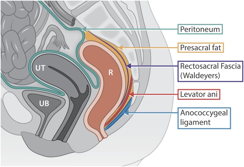
The presacral space is an extraperitoneal potential space between the upper two-thirds of the rectum and the sacrum. The retrorectal/presacral space is bounded anteriorly by the rectum and mesorectal fascia, superiorly by the peritoneal reflection of the rectosigmoid colon, inferiorly by the rectosacral/Waldeyer's fascia, posteriorly by the presacral fascia and laterally by the iliac vessels and ureters (Figure 1).1 The retrorectal space can be further divided into the anterior retrorectal and posterior presacral space, divided by the presacral fascia. Imaging allows limited differentiation of these spaces. A surgical approach to lesions in this region is discussed later.
Figure 1.
An lllustration demonstrating the presacral space. The boundaries are as follows: the rectum anteriorly, peritoneal reflection superiorly, levator ani muscle and anococcygeal ligament inferiorly, sacrum/coccyx posteriorly. R, rectum; UB, urinary bladder; UT, uterus.
A classification of pathologies involving the presacral/retrorectal space based on the origin is presented in Table 1.
Table 1.
Classification scheme for presacral masses
| Origin | Benign | Malignant |
|---|---|---|
| Congenital | Cystic hamartoma | Immature teratoma |
| Duplication cyst | Yolk sac tumour | |
| Dermoid cyst (mature teratoma) | ||
| Anterior sacral myelomeningocele | ||
| Osseous | Aneurysmal bone cyst | Osteosarcoma |
| Giant-cell tumour | Ewing's sarcoma | |
| Chondrosarcoma | ||
| Plasmacytoma | ||
| Metastasis | ||
| Mesenchymal | Myelolipoma | Fibrosarcoma |
| Haemangioma | Gastrointestinal stromal tumour | |
| Fibroma | Lymphoma | |
| Hibernoma | ||
| Castleman disease | ||
| Neurogenic | Neurofibroma | Chordoma |
| Schwannoma | Malignant schwannoma | |
| Ependymoma | ||
| Dural ectasia | ||
| Miscellaneous | Infectious | Desmoplastic round-cell tumour |
| Inflammatory | Metastasis | |
| Post traumatic |
CONGENITAL
Cystic lesions
Congenital cystic lesions are commonly encountered presacral masses with a female predilection.2 The majority of these lesions are benign, including developmental cysts (tailgut, rectal duplication, dermoid and epidermoid cysts) or anterior sacral meningoceles. Developmental cysts constitute approximately two-thirds of congenital presacral masses.3,4 Among these, tailgut cysts, also known as retrorectal cystic hamartoma, are the most common asymptomatic retrorectal masses found in adults.5 Tailgut cysts are often multiloculated cysts containing mucin and lack a muscular layer, a differentiating feature from rectal duplication cysts, which can be confirmed on endorectal ultrasound.6–8 Up to 13% of these cysts may undergo malignant transformation, and for this reason, they are removed.9 Rectal duplication cysts may be associated with other congenital abnormalities of the anorectal region and bladder/urethra.7 Sacrococcygeal teratoma is the most common presacral mass in children containing all three germ-cell lineages.10 Benign mature teratomas tend to be predominantly cystic containing fat, sebum, calcification and soft tissue from dermoid plugs.
On imaging, congenital developmental cysts are seen as well defined, unilocular or multilocular, cystic masses ranging from simple to complex in their internal contents (Figure 2). Thin wall calcifications may be seen with tailgut cysts. MRI is helpful in defining the anatomic relationship to adjacent structures and assessing for the presence of an enhancing or necrotic soft tissue favouring malignant transformation. Increased T1 weighted signal intensity on fat-saturated images represents haemorrhage, mucin or proteinaceous content, suggesting complicated cysts (Figure 3). The presence of mural nodularity and post-contrast enhancement should be viewed with suspicion for malignant transformation in congenital cystic masses.
Figure 2.
A 45-year-old-female with tailgut cyst. (a) Axial T2 weighted, (b) fat-saturated pre-contrast and (c) fat-saturated post-contrast subtraction T1 weighted images (T1WI) through the pelvis showing a presacral multiloculated cystic mass posterior to the rectum without post-contrast enhancement (arrows in a, c). High signal intensity on pre-contrast T1WI (arrow in b), suggesting haemorrhage or proteinaceous debris. Surgical resection confirmed tailgut cyst.
Figure 3.
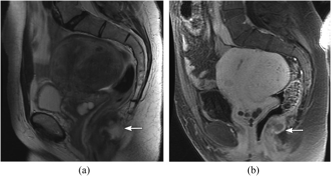
A 48-year-old female with rectal duplication cyst. (a) Sagittal T2 weighted and (b) sagittal post-contrast fat-saturated T1 weighted images show an incidentally detected small cystic mass (arrows) just posterior to the anorectal region without post-contrast enhancement.
Anterior sacral meningocele
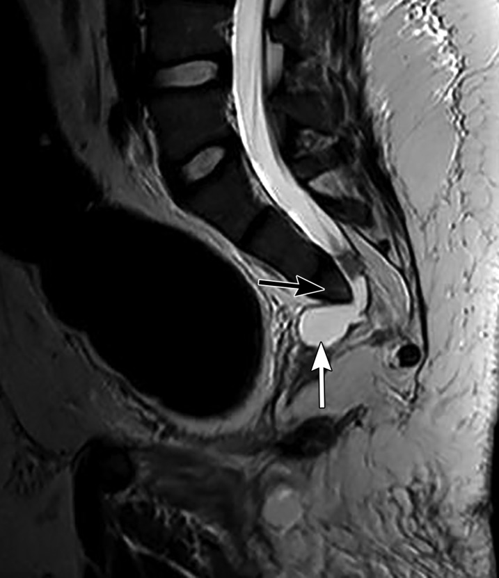
These are rare congenital lesions with female predominance and are associated with other congenital anomalies in 50% of cases.11 Such anomalies include the spina bifida, tethered spinal cord, imperforate anus, uterine/vaginal duplications and presacral lipomas.3,12,13 These lesions have a classic clinical presentation of headache during valsalva owing to increased cerebrospinal fluid (CSF) pressure transmitted via the connection between the meningocele and subdural space.7,8 Identification of this connection is important to imaging diagnosis and is critical to report because this defect must be closed during surgical resection.14 On pelvic radiographs, a unilateral anterior sacral defect may be seen with a rounded, concave border with scalloping/destruction of the surrounding bone, often referred to as the “scimitar sign”.14 On MRI, the signal intensity of the content within the meningocele should parallel the CSF signal on all sequences (Figure 4). Biopsy and aspiration should not be performed, given the risk of introduction of pathogens directly into the spinal meninges, which can result in meningitis.15,16 Surgical resection is curative as long as the dural defect is closed, and a posterior approach is generally taken, although large lesions may require an anterior approach.11,14
Figure 4.
A 17-year-old-male with anterior sacral meningocele. Sagittal T2 weighted image showing a sacral defect (black arrow) and a small anterior cystic mass with demonstrable direct communication with the dural sac (white arrow).
OSSEOUS
Aneurysmal bone cysts
Approximately 20% of aneurysmal bone cysts (ABCs) are located in the spine, and <20% of these spinal ABCs are seen in the sacrum.17 Imaging appearance is that of an expansile lytic lesion sometimes with a thin calcific rim representing the surrounding bone. Multiloculated spaces with fluid–fluid levels seen on CT and MR are characteristic of ABCs corresponding to the blood-filled spaces.18 Although these are benign lesions, they can be locally aggressive or highly symptomatic, given their spinal location via mass effect or pathologic fracture. While wide local resection has been described as a definitive treatment, embolization and sclerotherapy using alcohol-based sclerosants have been successful in treating the lesion.19–21
Giant-cell tumour
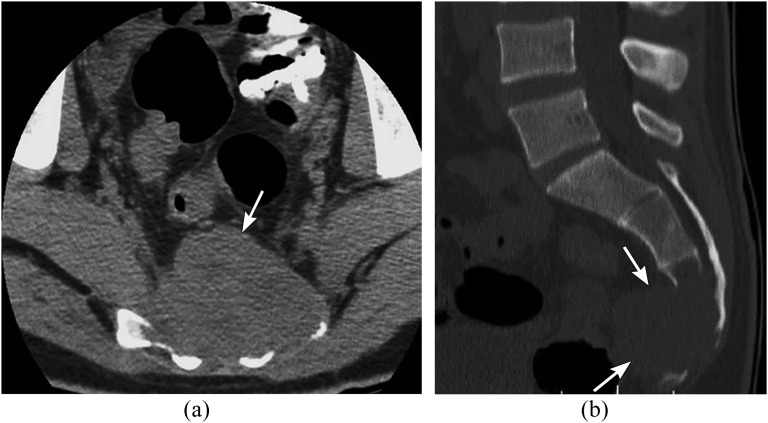
Giant-cell tumour (GCT) is uncommon in the spine, but is the second most common sacral tumour after chordoma. GCT has a female predilection and generally presents between 15 and 40 years of age.18,22,23 GCT is seen as an expansile, locally aggressive, lytic mass with internal haemorrhage and necrosis (Figure 5). A sclerotic rim, when present, is better appreciated on CT. MRI shows heterogeneous intermediate signal intensity on T1 weighted and T2 weighted imaging. GCTs are vascular tumours showing avid post-contrast enhancement.2 Complete resection is the preferred treatment unless location precludes this, in which case, partial curettage and radiation therapy can be used.23,24 Embolization has been used prior to surgery or in patients who cannot undergo surgery.25 Recurrence rates of 40–60% have been reported for incompletely resected tumours.25 Recently, denosumab (monoclonal antibody against receptor activator of nuclear factor k ligand) has been approved for use in patients with unresectable or metastatic GCT tumour of the bone with encouraging results.26
Figure 5.
A 45-year-old male with sacral giant-cell tumour. (a) Axial non-contrast and (b) sagittal bone window CT image through the pelvis showing a large, expansile, lytic mass (arrows) with presacral soft-tissue extension.
Osteosarcoma
Approximately 30% of the spinal osteosarcoma occurs in the sacrum, with male predominance and bimodal age distribution in the third and seventh decade.2,27–29 Occasionally, these tumours may arise in the pagetoid bone or in previously irradiated bones.2,4 Imaging is usually complementary between CT and MRI as the osseous matrix is best seen on CT, while MRI can better define the extent of soft-tissue tumour involvement. These neoplasms have a very aggressive appearance with bone destruction and periosteal new bone formation as well as surrounding soft-tissue mass. Total resection may be difficult owing to the large size at presentation and may have poor prognosis with 30–40% 5-year survival, despite the use of neoadjuvant chemoradiation therapy.25,28,30,31
Chondrosarcoma
While mostly seen in the thoracic spine, sacral involvement is rarely seen.25 Chondrosarcoma may arise from a pre-existing osteochondroma, most commonly in patients with multiple hereditary exostoses.2,4 Characteristic features of the chondroid matrix (“dense”, “stippled” or “rings and arcs”), cortical destruction with soft tissue extension, and fluid attenuation on CT/fluid signal on MRI in non-mineralized regions of the mass are features that aid in imaging diagnosis.2 Most lesions are low grade, allowing surgical cure and a mean survival of 5.9 years.32 Chemotherapy is used for high-grade lesions, and metastatic lesions to the lung are uncommon.2
Sacral plasmacytoma
Solitary osseous plasmacytoma is a unifocal form of multiple myeloma. In the spine, the common location is the thoracic spine, with rare involvement of the sacral spine. It has male predilection, with age distribution between the fourth and sixth decade.33 On imaging, they are seen as a expansile lytic mass with peripheral sclerosis. On MRI, plasmacytoma shows low signal intensity on the T1 weighted image and high signal on T2 weighted image with post-contrast enhancement (Figure 6). Treatment includes local excision with chemoradiation.
Figure 6.
A 49-year-old-male with biopsy confirmed plasmacytoma. (a) Axial contrast-enhanced CT, (b) axial T2 weighted and (c) fat-saturated post-contrast T1 weighted images through the pelvis showing an expansile lytic soft-tissue mass (arrows) showing mild post-contrast enhancement.
Small round-cell tumours
Ewing sarcomas, primitive neuroectodermal tumours and desmoplastic round-cell tumours are subsets of small round blue-cell tumours commonly occurring in older children and young adults between the first and third decade.34 These tumours have an aggressive and permeative appearance, with osseous expansion and associated soft-tissue mass, but often without frank cortical destruction. These are often large at presentation with poor prognosis, especially desmoplastic tumours, which have poor prognosis despite therapy.2 Up to 50% of the desmoplastic subtype manifest with metastases at the time of diagnosis to the peritoneum, lung, liver and bone.2 In fact, sacrococcygeal vs non-sacral ewing sarcomas have long-term survival rates of 25% vs 86%, respectively.2,34,35 The mainstay of therapy is chemoradiation, with local surgical debulking. Unfortunately, therapeutic options do not appear to improve survival rates.36
MESENCHYMAL
Myelolipoma
These are rare, benign retrorectal masses containing mature fat and haematopoietic cells similar to adrenal myelolipoma. These are usually incidentally detected but rarely manifest with pelvic or bowel symptoms owing to mass effect. Characteristic imaging features are the presence of macroscopic fat on CT or MR (Figure 7); however, this can sometimes be difficult to differentiate from other rare tumours like liposarcoma and histological confirmation from percutaneous or excisional biopsy may be required.37 Occasional presence of intralesional haemorrhage due to the haematopoietic component may be seen.
Figure 7.
A 58-year-old female with biopsy-proven presacral myelolipoma. (a) Axial T2 weighted and (b) axial fat-saturated post-contrast T1 weighted (T1W) images through the pelvis showing a presacral soft-tissue mass (white arrows) with intralesional macroscopic fat (black arrows) seen as loss of signal of the fat-saturated T1W image (b).
Solitary fibrous tumour
Solitary fibrous tumour is a rare, slow-growing tumour of fibroblastic and myofibroblastic origin, usually seen in middle-aged adults.38 CT shows a well-defined soft-tissue mass with intense post-contrast enhancement. MR shows a low-to-intermediate signal intensity on T1 weighted and T2 weighted images, with decreased T2 weighted signal intensity isointense to the muscle, helpful in differentiating this lesion as a fibrous tumour. Internal heterogeneity may be present secondary to necrosis, haemorrhage and cystic or myxoid degeneration.36,38
Gastrointestinal stromal tumour
Gastrointestinal stromal tumour is the most common mesenchymal neoplasm of the gastrointestinal tract, most frequently seen in the stomach and small bowel, with rare occurrence in the anorectal region.6 These neoplasms arise from the outer muscular layer and are typically exophytic. They may present with obstructing symptoms. While small lesions may not be detected on CT, larger lesions are seen as hypodense soft-tissue attenuation masses arising from the rectal wall. MR has higher sensitivity in detecting the lesion, which is seen as intermediate-to-low signal intensity on T1 weighted imaging and heterogeneous increased signal intensity on T2 weighted imaging with heterogeneous post-contrast enhancement.2
Lymphoma
Lymphoma can involve both the presacral space and sacrum primarily, although often it is a spectrum of systemic disease. Involvement of the presacral space manifests as a confluent homogeneous lymph nodal mass, with soft-tissue attenuation on CT and homogeneous post-contrast enhancement. These lymph node masses show low signal on T1 weighted image and high signal on T2 weighted image. Identification of the surrounding and distant lymph nodes can help suggest the diagnosis of lymphoma. Treatment consists of chemotherapy or radiation therapy based on the and grade of tumour, which is determined by biopsy.2
NEUROGENIC
Nerve sheath tumours
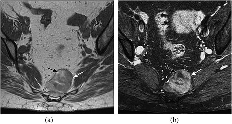
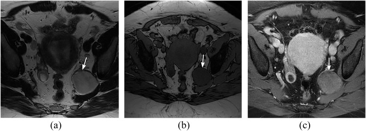
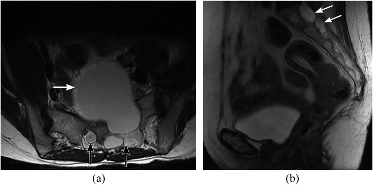
Schwannomas and neurofibromas are grouped together because they can be quite difficult to distinguish from one another by imaging. These tumours have a male predominance and generally present in the third–fifth decade, the exception being patients with neurofibromatosis-I in which they generally arise at a younger age.39,40 While the majority of these tumours are benign, rarely, transformation to malignant peripheral nerve sheath tumour is possible. Neurofibromas consist of fibroblasts, Schwann cells and other neural elements infiltrating their originating nerve, in contrast to schwannomas, which are nerve sheath tumours and lie along the peripheral aspect of the nerve. Neurofibromas are classically bilateral and symmetric in continuity with spinal nerve roots expanding the associated neural foramen. If there is asymmetry or rapid growth, the possibility of malignant transformation should be raised. Neurofibromas tend to have lower CT attenuation than surrounding soft tissues (typically compared with the psoas muscle) and may mimic enlarged lymph nodes.2 On MRI, they demonstrate high signal intensity on T2 weighted image with central low signal signifying the fibrous tissue surrounded by myxoid contents, referred to as the “target” sign (Figure 8). Schwannomas may be more heterogeneous on CT and MR with areas of cystic degeneration.2 A cystic structure with nodular enhancement arising from a peripheral nerve suggests schwannoma (Figure 9). Differential diagnosis includes a Tarlov or perineural cyst (Figure 10), which does not have enhancing elements and follows CSF signal on all sequences.
Figure 8.
A 39-year-old male with neurofibroma. (a) Axial contrast-enhanced CT, (b) axial T2 weighted image and (c) axial fat-saturated post-contrast T1 weighted image (T1WI) through the pelvis showing two soft-tissue masses in the presacral region (arrows). MR showing heterogeneous T2 weighted signal intensity with central hypointensity and peripheral hyperintensity (b). Mild enhancement of the central collagenous matrix is noted on post-contrast T1WI (c).
Figure 9.
A 49-year-old female with schwannoma. (a) Axial T2 weighted (T2W), (b) axial T1 weighted (T1W) and (c) fat-saturated post-contrast T1W images through the pelvis showing a well-defined mass arising from the left hemisacrum with presacral extension (arrows). The mass is hyperintense on T2W, hypointense on T1W and shows mild post-contrast enhancement.
Figure 10.
A 44-year-old female with Tarlov cysts. Axial (a) and sagittal T2 weighted image (b) showing a large well-defined left presacral cystic mass, isointense to the cerebrospinal fluid (white arrow in a) with widening of the multilevel sacral neural foramina (black arrows in a, white arrows in b).
Dural ectasia
This is a benign condition that occurs owing to the weakening of elastic fibres in the dural sac resulting in chronic expansion of the subdural space and osseous remodelling owing to increased CSF pressure.39 There is increased incidence of dural ectasia in patients with Marfan syndrome.39 On imaging, smooth remodelling and osseous expansion of the spinal canal is manifested as widening of the pedicular distance and posterior vertebral body scalloping.
Chordoma
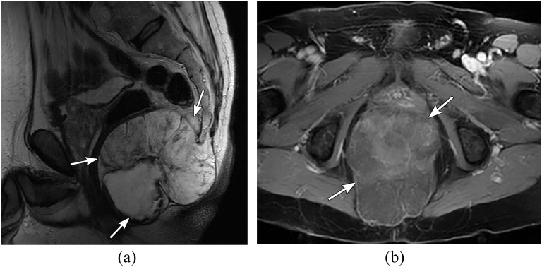
Chordoma is the most common primary sacral tumour that arises from the remnant of the embryologic notochord, presenting in the fourth–seventh decade with a male predilection.41 Chordomas are slow-growing tumours and often large at the time of presentation, given the slow onset of symptoms. On imaging, sacral chordomas are seen as midline large heterogeneous masses with variable post-contrast enhancement and osseous destruction. Calcifications are present up to 90% of the time on CT, and intralesional haemorrhage is not uncommon.25 MRI generally shows low T1 and high T2 signal intensity with internal haemorrhage rendering regions of high signal on the T1 weighted image (Figure 11). Given their local aggressive behaviour, chordomas are treated with resection and radiation therapy, which results in an 8–10-year average survival in sacrococcygeal lesions vs 4–5 years in other locations.42
Figure 11.
A 55-year-old female with sacral chordoma. (a) Sagittal T2 weighted and (b) axial fat-saturated post-contrast T1 weighted images through the pelvis showing a large lobulated mass in relation to the coccyx (arrows) with heterogeneous high signal on T2 weighted and heterogeneous post-contrast enhancement.
MISCELLANEOUS
Infectious and inflammatory
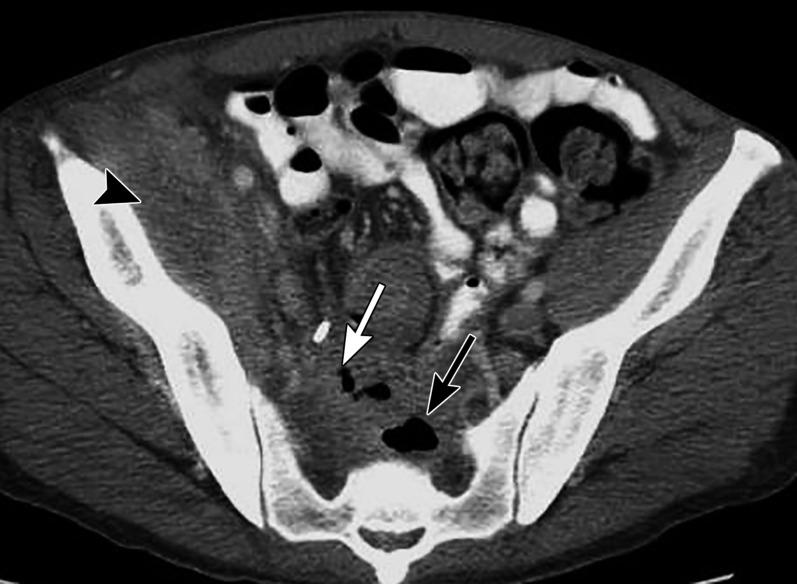
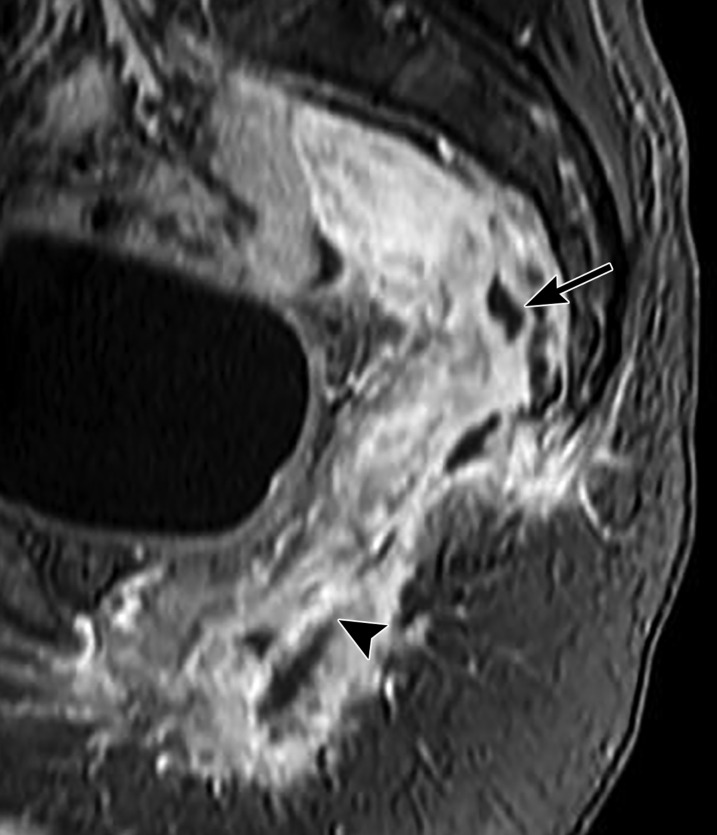
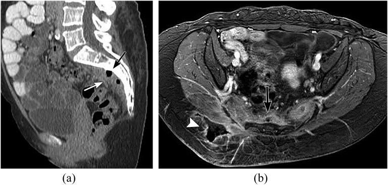
Inflammatory and infectious conditions involving presacral spaces are often an extension of the pelvic process (Figure 12). While reactive inflammatory changes are often seen as a sequela of therapy for pelvic malignancies, careful assessment for post-therapy complications like superadded infection with abscess formation or fistula should be made (Figure 13). A clinical history is the most helpful guide to this diagnosis. Osteomyelitis of the sacrum may be encountered in patients with decubitus sacral ulcers (Figure 14). MR is superior in the evaluation of suspected osteomyelitis for marrow changes, abscess and fistula, the given higher soft-tissue resolution.
Figure 12.
A 38-year-old female with confirmed pelvic actinomyces infection. Axial contrast-enhanced CT through the pelvis showing a complex collection involving the right iliacus muscle (black arrowhead) with gas and fluid collection extending to the presacral region (white and black arrows).
Figure 13.
A 61-year-old female status post abdominoperineal resection for rectal cancer showing presacral abscess. Sagittal fat-saturated post-contrast T1 weighted image showing heterogeneous enhancing soft tissue (arrow) and gas locules (arrowhead) in the presacral space consistent with abscess.
Figure 14.
A 51-year-old male with sacral osteomyelitis and presacral abscess. (a) Sagittal reformatted CT, (b) axial fat-saturated post-contrast T1 weighted image through the pelvis showing sclerosis of the sacrum (black arrow, a) with presacral soft tissue and gas (white arrow, a). MR showing phlegmonous changes in the presacral region (black arrow, b) with a fistulous track extending into the right gluteal region (white arrowhead, b).
Metastasis
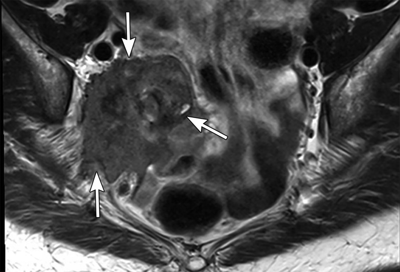
Metastasis in the presacral space may occur from direct, lymphogenous or haematogenous spread, typically from primary pelvic malignancy, with rectal cancer being the commonest. Local spread to the presacral space is most commonly seen, with primary rectal tumour extending beyond the mesorectal fascia (T3/T4).43 Post-operative fibrosis or radiation-associated changes may also manifest as a presacral soft-tissue mass and may be difficult to differentiate from recurrence (Figure 15). Tissue sampling or positron emission tomography-CT scan may be helpful in confirming the nature of these lesions.2,43
Figure 15.
A 48-year-old female with locally advanced cervical cancer. Axial T2 weighted image showing a recurrence of cervical cancer which is invading the piriformis muscle through the greater sciatic foramen (arrows). More caudal images (not shown here) demonstrated invasion of the bladder and encasement of the vagina.
Retroperitoneal fibrosis
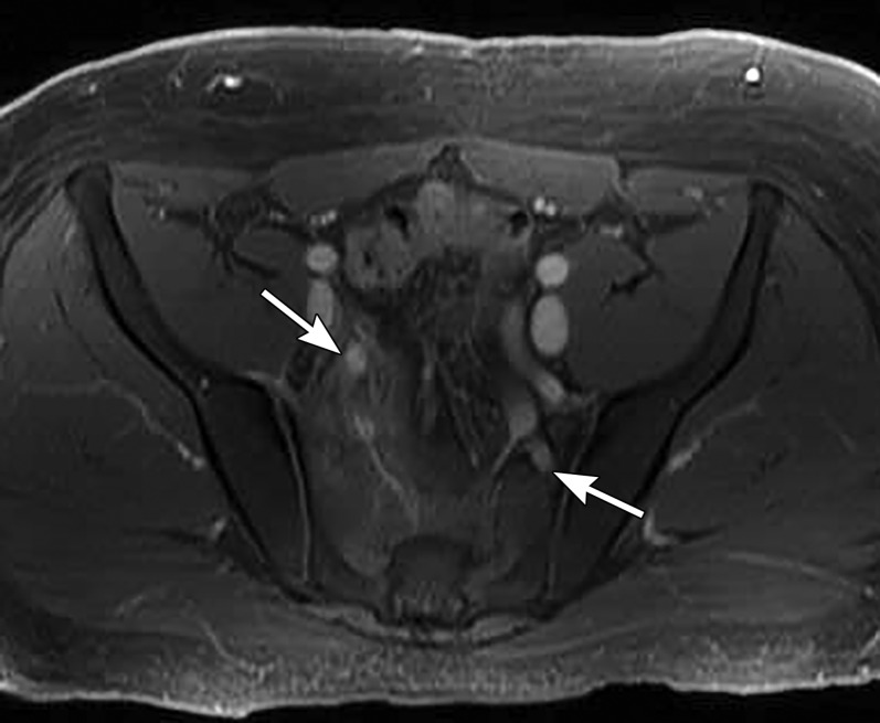
Retroperitoneal fibrosis is a chronic inflammatory process of the retroperitoneum, typically see in the lumbar region, but may extend caudally into the pelvis to involve the presacral region.44 While the majority of the cases are idiopathic, one-third of the cases occur secondary to underlying pathology. Ill-defined plaque-like soft tissue with variable degree of surrounding inflammatory stranding is the typical CT manifestation. MRI signal characteristics are variable but include low T1 weighted and T2 weighted signal intensity, as seen with the chronic fibrotic process (Figure 16), although the presence of high T2 WI and post-contrast enhancement suggest active inflammation.2
Figure 16.
A 42-year-old male with known retroperitoneal fibrosis. Axial fat-saturated post-contrast T1 weighted image through the pelvis showing a soft-tissue plaque-like mass in the presacral region (arrows) encasing the internal iliac vessels.
MANAGEMENT
The majority of the presacral/retrorectal mass will undergo surgical removal if the patient is able owing to the fact that this space is prone to contamination and even benign lesions may become superinfected and potentially lead to further complications such as inflammatory fistulae.
Three primary operative approaches have been described for managing presacral masses: anterior, posterior and combined.45,46 The anterior or abdominal approach is taken if the lesion is completely contained at or above the S3 vertebral level.47 This allows adequate visualization of the key anatomy (vascular and urinary) via mobilization of surrounding structures. Of note, the S3 level is the most cephalad palpable region able to be palpated during digital rectal examination. Anterior approach is only taken if there is no sacral invasion.
The posterior approach is taken for lesions below S3 (defined as S4 and below).48 Coccygectomy is almost always performed, and sacrectomy of variable extent is performed as well. Posterior approach increases the risk of haemorrhage, given the poor visualization of vascular structures and decreased ability to control inadvertent vascular injury. However, this approach allows for better preservation of neurological structures.
Combined abdominoperineal approaches are taken for masses extending craniocaudally above and below S3 or masses with sacral, vascular, pelvic sidewall, ureteral or rectal invasion. The recovery and operating times are increased in these cases.12,49
Reporting
The imaging approach to evaluation of these masses is not standardized. However, given the range of pathologies and multiplicity of soft-tissue structures in the presacral lesions, as well as common involvement of the spine and pelvic bones, MRI is the preferred modality. Tables 2 and 3 outline the protocols used at the authors' institution for the evaluation of rectal and presacral masses.
Table 2.
Suggested MRI protocol for rectal cancer imaging on 3.0-T magnets
| Sequence | Sagittal T2W TSE | Coronal T2W TSE | Axial T1W TSE | Axial DWI EPI | Oblique axial T2W TSE |
|---|---|---|---|---|---|
| TR/TE (msec) | 3000–5000/100 | 3000–6000/80 | 350/shortest | Shortest/shortest | 3000–5000/110 |
| Flip angle | 90 | 90 | 90 | 90 | 90 |
| FOV (cm) | 24 | 26 | 35 | 34 | 34 |
| Slice thickness (mm) | 3 | 3 | 1.5 | 3 | 3 |
DWI, diffusion-weighted imaging; EPI, echoplanar imaging; FOV, field of view; T1W, T1 weighted; T2W, T2 weighted; TE, echo time; TR, repetition time; TSE, turbo spin echo.
Table 3.
Suggested MRI protocol for pelvic mass imaging (including presacral masses) on 3.0-T magnets
| Sequence | Coronal T2W TSE | Axial DWI EPI | Sagittal T2W TSE | Axial IP/OP | Axial 3D TFE dynamic post gadolinium | Axial 2D TFE delayed post gadolinium |
|---|---|---|---|---|---|---|
| TR/TE (msec) | 3000–5000/100 | Shortest/shortest | 3000–6000/shortest | 180/1.5/2.3 | Shortest/shortest | Shortest/shortest |
| Flip angle | 90 | 90 | 90 | 55 | 7 | 10 |
| FOV (cm) | To fit | To fit | To fit | To fit | To fit | To fit |
| Slice thickness (mm) | 5 | 5.5 | 6 | 5.5 | 2 | 2 |
2D, two dimensional; 3D, three dimensional; DWI, diffusion-weighted imaging; EPI, echoplanar imaging; FOV, field of view; IP, in-phase; OP, opposed-phase; T2W, T2 weighted; TE, echo time; TR, repetition time; TFE, turbo fast echo; TSE, turbo spin echo.
The value of radiology to the surgeon is not always in 100% accuracy for diagnosis. In the case of presacral masses, identifying salient features which could change the surgical approach is essential. First and foremost, the surgeon needs to know whether a mass is benign or malignant. Although not always clear-cut, the majority of presacral masses can be placed easily into one of these two categories. The exception may be sarcomatous tumours with a predominant fatty component or lymphoma which has a homogeneous imaging appearance often associated with benignity.
Next, the origin of the mass should be elucidated. Masses of the presacral space originate from anterior structures—primary rectal origin; primary retrorectal area origin; or sacrum with anterior extension. Masses of rectal origin are most commonly gastrointestinal stromal tumour or rectal cancer, and colorectal surgeons should be the primary operators. Masses arising from the sacrum with close proximity to CNS structures require orthopaedic spinal or neurosurgery lead. Primary retrorectal origin is generally within the comfort zone of colorectal surgery, but the involvement of adjacent vascular, genitourinary or osseous structures warrants a multidisciplinary approach.
As with any mass, the extent of local invasion must be described. Invasion or contact (>180°) with adjacent structures indicates the need for the primary surgeon to consider collaboration with a surgeon specializing in the involved anatomy.45,46
Biopsy
In the past, biopsy was almost never undertaken because of risk of infection and injury to critical structures and generally poor sampling efficacy. However, the combination of improved technique and improved pre-operative treatment of several of the lesions outlined has increased the utility of tissue sampling prior to therapy. In particular, presacral lesions arising primarily from osseous structures will always undergo biopsy at our institution because their imaging appearance is not sufficiently specific. Ewing sarcoma, osteosarcoma, lymphoma and solitary fibrous tumour are some examples of lesions which could benefit from neoadjuvant therapy and require tissue sampling to determine malignant vs benign nature.47 Use of clinical and imaging parameters to suggest one of these lesions could point a radiologist towards suggesting tissue sampling. Three general approaches can be taken: transgluteal, anterolateral extraperitoneal and transsacral (Figures 17, 18).50,51
Figure 17.
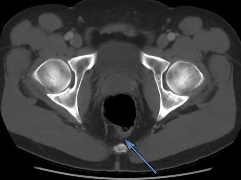
Transgluteal approach is preferred for the majority of presacral masses (arrow). The medial and inferior approach avoids injury to the sciatic nerve and superior and inferior gluteal vessels and avoids traversal of the piriformis muscle, which can be painful. This is ideally performed in the prone position.
Figure 18.
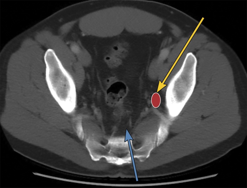
Transsacral (light blue arrow) approach is usually reserved for osseous lesions, which may extend to the presacral space. The approach is medial to the neural foramen and lateral to the spinal canal. The extraperitoneal anterolateral approach (yellow arrow) can target a pelvic sidewall lymph node or mass (red oval) with a course through the iliacus muscle. For colour image see online.
The transgluteal, approach is preferred by most radiologists, as this can be done by staying “low and medial” adjacent to the sacrum, below the superior gluteal artery, medial to the inferior gluteal artery and sciatic nerve. In addition, if seeding of the biopsy tract occurs, this is less of a problem because this region will likely be resected during surgery.12,51 Unfortunately, some patients cannot tolerate this approach because of the requirement for prone positioning.
Anterolateral extraperitoneal approaches essentially follow the course of the iliopsoas muscle to reach the presacral space, avoiding the bladder, bowel and vasculature, while remaining below the peritoneal lining to avoid infectious introduction.
The transsacral, transosseous approach commonly used for osseous lesions requires appropriate devices as well as adds the risk of neurologic injury; however, the bladder and bowel are well removed from the biopsy tract.
CONCLUSION
The presence of myriad embryologically distinct types of tissues in the retrorectal/presacral space accounts for the various lesions seen on imaging studies. While some masses have distinct clinical presentations or imaging appearances, it is more important for the radiologist to identify and report key findings that will affect management decisions than speculate on histology. Knowledge of the relevant anatomy and surgical approaches helps the radiologist make a clinical contribution to the care of patients with presacral pathology. In addition, if image-guided biopsy is required, the radiologist should be aware of critical anatomical and pathological considerations that may alter the biopsy approach.
Contributor Information
Nishant Patel, Email: pnishant@med.umich.edu.
Katherine E Maturen, Email: kmaturen@med.umich.edu.
Ravi K Kaza, Email: ravikaza@med.umich.edu.
Mahmoud M Al-Hawary, Email: alhawary@med.umich.edu.
Ashish P Wasnik, Email: ashishw@med.umich.edu.
REFERENCES
- 1.Schäfer A-O. Anorectal anatomy. In: MRI of rectal cancer. Schäfer A-O, ed. Berlin/Heidelberg, Germany: Springer-Verlag; 2010. pp. 5–13. [Google Scholar]
- 2.Hain KS, Pickhardt PJ, Lubner MG, Menias CO, Bhalla S. Presacral masses: multimodality imaging of a multidisciplinary space. Radiographics 2013; 33: 1145–67. doi: 10.1148/rg.334115171 [DOI] [PubMed] [Google Scholar]
- 3.Dahan H, Arrivé L, Wendum D, le Pointe HD, Djouhri H, Tubiana JM. Retrorectal developmental cysts in adults: clinical and radiologic-histopathologic review, differential diagnosis, and treatment. Radiographics 2001; 21: 575–84. doi: 10.1148/radiographics.21.3.g01ma13575 [DOI] [PubMed] [Google Scholar]
- 4.Hobson KG, Ghaemmaghami V, Roe JP, Goodnight JE, Khatri VP. Tumors of the retrorectal space. Dis Colon Rectum 2005; 48: 1964–74. doi: 10.1007/s10350-005-0122-9 [DOI] [PubMed] [Google Scholar]
- 5.Lev-Chelouche D, Gutman M, Goldman G, Even-Sapir E, Meller I, Issakov J, et al. Presacral tumors: a practical classification and treatment of a unique and heterogenous group of diseases. Surgery 2003; 133: 473–8. doi: 10.1067/msy.2003.118 [DOI] [PubMed] [Google Scholar]
- 6.Rouse HC, Godoy MC, Lee WK, Phang PT, Brown CJ, Brown JA. Imaging findings of unusual anorectal and perirectal pathology: a multi-modality approach. Clin Radiol 2008; 63: 1350–60. doi: 10.1016/j.crad.2008.06.008 [DOI] [PubMed] [Google Scholar]
- 7.Beattie CH, Garvey CJ, Hershman MJ. Endorectal magnetic resonance imaging of a rectal duplication cyst. Br J Radiol 1999; 72: 896–8. doi: 10.1259/bjr.72.861.10645197 [DOI] [PubMed] [Google Scholar]
- 8.Menassa-Moussa L, Kanso H, Checrallah A, Abboud J, Ghossain M. CT and MR findings of a retrorectal cystic hamartoma confused with an adnexal mass on ultrasound. Eur Radiol 2005; 15(2): 263–6. doi: 10.1007/s00330-004-2330-4 [DOI] [PubMed] [Google Scholar]
- 9.Mathis KL, Dozois EJ, Grewal MS, Metzger P, Larson DW, Devine RM. Malignant risk and surgical outcomes of presacral tailgut cysts. Br J Surg 2010; 97: 575–9. doi: 10.1002/bjs.6915 [DOI] [PubMed] [Google Scholar]
- 10.Wetzel LH, Levine E. Pictorial essay. MR imaging of sacral and presacral lesions. AJR Am J Roentgenol 1990; 154: 771–5. doi: 10.2214/ajr.154.4.2107674 [DOI] [PubMed] [Google Scholar]
- 11.Glasgow SC, Dietz DW. Retrorectal tumors. Clin Colon Rectal Surgery 2006; 19: 61–8. doi: 10.1055/s-2006-942346 [DOI] [PMC free article] [PubMed] [Google Scholar]
- 12.Neale JA. Retrorectal tumors. Clin Colon Rectal Surgery 2011; 24: 149–60. doi: 10.1055/s-0031-1285999 [DOI] [PMC free article] [PubMed] [Google Scholar]
- 13.Villarejo F, Scavone C, Blazquez MG, Pascual-Castroviejo I, Perez-Higueras A, Fernandez-Sanchez A, et al. Anterior sacral meningocele: review of the literature. Surg Neurol 1983; 19: 57–71. doi: 10.1016/0090-3019(83)90212-4 [DOI] [PubMed] [Google Scholar]
- 14.St Ville EW, Jafri SZ, Madrazo BL, Mezwa DG, Bree RL, Rosenberg BF. Endorectal sonography in the evaluation of rectal and perirectal disease. AJR Am J Roentgenol 1991; 157: 503–8. doi: 10.2214/ajr.157.3.1872236 [DOI] [PubMed] [Google Scholar]
- 15.Matsumoto T, Iida M, Suekane H, Tominaga M, Yao T, Fujishima M. Endoscopic ultrasonography in rectal carcinoid tumors: contribution to selection of therapy. Gastrointest Endosc 1991; 37: 539–42. doi: 10.1016/S0016-5107(91)70824-9 [DOI] [PubMed] [Google Scholar]
- 16.Prasad AR, Amin MB, Randolph TL, Lee CS, Ma CK. Retrorectal cystic hamartoma: report of 5 cases with malignancy arising in 2. Arch Pathol Lab Med 2000; 124: 725–9. doi: [DOI] [PubMed] [Google Scholar]
- 17.Capanna R, Van Horn JR, Biagini R, Ruggieri P. Aneurysmal bone cyst of the sacrum. Skeletal Radiol 1989; 18: 109–13. doi: 10.1007/BF00350658 [DOI] [PubMed] [Google Scholar]
- 18.Llauger J, Palmer J, Amores S, Bague S, Camins A. Primary tumors of the sacrum: diagnostic imaging. AJR Am Journal Roentgenol 2000; 174: 417–24. doi: 10.2214/ajr.174.2.1740417 [DOI] [PubMed] [Google Scholar]
- 19.Amendola L, Simonetti L, Simoes CE, Bandiera S, De Iure F, Boriani S. Aneurysmal bone cyst of the mobile spine: the therapeutic role of embolization. Eur Spine J 2013; 22: 533–41. doi: 10.1007/s00586-012-2566-7 [DOI] [PMC free article] [PubMed] [Google Scholar]
- 20.Adamsbaum C, Mascard E, Guinebretiere JM, Kalifa G, Dubousset J. Intralesional Ethibloc injections in primary aneurysmal bone cysts: an efficient and safe treatment. Skeletal Radiol 2003; 32: 559–66. doi: 10.1007/s00256-003-0653-x [DOI] [PubMed] [Google Scholar]
- 21.Varshney MK, Rastogi S, Khan SA, Trikha V. Is sclerotherapy better than intralesional excision for treating aneurysmal bone cysts? Clin Orthop Relat Res 2010; 468: 1649–59. doi: 10.1007/s11999-009-1144-8 [DOI] [PMC free article] [PubMed] [Google Scholar]
- 22.Brien EW, Mirra JM, Kessler S, Suen M, Ho JK, Yang WT. Benign giant cell tumor of bone with osteosarcomatous transformation (“dedifferentiated” primary malignant GCT): report of two cases. Skeletal Radiol 1997; 26: 246–55. doi: 10.1007/s002560050230 [DOI] [PubMed] [Google Scholar]
- 23.Eckardt JJ, Grogan TJ. Giant cell tumor of bone. Clin Orthop Relat Res 1986 (204): 45–58. [PubMed] [Google Scholar]
- 24.Randall RL. Giant cell tumor of the sacrum. Neurosurg Focus 2003; 15: E13. doi: 10.3171/foc.2003.15.2.13 [DOI] [PubMed] [Google Scholar]
- 25.Murphey MD, Andrews CL, Flemming DJ, Temple HT, Smith WS, Smirniotopoulos JG. From the archives of the AFIP. Primary tumors of the spine: radiologic pathologic correlation. Radiographics 1996; 16: 1131–58. doi: 10.1148/radiographics.16.5.8888395 [DOI] [PubMed] [Google Scholar]
- 26.Ueda T, Morioka H, Nishida Y, Kakunaga S, Tsuchiya H, Matsumoto Y, et al. : Objective tumor response to denosumab in patients with giant cell tumor of bone: a multicenter phase 2 trial. Ann Oncol 2015; 26: 2149–54. doi: 10.1093/annonc/mdv307 [DOI] [PMC free article] [PubMed] [Google Scholar]
- 27.Ozaki T, Flege S, Liljenqvist U, Hillmann A, Delling G, Salzer-Kuntschik M, et al. Osteosarcoma of the spine: experience of the Cooperative Osteosarcoma Study Group. Cancer 2002; 94: 1069–77. doi: 10.1002/cncr.10258 [DOI] [PubMed] [Google Scholar]
- 28.Schoenfeld AJ, Hornicek FJ, Pedlow FX, Kobayashi W, Garcia RT, DeLaney TF, et al. Osteosarcoma of the spine: experience in 26 patients treated at the Massachusetts General Hospital. Spine J 2010; 10: 708–14. doi: 10.1016/j.spinee.2010.05.017 [DOI] [PubMed] [Google Scholar]
- 29.Cade S. Osteogenic sarcoma; a study based on 133 patients. J R Coll Surgeons Edinb 1955; 1: 79–111. [PubMed] [Google Scholar]
- 30.Janinis J, McTiernan A, Driver D, Mitchell C, Cassoni AM, Pringle J, et al. ; London Bone and Soft Tissue Tumour Service. A pilot study of short-course intensive multiagent chemotherapy in metastatic and axial skeletal osteosarcoma. Ann Oncol 2002; 13: 1935–44. doi: 10.1093/annonc/mdf338 [DOI] [PubMed] [Google Scholar]
- 31.Schwab J, Gasbarrini A, Bandiera S, Boriani L, Amendola L, Picci P, et al. Osteosarcoma of the mobile spine. Spine 2012; 37: E381–386. doi: 10.1097/BRS.0b013e31822fb1a7 [DOI] [PubMed] [Google Scholar]
- 32.Katonis P, Datsis G, Karantanas A, Kampouroglou A, Lianoudakis S, Licoudis S, et al. Spinal osteosarcoma. Clin Med Insights Oncol 2013; 7: 199–208. doi: 10.4137/CMO.S10099 [DOI] [PMC free article] [PubMed] [Google Scholar]
- 33.Ooi GC, Chim JC, Au WY, Khong PL. Radiologic manifestations of primary solitary extramedullary and multiple solitary plasmacytomas. AJR Am J Roentgenol 2006; 186: 821–7. doi: 10.2214/AJR.04.1787 [DOI] [PubMed] [Google Scholar]
- 34.Pilepich MV, Vietti TJ, Nesbit ME, Tefft M, Kissane J, Burgert O, et al. Ewing's sarcoma of the vertebral column. Int J Radiat Oncol Biol Phys 1981; 7: 27–31. doi: 10.1016/0360-3016(81)90056-0 [DOI] [PubMed] [Google Scholar]
- 35.Bacci G, Boriani S, Balladelli A, Barbieri E, Longhi A, Alberghini M, et al. Treatment of nonmetastatic Ewing's sarcoma family tumors of the spine and sacrum: the experience from a single institution. Eur Spine J 2009; 18: 1091–5. doi: 10.1007/s00586-009-0921-0 [DOI] [PMC free article] [PubMed] [Google Scholar]
- 36.Shanbhogue AK, Prasad SR, Takahashi N, Vikram R, Zaheer A, Sandrasegaran K. Somatic and visceral solitary fibrous tumors in the abdomen and pelvis: cross-sectional imaging spectrum. Radiographics 2011; 31: 393–408. doi: 10.1148/rg.312105080 [DOI] [PubMed] [Google Scholar]
- 37.Itani M, Wasnik AP, Platt JF: Radiologic-pathologic correlation in extra-adrenal myelolipoma. Abdom Imaging 2014; 39: 394–7. doi: 10.1007/s00261-013-0062-0 [DOI] [PubMed] [Google Scholar]
- 38.Parida L, Fernandez-Pineda I, Uffman JK, Davidoff AM, Krasin MJ, Pappo A, et al. Clinical management of infantile fibrosarcoma: a retrospective single-institution review. Pediatr Surg Int 2013; 29: 703–8. doi: 10.1007/s00383-013-3326-4 [DOI] [PMC free article] [PubMed] [Google Scholar]
- 39.Ha HI, Seo JB, Lee SH, Kang JW, Goo HW, Lim TH, et al. Imaging of Marfan syndrome: multisystemic manifestations. Radiographics 2007; 27: 989–1004. doi: 10.1148/rg.274065171 [DOI] [PubMed] [Google Scholar]
- 40.Hirano K, Imagama S, Sato K, Kato F, Yukawa Y, Yoshihara H, et al. Primary spinal cord tumors: review of 678 surgically treated patients in Japan. A multicenter study. Eur Spine J 2012; 21: 2019–26. doi: 10.1007/s00586-012-2345-5 [DOI] [PMC free article] [PubMed] [Google Scholar]
- 41.Diel J, Ortiz O, Losada RA, Price DB, Hayt MW, Katz DS. The sacrum: pathologic spectrum, multimodality imaging, and subspecialty approach. Radiographics 2001; 21: 83–104. doi: 10.1148/radiographics.21.1.g01ja0883 [DOI] [PubMed] [Google Scholar]
- 42.Fourney DR, Gokaslan ZL. Current management of sacral chordoma. Neurosurg Focus 2003; 15: E9. doi: 10.3171/foc.2003.15.2.9 [DOI] [PubMed] [Google Scholar]
- 43.Metser U, You J, McSweeney S, Freeman M, Hendler A. Assessment of tumor recurrence in patients with colorectal cancer and elevated carcinoembryonic antigen level: FDG PET/CT versus contrast-enhanced 64-MDCT of the chest and abdomen. AJR Am Journal Roentgenol 2010; 194: 766–71. doi: 10.2214/AJR.09.3205 [DOI] [PubMed] [Google Scholar]
- 44.Park BK, Kim SH, Moon MH. Idiopathic presacral retroperitoneal fibrosis: report of two cases. Br J Radiol 2003; 76: 570–3. doi: 10.1259/bjr/61286585 [DOI] [PubMed] [Google Scholar]
- 45.Pappalardo G, Frattaroli FM, Casciani E, Moles N, Mascagni D, Spoletini D, et al. Retrorectal tumors: the choice of surgical approach based on a new classification. Am Surg 2009; 75: 240–8. [PubMed] [Google Scholar]
- 46.Woodfield JC, Chalmers AG, Phillips N, Sagar PM. Algorithms for the surgical management of retrorectal tumours. Br J Surg 2008; 95: 214–21. doi: 10.1002/bjs.5931 [DOI] [PubMed] [Google Scholar]
- 47.Reiter MJ, Schwope RB, Bui-Mansfield LT, Lisanti CJ, Glasgow SC. Surgical management of retrorectal lesions: what the radiologist needs to know. AJR Am J Roentgenol 2015; 204: 386–95. doi: 10.2214/AJR.14.12791 [DOI] [PubMed] [Google Scholar]
- 48.Buchs N, Taylor S, Roche B. The posterior approach for low retrorectal tumors in adults. Int J Colorectal Dis 2007; 22: 381–5. doi: 10.1007/s00384-006-0183-9 [DOI] [PubMed] [Google Scholar]
- 49.Hosseini-Nik H, Hosseinzadeh K, Bhayana R, Jhaveri KS. MR imaging of the retrorectal-presacral tumors: an algorithmic approach. Abdom Imaging 2015; 40: 2630–44. doi: 10.1007/s00261-015-0404-1 [DOI] [PubMed] [Google Scholar]
- 50.Gupta S, Nguyen HL, Morello FA, Jr, Ahrar K, Wallace MJ, Madoff DC, et al. Various approaches for CT-guided percutaneous biopsy of deep pelvic lesions: anatomic and technical considerations. Radiographics 2004; 24: 175–89. doi: 10.1148/rg.241035063 [DOI] [PubMed] [Google Scholar]
- 51.Merchea A, Larson DW, Hubner M, Wenger DE, Rose PS, Dozois EJ. The value of preoperative biopsy in the management of solid presacral tumors. Dis Colon Rectum 2013; 56: 756–60. doi: 10.1097/DCR.0b013e3182788c77 [DOI] [PubMed] [Google Scholar]


