

Abstract
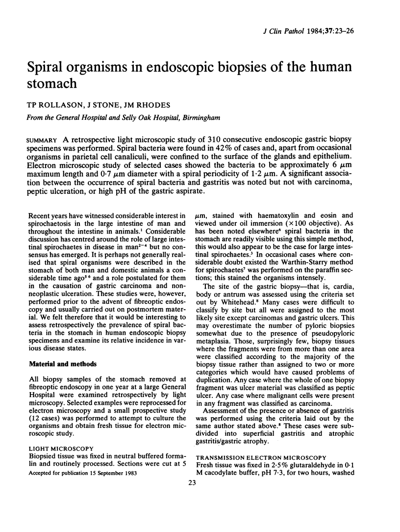
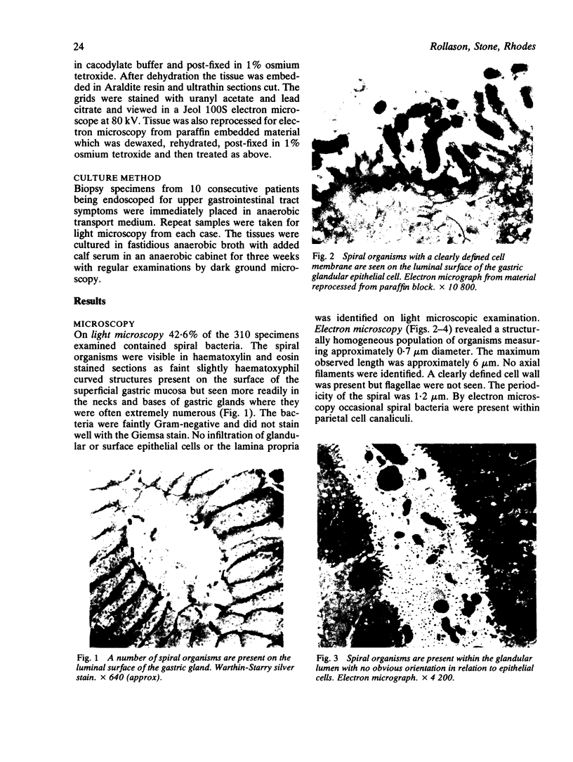
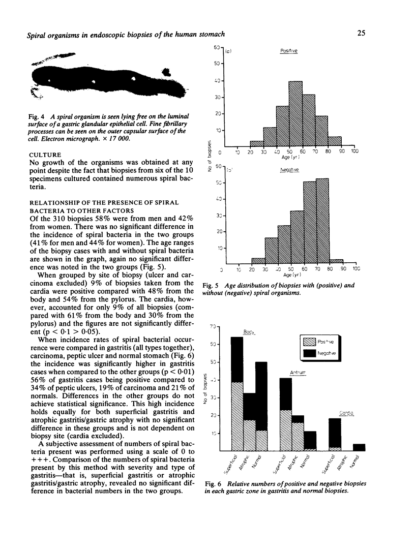
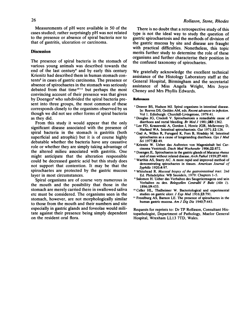
A retrospective light microscopic study of 310 consecutive endoscopic gastric biopsy specimens was performed. Spiral bacteria were found in 42% of cases and, apart from occasional organisms in parietal cell canaliculi, were confined to the surface of the glands and epithelium. Electron microscopic study of selected cases showed the bacteria to be approximately 6 micron maximum length and 0.7 micron diameter with a spiral periodicity of 1.2 micron. A significant association between the occurrence of spiral bacteria and gastritis was noted but not with carcinoma, peptic ulceration, or high pH of the gastric aspirate.
Full text
PDF
Images in this article
Selected References
These references are in PubMed. This may not be the complete list of references from this article.
- Douglas J. G., Crucioli V. Spirochaetosis: a remediable cause of diarrhoea and rectal bleeding? Br Med J (Clin Res Ed) 1981 Nov 21;283(6303):1362–1362. doi: 10.1136/bmj.283.6303.1362. [DOI] [PMC free article] [PubMed] [Google Scholar]
- Lee F. D., Kraszewski A., Gordon J., Howie J. G., McSeveney D., Harland W. A. Intestinal spirochaetosis. Gut. 1971 Feb;12(2):126–133. doi: 10.1136/gut.12.2.126. [DOI] [PMC free article] [PubMed] [Google Scholar]