

Abstract
Aneurysms and dissections of the right-sided aortic arch are rare and published data are limited to a few case reports and small series. The optimal treatment strategy of this entity and the challenges associated with their management are not yet fully investigated and conclusive. We performed a systematic review of the literature to identify all patients who underwent surgical or endovascular intervention for right aortic arch aneurysms or dissections. The search was limited to the articles published only in English. We focused on presentation and critically assessed different management strategies and outcomes. We identified 74 studies that reported 99 patients undergoing surgical or endovascular intervention for a right aortic arch aneurysm or dissection. The median age was 61 years. The commonest presenting symptoms were chest or back pain and dysphagia. Eighty-eight patients had an aberrant left subclavian artery with only 11 patients having the mirror image variant of a right aortic arch. The commonest pathology was aneurysm arising from a Kommerell's diverticulum occurring in over 50% of the patients. Twenty-eight patients had dissections, 19 of these were Type B and 9 were Type A. Eighty-one patients had elective operations while 18 had emergency procedures. Sixty-seven patients underwent surgical treatment, 20 patients had hybrid surgical and endovascular procedures and 12 had totally endovascular procedure. There were 5 deaths, 4 of which were in patients undergoing emergency surgery and none in the endovascular repair group. Aneurysms and dissections of a right-sided aortic arch are rare. Advances in endovascular treatment and hybrid surgical and endovascular management are making this rare pathology amenable to these approaches and may confer improved outcomes compared with conventional extensive repair techniques.
Keywords: Right-sided aortic arch, Kommerell's diverticulum, Aneurysm, Dissection
INTRODUCTION
A right-sided aortic arch was first described 250 years ago by Fioratti and Aglietti [1]. It results from alterations in the normal embryonic development with regression of the left fourth arch or the left dorsal aorta while the right dorsal aorta remains patent.
The commonest type of right-sided aortic arch is the mirror image type where the first branch of the aortic arch is the left brachiocephalic artery then the right common carotid artery (RCCA) and finally the right subclavian artery (RSCA) [2]. The other main type of right-sided aortic arch is an aberrant left subclavian artery (LSCA), originating from Kommerell's diverticulum (KD) which is a remnant of the left arch. This can run posteriorly to the oesophagus [2]. The branches originate from the aortic arch in the following order: left common carotid artery (LCCA), RCCA, RSCA and aberrant LSCA. If a left-sided ligamentum arteriosum is present, it will connect the LSCA to the left pulmonary artery which forms a vascular ring with the potential to compress mediastinal structures [2].
A right-sided aortic arch is found in 0.04–0.1% of autopsy studies [2]. There is an association with 22q11 deletion hence aortic arch laterality and branching can be a part of a spectrum of other cardiovascular anomalies [3]. Patients with right-sided aortic arches are normally asymptomatic; however, symptoms can occur either due to the aberrant anatomy of the right-sided aortic arch leading to compression of mediastinal structures or due to aneurysmal disease or dissection [4]. Compressive symptoms can lead to presentation, often in infancy, of dysphagia as a result of oesophageal compression or respiratory symptoms such as cough or stridor due to the compression of the trachea or bronchi.
Aneurysms of a right-sided aortic arch are rare with reports being limited to case reports and small case series. Aneurysmal disease often originates from KD resulting in Kommerell's aneurysm located at the origin of the aberrant LSCA. This can also be a site for dissections to originate from [4]. Distinguishing between a true KD that is an embryonic phenomenon and an aneurysm arising from the origin of the aberrant LSCA is difficult due to the atherosclerotic changes affecting the artery [5].
Given the rarity as well as the heterogeneity of right aortic arch aneurysms and dissections, there has been no accepted gold standard of treatment. Access to the right-sided aortic arch is difficult [6]. Many different surgical and more recently endovascular techniques have been described.
We performed a systematic review of the literature to study all patients who underwent surgical or endovascular intervention for right aortic arch aneurysms or dissections. We focus on patient presentation and management strategies for addressing the different pathologies which bridge competencies from aortic and congenital sub-specialization. Finally, we report short-term outcomes from different management strategies.
METHODS
Our search strategy was done in accordance with guidelines for the ‘Preferred Reporting Items for Systematic reviews and Meta-Analyses’ PRISMA [7]. We performed a Pubmed search with the search criteria of right aortic arch aneurysms. Articles were reviewed and if the article reported patient (s) with a right-sided aortic arch aneurysm or dissection who underwent surgery on the thoracic aorta, then it was included and the full text was retrieved. Our search was restricted to the articles published only in English. We performed the search in October 2014 and looked at all historical articles (see Fig. 1). We also reviewed the references of all relevant articles and reference lists of review articles. Patient characteristics and pathologies, different treatment modalities and outcomes were examined and critically analysed.
Figure 1:
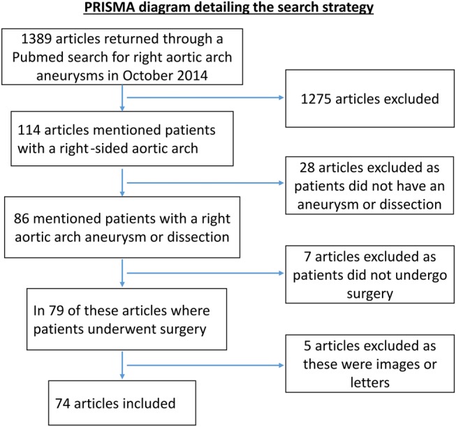
PRISMA diagram detailing the search strategy. PRISMA: Preferred Reporting Items for Systematic reviews and Meta-Analyses.
RESULTS
Reported studies and population characteristics
We identified 74 studies [4–6, 8–16, 18–79] that reported 99 patients undergoing surgical or endovascular intervention for a right aortic arch aneurysm or dissection. Most studies were case reports with some case series with the largest reporting 4 patients [8, 9]. Sixty-two patients were male, 21 were female and for 16 patients the gender was not reported. The median age at presentation was 61 years (range 21–85 years).
The commonest presenting symptoms were chest or back pain in 31% (31 patients), dysphagia in 20% (20 patients), respiratory symptoms such as cough, stridor or haemoptysis in 10% (10 patients). Eighteen patients (18%) were asymptomatic and the finding was incidental (Fig. 2). The combination of dysphagia and cough is as frequent as the ‘typical’ symptoms of aneurysms, therefore high level of suspicion in this subgroup.
Figure 2:

Presenting symptoms for all patients in whom this information was available.
Diagnosis of a right-sided aortic arch was most commonly made with a chest radiograph which was then confirmed with computed tomography (CT).
A total of 88 patients had an aberrant LSCA with only 11 patients having the mirror image variant. The commonest pathology was an aneurysm arising from KD which occurred in 54 patients. Twenty-eight patients had dissections, 19 of these were Type B and 9 were Type A (Fig. 3). Some of these Type B dissections originated from KD. Other pathologies were aneurysms arising from elsewhere in the thoracic aorta.
Figure 3:
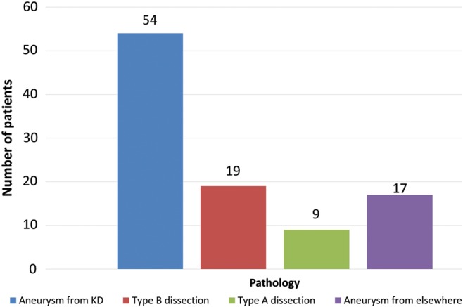
Different pathologies affecting patients with a right-sided aortic arch. KD: Kommerell's diverticulum.
Interventional approaches
Eighty-one patients had elective operations while 18 had emergency procedures. Sixty-seven patients underwent surgical treatment, 20 patients had a hybrid surgical and endovascular procedures and 12 had a totally endovascular procedure.
Of the hybrid procedures, 6 patients had a total arch replacement with an elephant trunk graft followed by a thoracic endovascular aortic repair (TEVAR) with an endograft inserted into the elephant trunk [10]. Five patients had a frozen elephant trunk and 1 patient had a modified elephant trunk. Eight patients had bypass procedures of the arch vessels followed by a TEVAR procedure [11, 12].
For the surgical group, the commonest way of accessing the aorta was via a median sternotomy or a right thoracotomy. Of the patients with information on their incision, 32 patients had a sternotomy, 4 of whom had mini sternotomies. Twenty-two patients had a right thoracotomy; this included 1 patient who had a double right thoracotomy through the fourth and seventh intercostal spaces. Additionally, 5 patients had a combined median sternotomy and right thoracotomy. Other incisions were a left thoracotomy (8 patients), left thoracotomy and sternotomy (2 patients), bilateral thoracotomy (2 patients) and left spiral thoraco-abdominal incision (1 patient). For patients undergoing hybrid procedure involving bypass of the arch vessels, this was achieved with J stick supraclavicular neck incisions.
In a group of 67 patients undergoing surgical treatment, 64 patients were put on cardiopulmonary bypass (CPB) or partial CPB. Circulatory arrest was needed in 37 of these patients.
Complications
The commonest complications reported were hoarse voice as a result of injury to the recurrent laryngeal nerve (6 patients, 6%) and respiratory complications [prolonged respiratory wean and chest infection (7 patients, 7%)]. There were 5 deaths, all of these except 1 were in patients undergoing emergency surgery and none in the endovascular repair group. The type of treatment and hospital survival based on the type of pathology is described in Table 1.
Table 1:
Type of treatment and hospital survival based on the pathology in patients with a right aortic arch
| Pathology | Number of patients | Treatment (number of patients/hospital death(s)) |
||
|---|---|---|---|---|
| Surgery | Endovascular | Hybrid | ||
| Type A dissection | 9 | 7/1 | 0/0 | 2/0 |
| Type B dissection | 19 | 13/1 | 6/0 | 0/0 |
| Aneurysm arising from KD | 54 | 38/2 | 3/0 | 13/0 |
| Aneurysm from elsewhere | 17 | 9/1 | 3/0 | 5/0 |
KD: Kommerell's diverticulum.
The first patient presented with a Type B dissection underwent emergency surgery with an interposition graft performed via a right thoracotomy. The LSCA was ligated. The patient never regained consciousness and died 3 days postoperatively [13].
The second patient presented acutely with haemoptysis and was found to have fistula formation between a proximal right aortic arch aneurysm and the right upper lobe of the lung. The patient underwent emergency surgery and a knitted polyester patch (Gelseal™) closure of the fistula was performed as well as an interposition graft for an additional aneurysm of the descending thoracic aorta (DTA). He subsequently developed a perigraft abscess that ruptured into the oesophagus that resulted in exsanguination [14].
The third patient died of a massive pulmonary embolism after undergoing elective LSCA to carotid transposition and interposition graft to treat an aneurysm arising from KD [4].
The fourth patient presented acutely and was found to have rupture of an aneurysm arising from KD. The patient underwent emergency LSCA to RSCA bypass followed by total arch replacement. The cause of death was reported as mediastinitis postoperatively [8].
The last patient presented acutely with a Type A dissection. Total arch replacement was performed; however, surgeons were unable to resect KD and so this was left. On postoperative day 30, the patient had massive haematemesis leading to death. Post-mortem examination revealed a fistula between KD and the oesophagus [15].
DISCUSSION
Our systematic review found 99 patients with right aortic arch aneurysm or dissection, highlighting the rarity of this condition. A higher proportion of patients were male and the median age of presentation was 61 years. Pain was the commonest presenting symptom and was either felt in the chest, epigastrium or back. Compression of the trachea, bronchi or the oesophagus leading to respiratory symptoms or dysphagia, respectively, was responsible for most of the other presentations. Eighty-nine percent of patients had an aberrant LSCA and only 11% had a mirror image variant. This differs from pathological studies of the right aortic arch where 85% of patients have the mirror image variant and 15% have an aberrant LSCA [2]. This difference is likely the result of KD predisposing to aneurysm formation as this area constitutes an area of weakness in the aortic wall that is a potential area for aneurysmal dilatation and dissections [16]. Additionally, the median age at presentation was 61 years which differs from patients with a left-sided aortic arch in whom the median age of presentation is about 65 years [17]. When there is a left-sided aortic arch, it is the ascending aorta that accounts for about 70% of the pathology [17]. These differences further highlight the significance of KD on causing aortic pathology in a right-sided arch.
CT was the commonest imaging modality for identifying right aortic arch pathology. This is due to the availability of CT scanning as well as the speed of scanning and high spatial resolution (see Fig. 4). Magnetic resonance imaging (MRI) has also been used for assessing aortic pathology in patients with right-sided aortic arches. It has the disadvantage of being less available than CT and the increased time taken to obtain images excludes its use in emergency settings. However, it has the utility in assessing the pulsatile nature of tracheal and oesophageal compression as well the advantage of not using ionizing radiation. A case report from almost 20 years ago in a patient with a Type B dissection in a right aortic arch found that differentiation between a dissection and an aortic aneurysm with mural thrombus may be difficult on CT compared with MRI; however, CT imaging had improved greatly during that time [18].
Figure 4:

CT images of a patient with a right-sided aortic arch with aberrant left subclavian artery and aneurysm of the descending aorta highlighting how the right-sided aortic arch passes posterior to the trachea and oesophagus. LCCA: left common carotid artery; ALSA: aberrant left subclavian artery.
The decision as to when to operate on a patient with an aneurysm arising from KD in patients with a right-sided aortic arch is not straightforward. Clearly, troublesome symptoms caused by the compression of mediastinal structures are an indication to intervene. Advising on surgical intervention based on size is hampered by the paucity of data. Balancing the risks of surgery against the risk of potential rupture if left untreated is difficult. Some researchers have advised to intervene when the diameter is greater than 3 cm while others recommended 5 cm as the point for intervention [4, 19]. However, rupture has been described in aneurysms measuring as little as 2 cm [4]. In the absence of significant comorbidities and with the advent of less invasive techniques, it may be that the size threshold for aneurysms of the right aortic arch is lower than the ones arising from the left.
The surgical or endovascular approach depends on the pathology and site of the aorta affected. We discuss the approaches used depending on the pathology.
Kommerell's aneurysm
Aneurysms arising from KD were the commonest pathology, occurring in 54% of patients (n= 54). The treatment varied depending upon the extent of the aneurysm.
For Kommerell's aneurysms that involved the arch and DTA repair could be performed as either a single procedure or a staged procedure. The frozen elephant trunk graft, which has been described previously, allows this to be performed as a single procedure (Fig. 5). Idrees et al. [20] reported doing this through a mini J sternotomy. The alternative is a two-stage procedure where the arch is replaced in the first instance with an elephant trunk graft followed by either a TEVAR endograft inserted into the elephant trunk [10] or surgical replacement of the DTA via a right thoracotomy [21].
Figure 5:
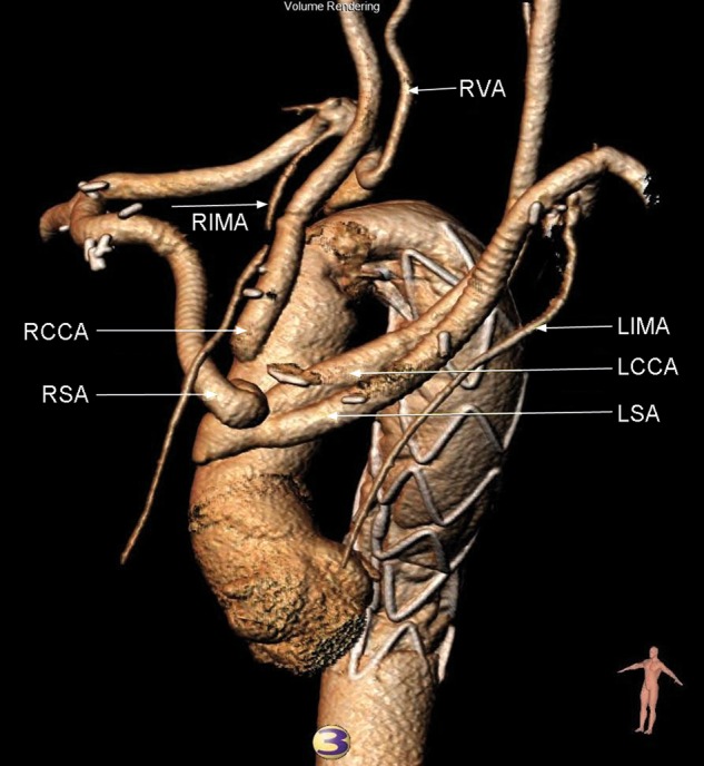
Frozen elephant trunk repair of a descending thoracic aortic aneurysm in a patient with a right-sided aortic arch aneurysm performed by our group. RCCA: right common carotid artery; RSA: right subclavian artery; LIMA: left internal mammary artery; LCCA: left common carotid artery; LSCA: left subclavian artery; RVA: right vertebral artery; RIMA: right internal mammary artery.
More localized aneurysmal dilatation of KD could be addressed by a number of surgical techniques. Options included transposition of the LSCA to the LCCA followed by endoaneurysmorrhaphy [4] or ligation of the proximal and distal ends of the aneurysm followed by bypass between the aorta and LSCA with a synthetic graft [22]. All of the described approaches persevered blood flow through the LSCA. Access was achieved via a median sternotomy, right or left thoracotomy. These procedures were performed either without bypass or with partial bypass from the left atrium to the femoral artery or to the DTA. Left heart bypass allows isolation of an aneurysm arising from KD as well as part of the aortic arch and DTA. An aortic cross is placed between either the RCCA or the RSCA and the aneurysm as well as a cross-clamp on the DTA distal to the aneurysm. Cinà et al. [4] used left heart bypass in 3 patients who had aneurysms arising from KD with or without arch involvement and required LSCA to LCCA transposition and endoaneurysmorrhaphy. If a cross-clamp can be placed between the aneurysm arising from KD and the aorta, then this could potentially be done without bypass; however, if there is more extensive involvement of the arch, then a period of circulatory arrest is required.
When the aneurysm affected the arch, it could be addressed either with a total arch replacement either by anastomosing all four branches separately to the graft [23], alternatively some surgeons transposed the LSCA to the LCCA or RSCA first and then performed total arch replacement [8]. All of these procedures were performed via a median sternotomy.
When aneurysmal dilatation was more localized to the distal arch ± the DTA, then this could be addressed with surgical, endovascular or hybrid procedures.
Surgical options were LSCA to LCCA transposition followed by the resection of Kommerell's aneurysm and the affected part of the aorta followed by an interposition graft [24]. If the DTA was also affected, then this was also replaced with a graft [24].
Endovascular options were TEVAR with the graft covering the LSCA [25] and endovascularly occluding the LSCA prior to TEVAR [26]. Alternatively to preserve the flow through the LSCA, the aortic graft could be deployed along with another LSCA covered stent directed parallel and extending more proximally to the main aortic graft (Chimney stent) [27].
Hybrid options depended on the extent that the aneurysm affected the aortic arch. Aneurysms more localized around the LSCA could be treated with LSCA to LCCA bypass with ligation of the proximal LSCA followed by TEVAR with the proximal land zone just distal to the RSCA [11]. If the aneurysm extended towards the origin of the RSCA, then RSCA to RCCA and LSCA to LCCA bypass with ligation of the proximal ends of both subclavian arteries (SCAs) followed by TEVAR with the proximal land just distal to the RCCA could be performed [12].
The options for managing a patient with an aneurysm arising from KD are shown in Fig. 6.
Figure 6:

Proposed surgical treatment algorithm for patients with aneurysm originating from KD of patients with right aortic arch based on our review of the literature. LCCA: left common carotid artery; DTA: descending thoracic aorta; TEVAR: thoracic endovascular aortic repair; LSCA: left subclavian artery; KD: Kommerell's diverticulum.
Type B dissection
Eighteen patients had a Type B dissection. Type B dissections could originate from distal or proximal to the LSCA. In some patients, KD was the area from which a tear occurred resulting in a Type B aortic dissection. Management can be surgical or endovascular. Surgical options if the dissection originates distally to the LSCA are with an interposition graft from the distal arch to the DTA [28]. If the dissection was proximal to the LSCA, then an interposition graft between aorta just distal to the origin of the RSCA and DTA can be placed. Either the LSCA can be ligated [13], or an interposition graft with a side branch anastomosed to the LSCA to preserve blood flow can be used [29]. These procedures were performed via a right thoracotomy using CPB via the femoral artery and vein.
Endovascular treatment of Type B dissection was performed in 6 patients and 3 of these patients had the mirror image variant. Three patients had TEVAR procedures with an aortic stent alone while the other 3 had an aortic stent followed by a Chimney stent. One patient had the chimney stent for the RSCA into the aortic stent, one patient had a Chimney stent deployed in the RCCA into the aortic stent with no stent for the RSCA [30] and the last patient had chimney stents in both the LSCA and RSCA [31].
Type A dissection
Nine patients had a Type A dissection, 8 acute and 1 chronic [32]. Surgical strategies were similar to a Type A dissection of a left-sided aortic arch. They included: aortic root replacement [33], interposition graft of the ascending aorta [34], total arch replacement [35], total arch replacement with elephant trunk and TEVAR into the elephant trunk [36] and frozen elephant trunk [32]. There was 1 death in this group that has been described above [15].
Descending thoracic aorta aneurysms
The aneurysms were managed surgically either using a left thoracotomy or a right thoracotomy and placing an interposition graft under circulatory arrest [24] or using left heart bypass [37] or endovascularly with a TEVAR stent deployed distally to the LSCA [38].
Specific surgical considerations in the right arch
Division of ligamentum arteriosum is required in all cases. The right aortic arch can be retro-oesophageal where the right aortic arch passes behind the trachea and oesophagus as opposed to the standard configuration where it is in front of them [2]. This requires careful mobilization of the retro-oesophageal portion, an area which is often less familiar for the general cardiac surgeon.
The means of accessing the right-sided aortic arch depends upon the pathology being addressed. A right thoracotomy gives better access to the right arch behind the oesophagus but hinders complex distal arch and descending repair (where a left thoracotomy is the preferred approach). Hybrid approaches allow for median sternotomy where central bypass techniques can also be safely utilized.
Other cardiac anomalies are commoner in patients with a right-sided aortic arch. Focused echocardiography and coronary angiography are required due to the association with septal defects and coronary anomalies (high take off of the right coronary artery which is very relevant in the case of Type A dissection) [2, 39].
Limitations
Given the rarity of aneurysms and dissections of the right aortic arch, the evidence is limited to case reports and small case series with short follow-up. The lack of available long-term data hinders to recommend what is the optimal approach for managing these pathologies.
CONCLUSIONS
Aneurysms and dissections of a right-sided aortic arch are rare. The anatomy of the right-sided aortic arch means that symptoms can be atypical for aneurysms as the predominant symptoms can be due to compression of the oesophagus or the airways. KD appears to be significant in causing aortic pathology in a right-sided arch and is the reason for a younger age of presentation in patients with a right aortic arch. Advances in endovascular treatment and hybrid surgical and endovascular management make more pathology amendable to these approaches and could reduce morbidity. Preoperative planning and careful delineation of anatomy and possible associated cardiac anomalies is essential.
Funding
This project was jointly funded by the National Institute for Health Research (NIHR) Biomedical Research Centre (based at Imperial College London and Imperial College Healthcare NHS Trust) and the NIHR Cardiovascular Biomedical Research Unit (based at Imperial College London and the Royal Brompton and Harefield NHS Foundation Trust).
Conflict of interest: none declared.
REFERENCES
- 1.Fioratti F, Aglietti F. A case of human right aorta. Anatom Rec 1763;45:365. [Google Scholar]
- 2.Knight L, Edwards JE. Right aortic arch. Types and associated cardiac anomalies. Circulation 1974;50:1047–51. [DOI] [PubMed] [Google Scholar]
- 3.Marino B, Digilio MC, Toscano A, Anaclerio S, Giannotti A, Feltri C et al. Anatomic patterns of conotruncal defects associated with deletion 22q11. Genet Med 2001;3:45–8. [DOI] [PubMed] [Google Scholar]
- 4.Cinà CS, Althani H, Pasenau J, Abouzahr L. Kommerell's diverticulum and right-sided aortic arch: a cohort study and review of the literature. J Vasc Surg 2004;39:131–9. [DOI] [PubMed] [Google Scholar]
- 5.Cinà CS, Arena GO, Bruin G, Clase CM. Kommerell's diverticulum and aneurysmal right-sided aortic arch: a case report and review of the literature. J Vasc Surg 2000;32:1208–14. [DOI] [PubMed] [Google Scholar]
- 6.Belov YV, Abugov SA, Komarov RN, Puretskiĭ MV, Stepanenko AB, Stogniĭ NY et al. Hybrid surgical management of a patient with an aneurysm of the arch and descending portion of the right-sided aorta combined with type B dissection and decompensated tracheal stenosis. Angiol Sosud Khir 2011;17:131–41. [PubMed] [Google Scholar]
- 7.Moher D, Liberati A, Tetzlaff J, Altman DG, PRISMA Group. Preferred reporting items for systematic reviews and meta-analyses: the PRISMA statement. BMJ 2009;339:b2535. [DOI] [PMC free article] [PubMed] [Google Scholar]
- 8.Hosoba S, Suzuki T, Asai T, Nota H, Kuroyanagi S, Kinoshita T et al. Surgical repair of Kommerell's diverticulum and an aberrant subclavian artery. Surg Today 2014;44:247–51. [DOI] [PubMed] [Google Scholar]
- 9.Tsukui H, Aomi S, Yamazaki K. Surgical strategy for Kommerell's diverticulum: total arch replacement. J Thorac Cardiovasc Surg 2014;148:1423–7. [DOI] [PubMed] [Google Scholar]
- 10.Kawajiri H, Shimizu H, Yoshitake A, Yozu R. Hybrid repair of a Kommerell diverticulum associated with a right aortic arch and a left descending aorta. J Vasc Surg 2012;56:1727–30. [DOI] [PubMed] [Google Scholar]
- 11.Naoum JJ, Parenti JL, LeMaire SA, Coselli JS. Endovascular repair of a right-sided descending thoracic aortic aneurysm with a right-sided aortic arch and aberrant left subclavian artery. Ann Thorac Surg 2008;85:1074–6. [DOI] [PubMed] [Google Scholar]
- 12.Frigatti P, Grego F, Deriu GP, Lepidi S. Hybrid endovascular treatment of aneurysm degeneration in a rare right-aortic arch anomaly with Kommerell diverticulum. J Vasc Surg 2009;50:903–6. [DOI] [PubMed] [Google Scholar]
- 13.Roan P, Parish S, Buja LM, Estrera A, Mills L, Atkins J et al. Dissecting aortic aneurysm involving a right-sided aortic arch. Am J Cardiol 1979;44:381–4. [DOI] [PubMed] [Google Scholar]
- 14.Matsuno O, Matsumoto T, Tsuda T. Aortic aneurysm involving a right-sided arch complicating aortobronchopulmonary and aortoesophageal fistula. Intern Med 2001;40:722–5. [DOI] [PubMed] [Google Scholar]
- 15.Agematsu K, Ueda T, Hoshino S, Nishiya Y. Rupture of Kommerell diverticulum after total arch replacement. Interact CardioVasc Thorac Surg 2010;11:800–2. [DOI] [PubMed] [Google Scholar]
- 16.Maxwell BG, Harrington KB, Beygui RE, Oakes DA. Congenital anomalies of the aortic arch in acute type A aortic dissection: implications for monitoring, perfusion strategy, and surgical repair. J Cardiothorac Vasc Anesth 2014;28:467–72. [DOI] [PubMed] [Google Scholar]
- 17.Davies RR, Goldstein LJ, Coady MA, Tittle SL, Rizzo JA, Kopf GS et al. Yearly rupture or dissection rates for thoracic aortic aneurysms: simple prediction based on size. Ann Thorac Surg 2002;73:17–27; discussion 27–8. [DOI] [PubMed] [Google Scholar]
- 18.Ko SF, Ng SH, Fu M, Lo PH, Cheng YF, Lee TY. Dissection of retroesophageal aortic diverticulum and descending aorta in a patient with right aortic arch: magnetic resonance demonstration. Cardiovasc Intervent Radiol 1996;19:438–41. [DOI] [PubMed] [Google Scholar]
- 19.Ota T, Okada K, Takanashi S, Yamamoto S, Okita Y. Surgical treatment for Kommerell's diverticulum. J Thorac Cardiovasc Surg 2006;131:574–8. [DOI] [PubMed] [Google Scholar]
- 20.Idrees J, Keshavamurthy S, Subramanian S, Clair DG, Svensson LG, Roselli EE. Hybrid repair of Kommerell diverticulum. J Thorac Cardiovasc Surg 2014;147:973–6. [DOI] [PubMed] [Google Scholar]
- 21.Smith R, Attaran S, Field M, Oo A. Management of a chronic Stanford type B dissection in a patient with a right-sided aortic arch. Interact CardioVasc Thorac Surg 2011;13:523–5. [DOI] [PubMed] [Google Scholar]
- 22.Muraoka M, Uchiyama Y, Yamaoka N, Yamauchi H, Hashiyada H, Nakamura A et al. An aberrant left subclavian artery aneurysm with right aortic arch: report of a case. Surg Today 1999;29:675–8. [DOI] [PubMed] [Google Scholar]
- 23.Kai M, Okabayashi H, Soga Y, Hanyu M, Nomoto T, Nakano J et al. Total arch replacement through a midsternotomy for a right-sided aortic arch aneurysm with an aberrant left subclavian artery. J Thorac Cardiovasc Surg 2006;132:1473–5. [DOI] [PubMed] [Google Scholar]
- 24.Robinson BL, Nadolny EM, Entrup MH, Svensson LG. Management of right-sided aortic arch aneurysms. Ann Thorac Surg 2001;72:1764–5. [DOI] [PubMed] [Google Scholar]
- 25.Midorikawa H, Kannno M, Ishikawa K, Takano T, Ono T, Morishima S. Endovascular repair of a Kommerell's Diverticulum and aneurysmal right-sided aortic arch: a case report. Ann Vasc Dis 2009;2:54–7. [DOI] [PMC free article] [PubMed] [Google Scholar]
- 26.Monaco M, Lillo S, La Marca Giordano A, Contaldo A, Schiavone V. Endovascular repair of a right-sided thoracic aortic aneurysm with Kommerell diverticulum and aberrant left subclavian artery. Ann Vasc Surg 2014;28:1323.e1–5. [DOI] [PubMed] [Google Scholar]
- 27.Silveira PG, Franklin RN, Cunha JR, Neves TT, Nascimento GG, Bortoluzzi CT. Total endovascular repair of aberrant left subclavian artery with Kommerell's diverticulum using a customized branched device. J Vasc Surg 2013;57:1123–5. [DOI] [PubMed] [Google Scholar]
- 28.Minato N, Rikitake K, Murayama J, Ohnishi H, Takarabe K. Surgery of the dissecting aneurysm involving a right aortic arch. J Cardiovasc Surg (Torino) 1999;40:121–5. [PubMed] [Google Scholar]
- 29.Kim SY, Lee YS, Bae KR, Lee JB, Lee S, Kwon OC. A case of type B dissecting aneurysm involving right sided aorta with Kommerell's diverticulum. Korean J Intern Med 2010;25:327–30. [DOI] [PMC free article] [PubMed] [Google Scholar]
- 30.Ma H, Yang H, Xu W, Zou J, Jiang J, Jiao Y et al. Endovascular repair with the chimney technique for Stanford type B aortic dissection involving right-sided arch with mirror image branching. J Endovasc Ther 2013;20:283–8. [DOI] [PubMed] [Google Scholar]
- 31.Zhang M, Yuan Y, Hu Y, Zhao Y, Liu H, Ma B et al. Endovascular repair with the chimney technique for stanford type B aortic dissection involving right-sided arch with aberrant left subclavian artery. Ann Vasc Surg 2014;28:1798.e7–1798.e10. [DOI] [PubMed] [Google Scholar]
- 32.Easo J, Hölzl P, Dikov V, Chavan A, Dapunt O. Frozen elephant trunk procedure in a dextropositioned arch and descending aorta. J Endovasc Ther 2010;17:751–4. [DOI] [PubMed] [Google Scholar]
- 33.Nomoto T, Ueda Y, Sugita T, Izumi C. A case of type A dissection involving right aortic arch. Eur J Cardiothorac Surg 1997;12:922–4. [DOI] [PubMed] [Google Scholar]
- 34.Moizumi Y, Komatsu T, Nagaya K, Sawamura Y, Sakurai M, Tabayashi K. Type A aortic dissection involving a right-sided aortic arch. J Cardiovasc Surg (Torino) 1999;40:117–9. [PubMed] [Google Scholar]
- 35.Hori D, Tanaka M, Yamaguchi A, Adachi H. Type A aortic dissection, right-sided aortic arch, and thoracic aortic aneurysm. Asian Cardiovasc Thorac Ann 2009;17:640–2. [DOI] [PubMed] [Google Scholar]
- 36.Sato S, Matsuda H, Fukuda T, Domae K, Iba Y, Tanaka H et al. Hybrid repair combined with open surgery and endografting for lesions in right aortic arch: report of three cases. Ann Vasc Dis 2012;5:61–4. [DOI] [PMC free article] [PubMed] [Google Scholar]
- 37.Oberwalder PJ, Bergmann P, Tillich M, Rigler B. Aneurysm of a right-sided aortic arch and right descending aorta: three-dimensional volume rendering of multislice computed tomographic aortography facilitates surgical planning and management. J Thorac Cardiovasc Surg 2005;129:953–4. [DOI] [PubMed] [Google Scholar]
- 38.Klonaris C, Avgerinos ED, Katsargyris A, Matthaiou A, Georgopoulos S, Psarros V et al. Endovascular repair of a right-sided descending thoracic aortic aneurysm associated with a right aortic arch and a left subclavian artery arising from a Kommerell's diverticulum. Cardiovasc Intervent Radiol 2009;32:758–61. [DOI] [PubMed] [Google Scholar]
- 39.Hu Y, Li Z, Chen J, Zhong Q. High takeoff of the right coronary artery associated with ventricular septal defect, right aortic arch, and bridging bronchus. J Card Surg 2014;29:829–31. [DOI] [PubMed] [Google Scholar]
- 40.Floten HS, Rose DM, Cunningham JN Jr. Surgical therapy of a dissecting aortic aneurysm involving a right-sided aortic arch. J Am Coll Cardiol 1984;4:1058–61. [DOI] [PubMed] [Google Scholar]
- 41.Ohteki H, Itoh T, Watanabe Y, Ueno T, Minato N, Natsuaki M. A dissecting aneurysm involving a right-sided arch, right descending aorta and mitral regurgitation. J Thorac Cardiovasc Surg 1987;35:235–7. [DOI] [PubMed] [Google Scholar]
- 42.Patiniotis TC, Mohajeri M, Hill DG. Right aortic arch with aberrant left subclavian artery: aneurysmal dilatation causing symptomatic compression of the right main bronchus in an adult. Aust N Z J Surg 1995;65:690–2. [DOI] [PubMed] [Google Scholar]
- 43.Masiello P, Mastrogiovanni G, Santoro G, Triumbari F, Naimoli G, Di Benedetto G. Type B aortic dissection involving an isolated right-sided aortic arch. Ann Thorac Surg 1996;62:887–8. [PubMed] [Google Scholar]
- 44.Imagawa H, Kadoba K, Taniguchi K, Sawa Y, Takahashi T, Fukushima N et al. Saccular aneurysm in the right-sided aortic arch: a successfully corrected case. J Vasc Surg 1997;25:941–4. [DOI] [PubMed] [Google Scholar]
- 45.Kuki S, Taniguchi K, Miyagawa S, Takano H. Dissecting thoracoabdominal aortic aneurysm associated with an isolated right-sided aortic arch. Eur J Cardiothorac Surg 2000;17:614–6. [DOI] [PubMed] [Google Scholar]
- 46.Tsukube T, Ataka K, Sakata M, Wakita N, Okita Y. Surgical treatment of an aneurysm in the right aortic arch with aberrant left subclavian artery. Ann Thorac Surg 2001;71:1710–1. [DOI] [PubMed] [Google Scholar]
- 47.Kaneda T, Lemura J, Zhang Z, Inoue T, Onoe M, Kitayama H et al. A case of Stanford type B aortic dissection involving a right-sided aortic arch with mirror-image branching and right-sided descending aorta. Thorac Cardiovasc Surg 2001;49:51–3. [DOI] [PubMed] [Google Scholar]
- 48.Juez M, Martínez León J, Haba J, Otero Coto E. Surgical treatment of aneurysm of right aortic arch with cervical aorta. Interact CardioVasc Thorac Surg 2006;5:18–9. [DOI] [PubMed] [Google Scholar]
- 49.Kiokawa K, Goh K, Akasaka N, Azuma N, Inaba M, Sasajima T. Total arch replacement for a distal aortic arch aneurysm with right aortic arch. Ann Thorac Surg 2007;83:e3–5. [DOI] [PubMed] [Google Scholar]
- 50.Munakata M, Itaya H, Fukui K, Ono Y. One stage repair for aortic regurgitation and Kommerell diverticulum with aneurysmal right aortic arch. J Thorac Cardiovasc Surg 2007;133:798–9. [DOI] [PubMed] [Google Scholar]
- 51.Follis F, Filippone G, Montalbano G, LoBianco E, Finazzo M, Follis M. Repair of postdissection descending thoracic aneurysm with right-sided aortic arch and aberrant left subclavian artery. J Thorac Cardiovasc Surg 2010;139:1086–7. [DOI] [PubMed] [Google Scholar]
- 52.Yang MH, Weng YG, Chang HH. A right-sided aortic arch with Kommerell's diverticulum of the aberrant left subclavian artery presenting with syncope. J Chin Med Assoc 2009;72:275–7. [DOI] [PubMed] [Google Scholar]
- 53.Karthekeyan BR, Sundar S, Rao S, Vakamudi M. Management of a patient with Kommerrell's aneurysm causing tracheal and esophageal compression. Indian J Anaesth 2009;53:358–61. [PMC free article] [PubMed] [Google Scholar]
- 54.Obitsu Y, Koizumi N, Iwahashi T, Saiki N, Shigematsu H. Surgical repair for aortic dissection accompanying a right-sided aortic arch. J Cardiothorac Surg 2010;5:35. [DOI] [PMC free article] [PubMed] [Google Scholar]
- 55.Shakil O, Chaudhry M, Tsai L, Mahmood B, Gerstle JR, Mahmood F et al. Endovascular repair of a right-sided aortic arch aneurysm and tracheal injury. J Bronchol Interv Pulmonol 2014;21:65–7. [DOI] [PubMed] [Google Scholar]
- 56.Thors A, Haurani MJ, Nelson KC, Crestanello JA. Aortic arch replacement through a left thoracotomy for right-sided aortic arch aneurysm with complete vascularring. Ann Thorac Surg 2014;97:317–9. [DOI] [PubMed] [Google Scholar]
- 57.Mavroudis CD, Copelan A, Sokhandon F, Altshuler J. Hybrid repair of a ruptured right-sided aortic arch with an aberrant left subclavian artery arising from a diverticulum of Kommerell: a case report. World J Pediatr Congenit Heart Surg 2014;5:623–6. [DOI] [PubMed] [Google Scholar]
- 58.Onohara T, Nakamura Y, Kishimoto Y, Harada S, Fujiwara Y, Saiki M et al. Two cases of thoracic aortic aneurysm with right aortic arch: comparison of two operative strategies for hybrid thoracic endovascular repair. Ann Vasc Dis 2014;7:343–6. [DOI] [PMC free article] [PubMed] [Google Scholar]
- 59.Patangi SO, Singh RK, Pauli H. Right-sided aortic arch with Kommerell's aneurysm. Ann Card Anaesth 2014;17:311–3. [DOI] [PubMed] [Google Scholar]
- 60.Tanaka K, Yoshitaka H, Chikazawa G, Sakaguchi T, Totsugawa T, Tamura K. Hybrid repair of right aortic arch aneurysm with a Kommerell's diverticulum. Asian Cardiovasc Thorac Ann 2013;22:725–7. [DOI] [PubMed] [Google Scholar]
- 61.Ishikawa N, Oi M, Maruta K, Iizuka H, Kawaura H. Surgical treatment for right aortic arch with Kommerell's diverticulum. Asian Cardiovasc Thorac Ann 2013;21:724–6. [DOI] [PubMed] [Google Scholar]
- 62.Nonaka M, An K, Nakatsuka D, Okada T, Sekine Y, Iwakura A et al. Hybrid procedure for a Kommerell's diverticulum in a right-sided aortic arch. Ann Thorac Cardiovasc Surg 2014;20 Suppl:821–4. [DOI] [PubMed] [Google Scholar]
- 63.Zhou W. Endovascular repair of a type B aortic dissection with a right-sided aortic arch: case report. J Cardiothorac Surg 2013;8:18. [DOI] [PMC free article] [PubMed] [Google Scholar]
- 64.Motoki M, Hattori K, Kato Y, Takahashi Y, Kotani S, Nishimura S et al. Endovascular repair of ruptured aberrant left subclavian artery with right aortic arch. Ann Thorac Surg 2013;95:699–701. [DOI] [PubMed] [Google Scholar]
- 65.Croccia MG, Levantino M, Cioni R, Bortolotti U. Endovascular stenting for type B dissection involving a right-sided aortic arch. Interact CardioVasc Thorac Surg 2012;15:304–6. [DOI] [PMC free article] [PubMed] [Google Scholar]
- 66.Panduranga P, Al-Delamie T, Ratnam L, Al-Mukhaini M, Zachariah S. Repair of Kommerell's diverticulum with aberrant left subclavian artery in an elderly patient with right aortic arch and dysphagia lusoria. J Card Surg 2011;26:637–40. [DOI] [PubMed] [Google Scholar]
- 67.Spina A, Gatti G, Belgrano M, Zingone B. Clamshell approach and partial cardiopulmonary bypass to repair a right aortic arch aneurysm. J Cardiovasc Med (Hagerstown) 2009;10:859–60. [DOI] [PubMed] [Google Scholar]
- 68.Murzi M, Mariani M, Karimov JH, Gilmanov D, Berti S, Glauber M. Hybrid repair of a Kommerell's diverticulum aneurysm. J Card Surg 2010;25:67–9. [DOI] [PubMed] [Google Scholar]
- 69.Dainese L, Spirito R, Barili F, Fusari M, Trabattoni P, Sommaruga S et al. Right aortic arch related to Kommerell diverticulum and internal carotid artery agenesis. Circ Cardiovasc Imaging 2009;2:e6–7. [DOI] [PubMed] [Google Scholar]
- 70.Tjang YS, Aramendi JI, Crespo A, Hamzeh G, Voces R, Rodríguez MA. Right cervical aortic arch with aberrant left subclavian artery. Asian Cardiovasc Thorac Ann 2008;16:e37–9. [DOI] [PubMed] [Google Scholar]
- 71.Chiesa R, Melissano G, Bertoglio L, Calliari F. Hybrid repair of an aortic arch aneurysm with complex anatomy: right aortic arch and anomalous origin of supra-aortic vessels. J Vasc Surg 2007;46:128–30. [DOI] [PubMed] [Google Scholar]
- 72.Nassar MI, de la Llana R, Diaz-Romero F, Garrido P, Martinez-Sanz R. Multiple overlapped conical endoprostheses in a patient with aneurysmatic right aortic arch and aorticcoarctation. Ann Thorac Surg 2007;83:663–4. [DOI] [PubMed] [Google Scholar]
- 73.Agostini M, Priotto R, Feola M, Losardo L, Grosso M, Grossi C. Surgical treatment of an aneurysm originating from a Kommerell's diverticulum in the right-sided aortic arch with retroesophageal component. Ital Heart J 2005;6:977–80. [PubMed] [Google Scholar]
- 74.Slisatkorn W, Laksanabunsong P, Thongcharoen P. Ruptured right aortic arch aneurysm. Asian Cardiovasc Thorac Ann 2004;12:360–2. [DOI] [PubMed] [Google Scholar]
- 75.Miyamoro S, Hadama T, Anai H, Sako H, Shigemitsu O, Iwata E et al. Successful open stent grafting of a right aortic arch and a descending aortic aneurysm originating from a Kommerell's diverticulum: report of a case. Surg Today 2002;32:359–61. [DOI] [PubMed] [Google Scholar]
- 76.Okada K, Sueda T, Orihashi K, Watari M, Naito A. Endovascular stent-graft repair for thoracic aortic aneurysm associated with right-sided aortic arch. J Thorac Cardiovasc Surg 2001;122:185–6. [DOI] [PubMed] [Google Scholar]
- 77.Tominaga R, Nishida T, Morita S, Masuda M, Yasui H. Atherosclerotic aneurysm in the circumflex retroesophageal right aortic arch. Ann Thorac Surg 2001;71:2030–2. [DOI] [PubMed] [Google Scholar]
- 78.Senthil V, Treasure T. Type B dissection involving a right-sided aortic arch. Eur J Cardiothorac Surg 1996;10:477–9. [DOI] [PubMed] [Google Scholar]
- 79.Caus T, Gaubert JY, Monties JR, Moulin G, Mouly A, Cornen A et al. Right-sided aortic arch: surgical treatment of an aneurysm arising from a Kommerell's diverticulum and extending to the descending thoracic aorta with an aberrant left subclavian artery. Cardiovasc Surg 1994;2:110–3. [PubMed] [Google Scholar]